271 Copyright © 2014 The Korean Society of Cardiology
Korean Circulation Journal
Introduction
In a differential diagnosis of a wide complex tachycardia (WCT), prior to which a 12-lead surface electrocardiogram (ECG) was taken, a comparison of QRS morphology between the sinus rhythm and the WCT is helpful. An identical QRS morphology between sinus rhythm and WCT strongly suggests supraventricular tachycardia (SVT) whereas a contralateral bundle branch block (BBB) morphol- ogy during tachycardia with a preexisting BBB strongly suggests ventricular tachycardia (VT).
1)Contrary to this rule, we present a case of SVT with left BBB (LBBB) aberration but right BBB (RBBB) during sinus rhythm.
Case
A 45-year-old woman with a 30-year history of palpitations pre-
Case Report
http://dx.doi.org/10.4070/kcj.2014.44.4.271 Print ISSN 1738-5520 • On-line ISSN 1738-5555
Supraventricular Tachycardia and Sinus Rhythm with Contralateral Bundle Branch Block Patterns
Seongwook Han, MD, John M Miller, MD, and Mithilesh Kumar Das, MD
Krannert Institute of Cardiology, Indiana University School of Medicine, Indianapolis, IN, USA
A contralateral bundle branch block (BBB) aberration during tachycardia with a preexisting BBB strongly suggests the presence of ven- tricular tachycardia. We report on a middle-aged, female patient presented with wide QRS tachycardia. The patient had orthodromic atrioventricular tachycardia with a left BBB aberration in the presence of a preexisting right BBB due to an abnormal His-Purkinje system.
We learned that the contralateral BBB aberration with supraventricular tachycardia could be seen when the His-Purkinje system was ab- normal. (Korean Circ J 2014;44(4):271-273)
KEY WORDS: Bundle-branch block; Tachycardia, supraventricular; Bundle of His; Purkinje fibers.
Received: June 18, 2013
Revision Received: September 15, 2013 Accepted: November 18, 2013
Correspondence: Mithilesh Kumar Das, MD, Krannert Institute of Cardiol- ogy, Indiana University School of Medicine, 1800 North Capitol Avenue, Indianapolis, IN 46202, USA
Tel: 1-317-962-0101, Fax: 1-317-962-0100 E-mail: [email protected]
• The authors have no financial conflicts of interest.
This is an Open Access article distributed under the terms of the Creative Commons Attribution Non-Commercial License (http://creativecommons.
org/licenses/by-nc/3.0) which permits unrestricted non-commercial use, distribution, and reproduction in any medium, provided the original work is properly cited.
sented herself for an electrophysiologic (EP) study and a catheter ablation. Her present arrhythmia was a LBBB morphology tachycar- dia (LBBB-T) at 200 beats/min which was converted into a sinus rhythm with intravenous adenosine. During the EP study, multipolar catheters were placed in the high right atrium (HRA), His-bundle region (His), right ventricle (RV), and coronary sinus (CS).
At the beginning of the EP study, the surface ECG did not show any BBB or intraventricular conduction delays (Fig. 1A), and the baseline conduction intervals were within normal limits. The AH in- terval was 72 msec, and the HV interval was 54 msec. However, an RBBB occurred due to mechanical trauma during His-bundle cath- eter placement (Fig. 1B). The baseline EP study showed ventriculo- atrial (VA) conduction was present through both the atrioventricu- lar (AV) node and the concealed accessory pathway (AP) in the left posterior, which was blocked by 250 msec pacing with an AV Wenckebach cycle length of 310 msec. The clinical LBBB-T (cycle length: 330–350 msec) was easily induced by catheter-manipula- tion and spontaneous premature atrial complexes (Fig. 1C). LBBB-T has several possible mechanisms including orthodromic atrioven- tricular reentrant tachycardia (ORT) with an LBBB aberration using a concealed AP, antidromic atrioventricular reentrant tachycardia (ART) using various types of right-sided APs anterogradely, RV VT, and bundle branch reentry VT.
Fig. 2 shows that the LBBB-T terminated spontaneously and was followed by a sinus beat with a RBBB.
The LBBB-T terminated without any following atrial activation;
therefore, VT with 1:1 retrograde conduction was ruled out. The His-
bundle activation during the tachycardia preceded the beginning
272 SVT with Contralateral BBB Aberration
http://dx.doi.org/10.4070/kcj.2014.44.4.271 www.e-kcj.org
of the QRS complex, which ruled out an ART. The site of the earliest atrial activation during the LBBB-T was in the mid-CS rather than in the His-bundle. Delivery of premature ventricular stimulus dur- ing the His-bundle refractory terminated tachycardia without atri- al activation. This confirmed that the tachycardia was an ORT us- ing a concealed left posterior AP. Ablation was successful and the tachycardia could no longer be induced. After ablation, VA con- duction was present through the AV node only, but a VA conduc- tion block occurred at the VH level with a Wenckebach pattern (Fig. 3),
which suggested a conduction abnormality in the His-Purkinje system or between the His-Purkinje system and the ventricle.
Discussion
Bundle branch block aberrancy during SVT in the presence of pre- existent contralateral BBB is extremely rare, even in the presence of a His-Purkinje system dysfunction. A functional or anatomical block at one or more levels in the conduction system can cause LBBB.
2)It can occur at the His bundle level (dedicated fibers to LB) before its bifurcation, the left bundle branch level, the left fascicle level (due to variation in the anatomy of fascicles
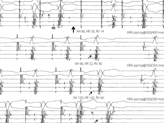
3)), or diffuse disease of the very distal ramifications of the left bundle. Fig. 4 shows conduction patterns during programmed and burst stimulation from the HRA.
Burst pacing at 280 msec revealed a conduction delay between the His and BB resulting in an infra/intra Hisian block (Wenckebach type). The interval following a non-captured beat allowed recovery of the left bundle, and this resulted in the re-appearance of the RBBB QRS morphology similar to that of baseline conduction. Early- coupled atrial extrastimuli caused a progressive delay within the His bundle or between the His and BB, and the RB potential to the QRS interval became longer and fixed during the LBBB morphology. The lower tracing in Fig. 4 showed that the LBBB did not recover from previous stimulation, but the interval from His to RB became longer by a premature stimulation without a change in LBBB morphology.
This implies that a certain amount of conduction delay between the His and bundle branch may be the cause of manifestation of LBBB in this case.
We propose that conduction delay between His and bundle
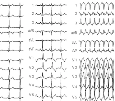
1 2 3 aVR aVL aVF V 1 V 2 V 3 V 4 V 5 V 6
1 2 3 aVR aVL aVF V 1 V 2 V 3 V 4 V 5 V 6
1 2 3 aVR aVL aVF V 1 V 2 V 3 V 4 V 5 V 6
A B C
Fig. 1. 12-lead surface electrocardiograms. (A) shows the baseline normal sinus rhythm. (B) shows sinus rhythm with right bundle branch block after mechanical trauma. (C) shows a left bundle branch block morphology tachycardia with a rate of 200 beats/min.
12 3 V 1V 6 HRA His-p His-d CS-p
CS-d RV
Fig. 2. The surface electrocardiogram and the intracardiac electrograms during the tachycardia. Tachycardia terminated spontaneously without atrial activation. The earliest retrograde atrial (A) activation site was in the mid-CS (arrow). The His (H) activation followed the atrial activation and occurred before the beginning of the QRS. p: proximal, d: distal, CS: coro- nary sinus, HRA: high right atrium, RV: right ventricle.
12 3 V 1V 6 HRA His-p His-d CS-p
CS-d RV
Fig. 3. Right ventricular burst pacing with a cycle length of 420 msec after AP ablation. His potential was obtained from left ventricle. During RV pac- ing, there was progressive prolongation of VH interval (arrows) that ended up with VA conduction block (Wenckebach VA block at below the His). p:
proximal, d: distal, HRA: high right atrium, CS: coronary sinus, AP: accessory
pathway, RV: right ventricle, VH: ventriculo-Hisian, VA: ventriculo-atrial.
273 Seongwook Han, et al.
http://dx.doi.org/10.4070/kcj.2014.44.4.271 www.e-kcj.org
branch may be the possible mechanism of the LBBB aberration with SVT during preexisting RBBB.
References
1. Approach to wide QRS tachycardias. In: Issa Z, Miller J, Zipes D. Clini- cal arrhythmology and electrophysiology. 1st ed. Philadelphia: WB
Saunders;2009. p.398.
2. de P.dua F, Pereirinha A, Marques N, Lopes MG, Macfarlane PW. Chap- ter 14. Conduction defect. In: Macfarlane PW, Oosterom A, Pahlm O, Kligfield P, Janse M, Camm J, editors. Comprehensive Electrocardiolo- gy. 2nd ed. London: Springer;2011. p.554.
3. Demoulin JC, Kulbertus HE. Histopathological examination of con- cept of left hemiblock. Br Heart J 1972;34:807-14.
12 V 13 V 6 HRA His-p His-d
V 1V 6 HRA His-p His-d
V 1 V 6 HRA His-p His-d
V 1V 6 His-pHRA
His-d
HRA pacing@280 msec
HRA pacing@500/400 msec
HRA pacing@500/350 msec
HRA pacing@500/280 msec AH 126, HR 142, RV 40
AH 98, HR 52, RV 40 AH 86, HR 38, RV 14