급성 심근경색과 유사한 발현양을 보이는 새로운 형태의 심근병증 2예
5
0
0
전체 글
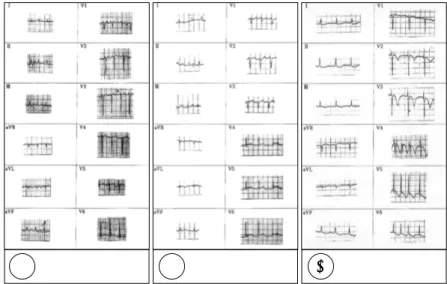
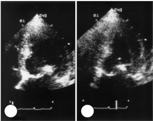
(2) 에도 불구하고 심근효소치가 미약하게 상승되는 경우가. IU/L), Troponin-T 0.106 ng/mL(정상치 0~0.1 ng/. 많고, 심전도의 변화가 미미한 경우도 많이 있으며 또한. mL), CK-MB 5.39 ng/mL(정상치 0~5.0 ng/mL)으. 흉통이나 심전도변화가 있을 때 시행한 관상동맥조영술. 로 경미한 상승을 보였다. Dopamine정주를 한 후 혈압. 상 정상소견을 보이고, 벽운동의 감소가 한 관상동맥혈. 은 정상화되었고, 다음날 시행한 심초음파에서는 좌심실. 관의 혈류공급부위에 국한되지 않고 좌심실기저부위를. 의 기저부위는 활발한 벽운동을 보인 반면에, 심첨부위. 제외한 심첨부위쪽의 모든 부위에서 감소되어 있다는 점. 는 관상동맥분포영역과 관계없이 모든 부위에서 무운동. 들이 급성심근경색과 차이를 보이는 점이다. 이와 같은. 을 보였으며(심초음파는 흉통 발생 16시간후에 시행되. 2)3). 형태의 심첨부 벽운동이상은 다혈관 관상동맥연축후,. 었고, 심초음파시행시 환자상태는 안정되어 혈압은 100/. 지주막하출혈후,4)5) 혹은 갈색세포종의 발작성위기시에. 70 mmHg, 맥박은 심방세동으로 불규칙하였으나 평균. 6)7). 도. 드물게 관찰되고 있다. 저자들은 다양한 스트레스. 분당 93회이었음), 좌심실구혈율은 29%이었다(Fig. 2).. 후 발생한 새로운 형태의 심근병증 환자 2예를 경험하였 기에 문헌고찰과 함께 보고하는 바이다.. 증. 례. 증 례 1: 환 자:이○례, 여자 77세. 임상경과:환자는 급성신우신염에 의한 패혈증과 파 종성 혈관내응고증후군으로 본원에 입원하였다. 3병일 째 환자는 저녁식사후 안정을 취하고 있던 중 갑작스런 흉통을 호소하였고 당시 혈압은 80/40 mmHg이었으며 맥박은 분당 130회로 갑작스런 상승을 보였다. 심전도 에서는 내원시의 심전도와 비교하여 V5와 V6유도에서 ST분절의 상승이 관찰되었다(Fig. 1). 흉통 호소시 시 행한 최고 심근효소치는 CPK 52 IU/L(정상치 26~200. A. B. A. B. Fig. 2. Apical four chamber view at the acute stage in case 1:diastole (A) and systole (B). The echocardiography revealed a balloon-like asynergy of the apical region with hypercontraction of the basal segment of the left ventricle.. C. Fig. 1. Electrocardiogram in case 1. The electrocardiogram during chest pain (B) demonstrated ST-segment elevation in leads V5 to V6 compared with the electrocardiogram on the admission (A). The electrocardiogram on the next day showed deep T wave inversion on leads V2 to V6 (C).. 609.
(3) 이때의 심전도에서는 ST 분절의 상승이 소실되고 전흉. 임상경과:환자는 만기임신으로 개인의원에서 제왕절. 부유도에서 T파 역위소견이 관찰되었다(Fig. 1). 7일후. 개술을 시행받은후 자궁이완증이 발생하였으나 자궁수. 시행한 심초음파에서 좌심실의 운동이상소견은 완전히. 축제에 반응하지 않아 본원으로 전원되어 자궁적출술을. 소실되어 정상 벽운동을 보였으며 좌심실구혈율은 55%. 시행받았다. 수술 8시간후에 환자는 흉부 불쾌감을 호. 이었다(Fig. 3). 25병일에 환자는 퇴원하였고 15일후 외. 소했으며 정신상태가 혼미해졌다. 이때 혈압은 70/40. 래에서 시행한 운동부하 핵의학검사(thallium 201)에서. mmHg로 낮아지고 맥박은 분당 133회였다. 당시 시행. 관류결손은 관찰되지 않았다.. 한 심전도에서는 유도 I, II, aVL, 그리고 aVF에서 T파 역위가 관찰되었고(Fig. 4), 최고심근효소치는 CPK 290. 증 례 2:. IU/L, troponin-T 0.5 ng/mL, CK-MB 5.82 ng/mL 로 경미한 상승을 보였다. 심초음파는 환자가 쇼크상태. 환 자:정○경, 여자 37세.. 일 때(혈압은 70/40 mmHg, 맥박은 분당 133회) 시행 되었고 안정을 찾은 이후인 3일후에도(혈압은 120/80 mmHg, 맥박은 분당 93회) 다시 시행되였는데, 두 차례 검사에서 모두 좌심실의 기저 부위는 활발한 벽운동을 보이는데 반해 심첨부위는 무운동상태를 보였으며 좌심 실구혈율은 34%이었다(Fig. 5). 자궁적출술 시행 후 30 일에 외래에서 시행한 심초음파에서는 정상적인 좌심실 벽운동을 보였으며 좌심실구혈율은 59%이었고(Fig. 6) 심전도에서도 정상소견을 보였다. 운동부하 핵의학검사 (thallium 201)에서도 관류결손은 관찰되지 않았다.. A. B. Fig. 3. Seven-day-later apical four chamber view in case 1:diastole (A) and systole (B). The echocardiography demonstrated complete recovery of left ventricular apical wall motion abnormalities.. A. B. 고. 찰. 본 연구에서는 다양한 스트레스와 관련되어 발생하며,. C. Fig. 4. Electrocardiogram in case 2. The electrocardiogram during chest pain showed T wave inversion in leads I, II, aVL and aVF, and poor R wave progression in leads V1 to V4 (A). The electrocardiogram on the next day revealed deep T wave inversion in leads I, aVL, and V2 to V6 (B). One month later, the electrocardiogram returned to normal without ST-T changes (C).. 610. Korean Circulation J 2002; 32(7):608-612.
(4) A. B. A. B. Fig. 6. One-month-later apical two chamber view in case 2:diastole (A) and systole (B). The echocardiography demonstrated complete recovery of left ventricular apical wall motion abnormalities.. 정 유발요인은 없었고, 환자에게 갑작스럽게 스트레스 로 작용했을 것으로 짐작되는 전신질환의 악화 혹은 예 측치 못한 수술등이 새로운 심근병증에 선행하였다. 본 질환의 정확한 발생기전은 아직 밝혀져 있지 않으. C. D. 나 추정되는 기전으로는 다혈관 관상동맥 연축,3)7)8) 미 세관상동맥연축,9) 그리고 증강된 교감신경 활성도등이 있다. 갈색세포종의 내분비적 위기시 발생한 심근병증이 이 새로운 질환과 유사하고, 이 질환을 가진 환자들에서 시행한 심장조직검사에서 카테콜라민에 의한 심근병증과 유사한 심근손상을 보였으며,10) 대부분의 환자에서 심한 스트레스후 발생한다는 점들을 종합해 보면 일시적인 카 테콜라민의 증가가 질환발생의 중요한 원인이 아닌가 보 여진다.. E. F. 활발한 벽운동을 보이는 좌심실 기저부위에 반해 심 첨부위가 왜 무운동을 보이는가도 이 질환의 발생원인을. Fig. 5. Apical two chamber view:diastole (A) and systole (B), parasternal short axis at basal level:diastole (C) and systole (D), and parasternal short axis at the papillary muscle level:diastole (E) and systole (F), 3 days after chest discomfort in case 2. The echocardiography showed hypercontraction of the basal segments of the left ventricle with akinesia and balloon-like dilation of the apical region.. 찾는데 중요한 점으로 생각된다. Tsuchihashi 등1)은 좌 심실 심첨부가 벽이 기저부위에 비해 얇고 과도한 팽창 후에는 쉽게 탄력성을 잃어 좌심실의 전체적인 운동저하 후 심첨부의 운동 이상이 지연되어 회복되기 때문이 아 닐까 추정했다. 본 저자들은 이외에도 좌심실의 심근에 서 베타 아드레날린 수용체 분포의 국소적인 차이가 중. 급성심근경색과 유사한 발현양상을 보이면서, 일시적으. 요한 요인이 아닐가 제안한다. 아직 사람에서는 증명되. 로 좌심실 심첨부위의 무운동을 보이는 새로운 형태의. 지 않았지만 고양이의 좌심실에서는 베타 아드레날린 수. 심근병증 2예를 보고하였다. Tsuchihashi 등1)의 보고. 용체의 밀도가 심첨부위에서 가장 높은 것으로 보고하. 를 살펴 보면 이 새로운 심근병증의 유발요인으로 어떤. 였다.11) 또한 이 질환이 고령의 여성에서 잘 발생한다는. 특정한 질환이 있는 것이 아니라 환자에게 상당한 정도. 점도 연령과 성별에 따른 수용체밀도의 차이로 심첨부위. 의 스트레스를 줄 수 있는 요인이면, 그것이 질환, 시술,. 의 무운동이 잘 발생하는 것이 아닐까 추정된다. 향후 더. 혹은 상황이나 정서적 문제등에 관계없이 유발요인으로. 많은 연구가 필요할 것으로 사료된다.. 작용하는 것으로 생각된다. 본 증례들에서도 공통된 특 611.
(5) 요. 약 4). 심리적 혹은 외인적 스트레스후 급성심근경색과 유사 한 흉통과 심전도 변화가 발생하고 이와 더불어 가역적 인 좌심실 심첨부위의 팽대와 심근운동 저하가 관찰되는. 5). 새로운 형태의 심근병증이 최근 보고되었다. 광범위한 벽운동 저하에도 불구하고 심근효소치의 상승과 심전도. 6). 의 변화가 미미한 경우가 많이 있으며 급성기에 시행한 관상동맥조영술상 정상소견을 보이는 점등이 급성심근. 7). 경색과 차이를 보이는 점들이다. 저자들은 다양한 스트 레스후 발생한 새로운 형태의 심근병증 환자 2예를 경험. 8). 하였기에 보고한다.. 중심 단어:스트레스;급성심근경색;심근병증.. 9). REFERENCES 1) Tsuchihashi K, Ueshima K, Uchida T, Oh-mura N, Kim-. ura K, Owa M, Yoshiyama M, Miyazaki S, Haze K, Ogawa H, Honda T, Hase M, Kai R, Morii I. Transient left ventricular apical ballooning without coronary artery stenosis: a novel heart syndrome mimicking acute myocardial infarction. J Am Coll Cardiol 2001;38:11-8. 2) Satoh H, Tateishi H, Uchida T. Takotsubo-type cardiomyopathy due to multivessel spasm. In: Kodama K, Haze K, Hon M, editor. Clinical aspect of myocardial injury: from ischemia to heart failure. Tokyo: Kagakuhyouronsya;1990. p.56-64. 3) Dote K, Sato H, Tateishi H, Uchida T, Ishihara M. Myo-. 612. 10). 11). cardial stunning due to simultaneous multivessel spasm: a review of five cases. J Cardiol 1991;21:203-14. Kono T, Morita H, Kuroiwa T, Onaka H, Takatsuka H, Fujiwara A. Left ventricular wall motion abnormalities in patients with subarachnoid hemorrhage: neurogenic stunned myocardium. J Am Coll Cardiol 1994;24:636-40. Ohtsuka T, Hamada M, Kodama K, Sasaki O, Suzuki M, Hara Y, Shigematsu S, Hiwada K. Neurogenic stunned myocardium. Circulation 2000;101:2122-4. Yamanaka O, Yasumasa F, Nakamura T, Ohno A, Endo Y, Yoshimi K, Miura K, Yamaguchi H. Myocardial stunning-like phenomenon during a crisis of pheochromocytoma. Jpn Circ J 1994;58:737-42. Shaw TRD, Bafferty P, Tait GW. Transient shock and myocardial impairment caused by pheochromocytoma crisis. Br Heart J 1987;57:194-8. Fournier C, Boujon B, Herbert JL, Zamani K, Grimon G, Blondeau M. Stunned myocardium following coronary spasm. Am Heart J 1991;121:593-5. Ito K, Sugihara H, Kawasaki T, Yuba T, Doue T, Tanabe T, Adachi Y, Katoh S, Azuma A, Nakagawa M. Assessment of ampulla (Takotsubo) cardiomyopathy with coronary angiography, two-dimensional echocardiography and 99mTc-tetrofosmin myocardial single photon emission computed tomography. Ann Nucl Med 2001;15:351-5. Kawa S, Suzuki H, Yamaguchi H, Tanaka K, Sawada H, Aizawa T, Watanabe M, Tamura T, Umawatari K, Kawada M, Nakamura T, Yamanaka O, Okada R, Kawada M. Ampulla cardiomyopathy (Takotsubo cardiomyopathy): reversible left ventricular dysfunction with ST segment elevation. Jpn Circ J 2000;64:156-9. Lathers CM, Levin RM, Spivey WH. Regional distribution of myocardial beta-adrenoceptors in the cat. Eur J Pharmacol 1986;130:111-7.. Korean Circulation J 2002; 32(7):608-612.
(6)
수치

관련 문서
This study examined the changes of kinetic expression factors of movement through art historical approach and as a result of analyzing internal and external
스파이럴 볼트는 현재 개발되고 있는 새로운 형태의 지보재로서 판상의 강판에 비틀림 힘을 작용시켜 만든 나선형 형태의 지보재다(Hi rataetal. ,2003,2005).그 라운드
STEAM은 실생활 문제 해결을 위해 여러 과목 요소를 융합시켜서 기존 교육 방식으로 진행되던 학교 현장에서 적용하기가 쉽지 않습니다.. 시대적인 필요성과
Efficacy of syringe irrigation, RinsEndo and passive ultrasonic irrigation in removing debris from irregularities in root canals with different apical
As the number of clinical cases requiring supracrestal apical-coronal positioning is increasing, we examined the utility of FEA in such cases. In this study,
In these 3 cases common factors were searched, 1) upstair jet stream which passaged from Sandung peninsula to Yodong peninsula helped the low stair conversion and an
Absolute and Relative Velocity in Plane Motion Instantaneous Center of Rotation in Plane Motion Absolute and Relative Acceleration in Plane Motion... Analysis of Plane
Pilot Confirm runway 09 left for landing. ATC gives you further instructions to turn right heading 310. On the radar display, there was an area to avoid which requires a
