Original Article
Comparison of Outcomes of Unilateral Lateral Rectus Recession for Exotropia between First and Second Operations
Kyoungsook Lee
1, Kyeong Seob Shin
2, Yongjune Kim
3, Mi Young Choi
11
Department of Ophthalmology, Chungbuk National University College of Medicine, Cheongju, Korea
2
Department of Laboratory Medicine, Chungbuk National University College of Medicine, Cheongju, Korea
3
Department of Urology, Chungbuk National University College of Medicine, Cheongju, Korea
Purpose: To compare the outcomes of unilateral lateral rectus recession between the first operation and second op- eration for exotropia.
Methods: Thirty-two patients who underwent unilateral lateral rectus recession for exotropia of 15 to 20 prism diop- ters (PD) were investigated. The follow-up period was at least 6 months. We classified 17 patients without a surgi- cal history for exotropia (first operation group) and 15 patients with a previous procedure (second operation group). Surgical success was defined as an exodeviation or esodeviation of less than 10 PD at the primary position. Postoperative deviation angles and success rates were compared between the two groups.
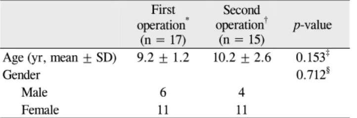
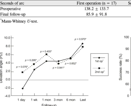
Results: There were no significant differences in the age, gender, visual acuity (logarithm of the minimal angle of resolution), preoperative deviation, and amount of recession between the two groups. Postoperative deviations were -1.7 ± 5.5:1.3 ± 5.1 PD at one day, 4.3 ± 3.8:5.6 ± 5.1 PD at 1 month, 4.3 ± 4.3:3.0 ± 8.1 PD at 3 months, and 5.0 ± 4.3:4.5 ± 7.2 PD at 6 months post-operation, and there was no statistically significant difference between the two study groups. Surgical success rate were 95.2:100% at one day, 95.2:92.9% at 1 month, 90.5:85.7% at 3 months, and 90.0:92.9% a 6 months post-operation.
Conclusions: In patients with exotropia of 15 to 20 PD, no significant difference was found in terms of the post- operative deviation angle and the surgical success rate between the first operation and the second operation groups. Unilateral lateral rectus recession can lead to similar results in 15 to 20 PD exotropia for the first oper- ation or recurrent exotropia.
Key Words: Exotropia, Recurrence, Surgery
ⓒ2011 The Korean Ophthalmological Society
This is an Open Access article distributed under the terms of the Creative Commons Attribution Non-Commercial License (http://creativecommons.org/licenses /by-nc/3.0/) which permits unrestricted non-commercial use, distribution, and reproduction in any medium, provided the original work is properly cited.
Received: July 13, 2010 Accepted: October 15, 2010
Reprint requests to Mi Young Choi, MD. Department of Ophthalmology, Chungbuk National University Hospital, #410 Seongbong-ro, Heungdeok- gu, Cheongju 361-711, Korea. Tel: 82-43-269-6335, Fax: 82-43-264- 5263, E-mail: [email protected]
* This article was presented at the Korean Ophthalmological Society 102nd annual meeting on November, 2009 in Goyang, Korea.
The classical operation methods for basic type exotropia are bilateral lateral rectus recession or unilateral recession of the lateral rectus and resection of the medial rectus (R & R) [1]. The use of unilateral lateral rectus recession (ULR) for small- to moderate-angle exotropia has become increasingly popular, although the surgical results have yielded varied outcomes [2-12].
Operating on one muscle instead of two muscles has the
advantages of shortening the anesthesia and operating time, diminishing the risk associated with muscle surgery, such as scleral perforation, retinal detachment, or endophthalmitis, and performing a second operation more easily if exotropia recurs [2]. In addition, it also has the advantage of reducing amblyopia associated with overcorrection and deterioration of stereopsis due to amblyopia [3]. However, undercorrection can appear after long-term follow-up [3], and it has been re- ported that a limitation of abduction could develop due to a large amount of recession in one muscle [4].
Deutsch et al. [5] performed ULR surgeries in 30 patients
with moderate-angle exotropia. All 30 patients had ortho-
phoria or deviations of 10 prism diopters (PD) or less after 21
months follow-up. In contrast to this study, Kim and Chang
[6] reported a poor prognosis for ULR patients in cases of re-
current exotropia. Among 40 patients with recurrent exo-
tropia who underwent ULR, the rate of success, defined as
Table 1. Patient demographics
First
operation
*(n = 17)
Second operation
†(n = 15) p-value Age (yr, mean ± SD) 9.2 ± 1.2 10.2 ± 2.6 0.153
‡Gender 0.712
§Male 6 4
Female 11 11
*
Exotropia without previous surgery;
†Recurred exotropia;
‡Mann- Whitney U-test;
§Fisher’s exact test.
stability in 10 PD, was 55.6% at 12 to 14 months post-operation.
We speculate that the reason for the difference of these re- sults may lie in their demographic characteristics, including the surgical history for exotropia. The surgical success rate of reoperation for exotropia was different from that of the first operation for exotropia due to a lesser change in postoperative divergence at reoperation than with the first operation [13].
However, a comparison of surgical results after ULR be- tween the first operation and the second operation for exo- tropia has not yet been evaluated. So, we decided to perform this study and compare the surgical results of ULR between the first operation group for exotropia and the second oper- ation group for recurring exotropia.
Materials and Methods
We retrospectively reviewed the medical records of pa- tients who underwent ULR for basic type exotropia and re- curred exotropia of 15 to 20 PD between March 2003 and January 2009. All patients had a minimum of 6 months of fol- low-up after surgery. The exclusion criteria were as follows:
1) amblyopia at surgery, 2) type A or type V exotropia, 3) pa- ralytic strabismus, 4) concurrent oblique or horizontal mus- cle surgery, 5) abnormal version and duction preoperatively, and 6) presence of systemic abnormalities such as Down syn- drome or cerebral palsy.
Fifteen patients that underwent ULR for recurred exo- tropia (15 to 20 PD) were included in second operation group. The mean age at the time of the second surgery was 10.2 ± 2.6 years (range, 5.5 to 16.0 years), and the average duration of postoperative follow-up was 22.9 ± 15.0 months (range, 7 to 53 months). Seventeen patients who underwent ULR for exotropia (15 to 20 PD) between 2005 and 2007 were assigned to first operation group based on consideration of preoperative bias (age of patients and duration of post- operative follow-up) for operative results. The mean age at the time of surgery was 9.2 ± 1.2 years (range, 8.1 to 12.4 years), and the duration of postoperative follow-up was 19.3
± 7.8 months (range, 7 to 39 months).
The following parameters were reviewed and analyzed:
gender, age at each operation, methods of first and second operations, preoperative best corrected visual acuity (BCVA), and history of amblyopia treatment. All examinations were performed under BCVA. The deviation angles were meas- ured in the primary position at distance and near fixation, and the secondary position at distance fixation using an alternate prism cover test. Stereopsis was measured using the Titmus stereo test (Stereo Optical Cl Inc., Chicago, IL, USA) at both preoperative and final follow-up examinations. All surgeries were performed by one surgeon. The surgery was performed on the basis of the largest angle of deviation during fol- low-up, according to the Wright table [14], and the operation was performed on the virgin muscle in both groups. The amount of recession was 7 to 9 mm according to their devia- tion angle.
Surgical success was defined as having a deviation angle less than 10 PD at each follow-up examination after the operation. We defined a recurrence as a 10 PD or more exotropia. The deviated angle and the success rate of surgery were compared at one day, one week, 1 month, 3 months, and 6 months post-operation, and at a final follow-up visit after surgery.
Statistical analysis was carried out using SPSS ver. 12.0 (SPSS Inc., Chicago, IL, USA), and a p-value of less than 0.05 was considered statistically significant.
Results
The mean age was 9.2 ± 1.2 years (range, 8.1 to 12.4 years) at the time of surgery in first operation group and 10.2 ± 2.6 years (range, 5.5 to 16 years) at the time of the second sur- gery in second operation group, and there was no significant difference between the two groups (Mann-Whitney U-test, p
= 0.153). There was also no difference in gender between the two groups (Fisher’s exact test, p = 0.712) (Table 1). The mean age at the time of the first surgery was 5.8 ± 1.7 years (range, 4.0 to 8.4 years) in the 15 patients of second operation group. In the first surgery of the patients in second operation group, 7 patients (46.7%) underwent ULR, while the remain- ing patients (53.5%) underwent R & R. The second surgery was performed at 53.8 ± 31.5 months (range, 18 to 111 months) after the first surgery.
There was no difference in the preoperative visual acuity of the better and worse eyes between the two groups (Mann-Whitney U-test, p = 0.411, p = 0.852), and the pre- operative deviation was 19.4 ± 1.7 PD in first operation group and 19.6 ± 1.3 PD in second operation group (Mann-Whitney U-test, p = 0.823). The average amount of ULR recession was 7.8 ± 0.4 mm in first operation group and 8.1 ± 0.4 mm in second operation group, and there was no significant difference between the two groups (Mann-Whitney U-test, p = 0.058). The average duration of follow-up after surgery was 19.3 ± 7.8 months (range, 7 to 39 months) in first operation group and 22.9 ± 14.8 months (range, 7 to 53 months) in second operation group. The duration was 3 months longer in second operation group, although there was no statistically significant difference (Mann-Whitney U-test, p = 0.852) (Table 2).
The mean deviation angles were -2.1 ± 5.7 PD and 1.6 ±
Table 2. Clinical characteristics
First operation (n = 17) Second operation (n = 15) p-value
*Visual acuity (logMAR)
Better eye 0.04 ± 0.07 0.01 ± 0.04 0.411
Worse eye 0.08 ± 0.11 0.12 ± 0.18 0.852
Hx of amblyopia treatment (no.) 0 2
Deviation angle (PD) 19.4 ± 1.7 19.6 ± 1.3 0.823
Recession (mm) 7.8 ± 0.4 8.1 ± 0.4 0.058
Postoperative follow-up (mon) 19.2 ± 7.8 22.9 ± 15.0 0.655
logMAR = logarithm of the minimal angle of resolution; PD = prism diopters.
*
Mann-Whitney U-test.
Table 3. Stereoacuity at preoperative and final follow-up examinations
Seconds of arc First operation (n = 17) Second operation (n = 15) p-value
*Preoperative 138.2 ± 133.7 188.3 ± 158.7 0.347
Final follow-up 85.9 ± 91.8 191.7 ± 218.2 0.066
*
Mann-Whitney U-test.
Fig. 1. Comparison of deviation angle at each follow-up examina- tion after surgery. PD = prism diopters.
*Mann-Whitney U-test;
†
Exotropia without previous surgery;
‡Recurred exotropia.
Fig. 2. Surgical success rate at each follow-up examination after surgery.
*