Extracellular Nucleotides Can Induce Chemokine (C-C motif) Ligand 2 Expression in Human Vascular Smooth Muscle Cells
6
0
0
전체 글
(2) 32. JI Kim, et al. cells (VSMCs). We found that nucleotides such as NAD+, + NADP , and ATP enhanced CCL2 expression in human aortic smooth muscle cells (AoSMCs). Moreover, we identified the cellular factors involved in nucleotide-mediated CCL2 expression, and found that mitogen-activated protein kinases (MAPKs), protein kinase C (PKC), Akt, and NADPH oxidase were involved in NAD+- and NADP+-mediated CCL2 up-regulation.. METHODS Cell culture and reagents Human AoSMCs were purchased from American Type Culture Collection (ATCC, Manassas, VA, USA), and main+ + tained as previously reported [14]. NAD , NADP , RO318220, GF109203X, LY294002, diphenyleneiodonium chloride (DPI), and SP600125 were purchased from SigmaAldrich Co. (St. Louis, MO, USA). U0126, SB202190, Akt inhibitor IV (Akti IV), and anti-phosphorylated Akt antibody were purchased from Cell Signaling Technology (Danvers, MA, USA). Antibodies against phosphorylated ERK and phosphorylated p38 MAPK were purchased from Santa Cruz Biotechnology (Santa Cruz, CA, USA) and R&D systems (Minneapolis, MN, USA), respectively. Anti phosphorylated JNK antibody was purchased from Stressgen Biotechnologies Corporation (Victoria, BC, Canada). CCL2 Enzyme-linked immunosorbent assay (ELISA) The amount of CCL2 released from AoSMCs was determined using a commercially available ELISA kit, according to the manufacturer’s instructions (BD Biosciences, San Diego, CA, USA). AoSMCs were incubated for 12 h in the presence of 1% fetal bovine serum (FBS) and exposed for + + 9 h to NAD , NADP , or ATP prior to collection of cell culture media. Cell culture media and standards for CCL2 were added to a microtiter plate pre-coated with monoclonal antibody against CCL2. After incubation for 2 h, the plate was washed and incubated with the enzyme-conjugated polyclonal antibody specific for CCL2. The substrate solution was added after several plate washes, and color intensity was measured. The amount of CCL2 present in the medium was determined from a standard curve. Data are expressed as mean±SD. Reverse transcription (RT) - polymerase chain reaction (PCR) Total RNAs were extracted from cells and reverse-trano scribed for an hour at 42 C with Moloney murine leukemia virus reverse transcriptase. PCR amplification was performed for 30 cycles (94oC for 30 sec; 55oC for 30 sec; 72oC for 30 sec) in the presence of primers. PCR primers for CCL2 were 5’-TGGCTGTGTTTGCTTCTGTC-3’ (forward) and 5’-TCTCACTGCCCTATGCCTCT-3’ (reverse). PCR primers for GAPDH were 5’-GAGTCAACGGATTTGGTCGT-3’ (forward) and 5’-TGTGGTCATGAGTCCTTCCA-3’ (reverse). Primers for P2 purinergic receptors were prepared as previously described [14]. PKC activity assay PKC activity was determined using a PKC activity assay. kit (BD Biosciences) according to the manufacturer’s instructions. Cells were collected and lysed in ice-cold lysis buffer containing 20 mM 3-morpholinopropanesulfonic acid, 50 mM β-glyceraldehyde phosphate, 50 mM sodium fluoride, 1 mM sodium vanadate, 5 mM EGTA, 2 mM EDTA 1% NP40, 1 mM dithiothreitol, 1 mM benzamidine, 1 mM phenylmethanesulphonyl fluoride, and 10 μg/ml each aprotinin and leupeptin. Cell lysate was collected and centrifuged to obtain supernatant. Purified active PKC provided in the kit and the supernatants were added to wells in a 96-well plate, and PKC reactions were initiated by adding ATP. After incubation for 90 min, the reactions were stopped by aspirating the wells. Phospho-specific substrate antibody was added to each well and incubated for 50 min at room temperature. HRP-conjugated secondary antibody was added and incubated for 30 min at room temperature. After washing, substrate solution was added and incubated at room temperature. Absorbance was measured at 450 nm. Western blot analysis Cell lysate was prepared using a lysis buffer [1% SDS, 1 mM NaVO3, 10 mM TrisHCl (pH 7.4)] containing protease inhibitors. The isolated cell lysate was separated by SDS-PAGE and transferred to PVDF membranes. After the membranes were blocked by 1 h incubation in 5% skim milk/0.1% Tween 20 in phosphate buffered saline (PBS), o they were incubated overnight at 4 C with appropriate primary antibodies. After three washes using 0.1% Tween20 in PBS, the membranes were incubated for 1 h at room temperature with horseradish peroxidase-conjugated secondary antibodies. Bands were visualized using a chemiluminescent reagent. Statistics Statistical analyses were performed using GraphPad PRISM, version 5.0 (GraphPad Software Inc., San Diego, CA, USA), and p<0.05 was considered statistically significant.. RESULTS Enhancement of CCL2 expression by NAD + , NADP+ , and ATP in human AoSMC We examined the P2 purinergic receptors found on human AoSMCs. The transcript of the following P2 receptors were readily detected in human AoSMC total RNA that was subjected to RT-PCR: P2Y1, P2Y2, P2Y4, P2Y6, P2Y11, P2X1, and P2X4 (Fig. 1A). Among these receptors, P2Y6, P2Y11, P2X1, and P2X4 appeared abundantly expressed. We inves+ tigated whether NAD , an agonist of P2Y11, affected expression of chemokines in parallel with NADP+ and ATP, an agonist of P2Y2, P2Y11, and several P2X receptors. + + RT-PCR analysis indicated that both NAD and NADP increased CCL2 expression, which was evident by 6 h post-treatment. CCL2 expression was also increased by ATP, but not by UTP, an agonist of P2Y 2 and P2Y 4 (Fig. 1B). When we examined the effects NAD+ and NADP+ on expression of the genes for CXCL8 and interleukin (IL)-6, the number of CXCL8 and IL6 transcripts were not influenced by treatment using 10−4 M NAD+ and NADP+, concentration which elevated the number of CCL2 transcripts (Fig. 1C)..
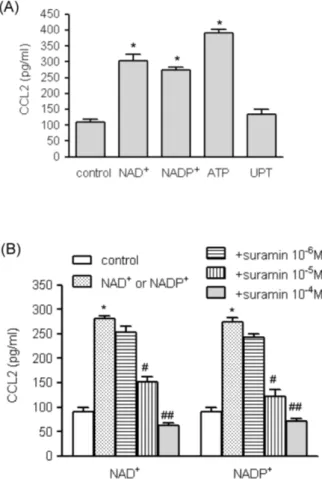
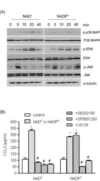
(3) +. NAD. 33. Induces CCL2 Expression in VSMCs. Fig. 1. The effects of extracellular nucleotides on CCL2 gene transcripts in human VSMCs. (A) Total RNA was isolated from human AoSMCs, and transcripts of the indicated P2 purinergic receptors were identified by RT-PCR. (B) Human AoSMCs were treated with nucleotides for the indicated time periods, and CCL2 transcripts were amplified by RT-PCR. (C) Human AoSMCs were + + treated for 6 h with the indicated concentrations of NAD or NADP , and induction of CCL2 gene transcripts was examined by RT-PCR.. We also determined the effects of the nucleotides on + + CCL2 protein expression. NAD , NADP , and ATP significantly elevated CCL2 release from AoSMCs, but UTP did not (Fig. 2A). Because the cellular and molecular effects of ATP are well documented, we focused on the mechanisms + + of action of NAD and NADP . We examined whether suramin, an antagonist of purinergic receptors, could modulate + + the induction of CCL2 expression by NAD and NADP . Suramin inhibited CCL2 secretion from AoSMCs in a concentration-dependent manner, and NAD+- and NADP+mediated CCL2 release was almost completely blocked in −4 M suramin (Fig. 2B). the presence of 10 Involvement of mitogen-activated protein kinases (M APKs) in NAD + - and NADP+ -mediated CCL2 up-regulation MAPKs play important roles in VSMC secretion of cyto-. Fig. 2. The effects of nucleotides on NAD+- and NADP+-mediated CCL2 release. (A) Human AoSMCs (1×10⁶ cells) were incubated −4 in the absence (control) or presence of indicated nucleotides (10 M for each nucleotide). Culture medium was collected and the amount of secreted CCL2 was measured by ELISA. *p<0.001 vs. + + control. (B). Human AoSMCs were incubated with NAD or NADP (10−4 M) in the absence or presence of indicated concentrations of suramin. CCL2 released into the medium was measured by ELISA. # + + ## *p<0.001 vs. control; p<0.01 vs. NAD or NADP , p<0.001 vs. + + NAD or NADP .. kines and chemokines [15,16]. We investigated whether MAPKs affected nucleotide-mediated CCL2 secretion. We + + found that NAD and NADP enhanced phosphorylation of MAPKs (Fig. 3A). Phosphorylated extracellular signal-regulated kinase (ERK) and p38 MAPK reached maximum levels 20 min post treatment, and c-jun N-terminal kinase (JNK) was phosphorylated 5 min post treatment + + with NAD or NADP . To assess the roles of MAPKs on + + NAD - and NADP -mediated CCL2 up-regulation, we applied following MAPK inhibitors; SP600125 (JNK inhibitor), U0126 (ERK inhibitor) and SB202190 (p38 MAPK inhibitor). + NAD -mediated CCL2 release was completely blocked by those inhibitors, and NADP+-induced release of CCL2 protein was inhibited by SP600125 and U0126 (Fig. 3B). Involvement of Akt, PKC, and NADPH oxidase in + + NAD - and NADP -mediated up-regulation of CCL2 +. +. We examined the effects of NAD and NADP on Akt phosphorylation using Western blot analysis. NAD+ and.
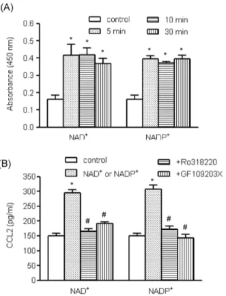
(4) 34. JI Kim, et al. Fig. 4. The roles of Akt pathways in NAD+- and NADP+-mediated CCL2 release. (A) Human AoSMCs were exposed to NAD+ or + NADP for the indicated time periods, after which an equal amount of protein was subjected to Western blot analysis using antibodies for Akt and phosphorylated Akt. (B) Human AoSMCs were incubated for 1 h with LY294002 and Akti IV (10 μM each) and stimulated with NAD+ or NADP+. The amount of secreted CCL2 was measured by ELISA. *p<0.001 vs. control, #p<0.001 vs. NAD+ or + NADP . +. +. Fig. 3. The roles of MAPKs in NAD - and NADP -mediated CCL2 release. (A) Human AoSMCs were exposed to NAD+ or NADP+ for the indicated time periods, after which an equal amount of protein was subjected to Western blot analysis using antibodies for α-tubulin and phosphorylated and unphosphorylated forms of ERK, p38 MAPK, and JNK. (B) Human AoSMCs were incubated for an hour with SP600125, U0126, and SB202190 (10 μM each) and stimulated with NAD+ or NADP+. Culture media were collected to measure the amount of secreted CCL2. *p<0.001 vs. # + + control, p<0.001 vs. NAD or NADP .. NADP+ enhanced Akt phosphorylation, which was maximum 10 min post treatment (Fig. 4A). To investigate whether the Akt pathway was involved in nucleotidemediated CCL2 expression, we used LY294002, a phosphoinositide 3-kinase (PI3K) inhibitor, and Akti IV. When the effects of LY294002 and Akti IV on CCL2 release were examined by ELISA, both inhibitors profoundly attenuated + + NAD and NADP -mediated CCL2 release (Fig. 4B). AoSMCs express PKC subtypes which play roles in chemokine and cytokine secretion [17]. Examining the effects + + + of NAD and NADP on PKC activity, we found that NAD + and NADP enhanced PKC activity (Fig. 5A). PKC activity was increased 5 min post treatment and remained en+ + hanced up to 30 min post treatment by NAD or NADP . To assess involvement of PKC in the nucleotide-mediated. up-regulation of CCL2, we used two PKC inhibitors, GF109203X and RO318220. Both inhibitors profoundly atte+ + nuated NAD and NADP -mediated CCL2 release (Fig. 5B). We also investigated whether reactive oxygen species (ROS) participated in nucleotide-mediated CCL2 expression, using DPI, an inhibitor of NADPH oxidase. DPI appeared to attenuate NAD+ and NADP+-mediated CCL2 expression at the messenger mRNA level and significantly inhibited CCL2 release (Fig. 6).. DISCUSSION Extracellular nucleotides such as ATP have long-term trophic effects, including smooth muscle cell proliferation via the P2Y2 and/or P2Y4 receptors, which transmit the signals through MAPK pathways [18,19]. In response to the MAPK cascade, two transcription factors, c-fos and c-myc, are synthesized in addition to expression of other immediate early genes prior to downstream gene expression of mitogenesis [20]. In this study, we investigated the short-term actions of extracellular nucleotides, and found that NAD+ + and NADP , in addition to ATP, promoted a VSMC proinflammatory phenotype by causing elevated CCL2 expression..
(5) +. NAD. +. Induces CCL2 Expression in VSMCs. +. Fig. 5. The roles of PKC in NAD - and NADP -mediated CCL2 + + release. (A) Human AoSMCs were exposed to NAD or NADP for the indicated time periods, after which PKC activity was determined. *p<0.01 vs. control. (B) Human AoSMCs were incubated for 1 h with GF109203X (3 μM) and RO318220 (1 μM) and stimulated with NAD+ or NADP+. The amount of secreted CCL2 was measured by ELISA. *p<0.001 vs. control, #p<0.001 + + vs. NAD or NADP .. +. +. NAD and NADP enhanced the activity of MAPKs, PKC, and Akt, and pharmacological inhibitors caused attenuated CCL2 expression, which indicates involvement of the signaling pathways in short-term activation of P2 purinergic receptors. We demonstrated that human AoSMCs abundantly expressed multiple P2 purinergic receptor subtypes, including P2Y11 in addition to P2X1, P2Y2 and P2Y6. This finding is consistent with the results of a previous study by Wang et al. Using real-time PCR, they found that P2 receptors were expressed by VSMCs isolated from internal mammary arteries [15]. The effects of P2Y2 and P2Y4 on vascular cells are understood somewhat [18,21]. However, it is not clear what cellular events are mediated and which signaling pathways are activated by P2Y6 and P2Y11 receptors on VSMCs. In the present study, we investigated whether P2Y11 is involved in the development of a VSMC proinflammatory phenotype of VSMCs using NAD+, a P2Y11 agonist. + + Stimulation of VSMCs with NAD , NADP , or ATP enhanced CCL2 expression, and CCL2 release from VSMCs by NAD+ and NADP+ was completely inhibited in a dosedependent manner by the P2 receptor-specific inhibitor, suramin. These results indicate that P2 purinergic receptors are responsible for NAD+- and NADP+-mediated CCL2 up-regulation.. 35. Fig. 6. The roles of NADPH oxidase in NAD+- and NADP+ -mediated CCL2 release. Human AoSMCs were incubated for 1 h + + with DPI (10 μM) and stimulated with NAD or NADP . CCL2 gene transcript was amplified by RT-PCR (A), and the amount of released CCL2 was measured by ELISA (B). *p<0.001 vs. control, # + + p<0.001 vs. NAD or NADP .. We determined which cellular factors were involved in + + CCL2 expression in response to NAD and NADP . Since MAPKs are responsible for the mitogenic effects of P2Y receptors [18,22], we investigated whether kinases are also + + involved in CCL2 expression. NAD and NADP treatment of human HAoSMCs led to phosphorylation of p38 MAPK, + + ERK1/2, and JNK, and NAD - and NADP -mediated CCL2 release was completely blocked by inhibitors of ERK2/1 and JNK. Inhibition of p38 MAPK affected CCL2 release in a + different pattern. SB202190 inhibited NAD -mediated, but + not NADP -mediated, CCL2 release, which suggests a different role for p38 MAPK in CCL2 release in response to extracellular nucleotides. Together, the results indicated that MAPKs are actively involved in the mechanisms of CCL2 expression by VSMCs responding to extracellular nucleotides. We have also identified other molecules involved in P2Y11 signaling pathways. In a study of the mechanisms involved in ATP-induced proliferation of VSMCs, it was shown that P2Y receptor activation is coupled to a pertussis toxin-insensitive Gq protein, triggering phosphoinositide hydrolysis and subsequent activation of PKC, Raf 1, and MAPK [22,23]. In addition, the mitogenic actions of ATP required PI3K signaling pathways [19]. Therefore, we investigated + the roles of PKC and PI3K in CCL2 expression. NAD and + NADP not only enhanced phosphorylation of Akt but also elevated PKC activity. PI3K, Akt, and PKC inhibitors + + blocked CCL2 secretion stimulated by NAD and NADP ..
(6) 36. JI Kim, et al. We believe that some PKC subtypes such as PKC-βI, λ, and μ are also involved in CCL2 secretion, because the AoSMCs used in this study have been reported to express those PKC subtypes, but not PKC- α/ δ/ ξ/ γ, βII, ζ, η, and θ subtypes [17]. NADPH oxidases are a major source of ROS, which are involved in atherogenesis, where they act as second messengers that mediate the signaling pathways in VSMC activation and proliferation [24]. We investigated the role of ROS in CCL2 expression, using DPI, an inhibitor of NADPH oxidase inhibitor. DPI attenuated CCL2 expression at both the mRNA and protein levels. However, we were not able to obtain conclusive evidence that NAD+ and NADP+ significantly enhanced ROS generation in VSMCs exposed to 2,7-dichlorofluorescin diacetate (data not shown). We showed that P2 purinergic receptor agonists induced expression of CCL2 gene transcripts and enhanced CCL2 release from VSMCs, and found that MAPKs, PKC, PI3K, Akt, and NADPH oxidase participated in that process. This study, however, did not specifically determined if these factors acted in an independent or cooperative manner to up-regulate CCL2 expression. Further investigation is necessary to elucidate the types of connections or crosstalk that may be occurring in the context of a possible signaling cascade.. ACKNOWLEDGEMENTS This study was supported by Medical Research Institute grant (2009-6) Pusan National University.. REFERENCES 1. Aukrust P, Halvorsen B, Yndestad A, Ueland T, Øie E, Otterdal K, Gullestad L, Damås JK. Chemokines and cardiovascular risk. Arterioscler Thromb Vasc Biol. 2008;28:1909-1919. 2. Hansson GK, Libby P. The immune response in atherosclerosis: a double-edged sword. Nat Rev Immunol. 2006;6:508-519. 3. Deshmane SL, Kremlev S, Amini S, Sawaya BE. Monocyte chemoattractant protein-1 (MCP-1): an overview. J Interferon Cytokine Res. 2009;29:313-326. 4. Ohtsuki K, Hayase M, Akashi K, Kopiwoda S, Strauss HW. Detection of monocyte chemoattractant protein-1 receptor expression in experimental atherosclerotic lesions: an autoradiographic study. Circulation. 2001;104:203-208. 5. Takeya M, Yoshimura T, Leonard EJ, Takahashi K. Detection of monocyte chemoattractant protein-1 in human atherosclerotic lesions by an anti-monocyte chemoattractant protein-1 monoclonal antibody. Hum Pathol. 1993;24:534-539. 6. Boisvert WA, Santiago R, Curtiss LK, Terkeltaub RA. A leukocyte homologue of the IL-8 receptor CXCR-2 mediates the accumulation of macrophages in atherosclerotic lesions of LDL receptor-deficient mice. J Clin Invest. 1998;101:353-363. 7. Gosling J, Slaymaker S, Gu L, Tseng S, Zlot CH, Young SG, Rollins BJ, Charo IF. MCP-1 deficiency reduces susceptibility to atherosclerosis in mice that overexpress human apolipo-. protein B. J Clin Invest. 1999;103:773-778. 8. Burnstock G. The past, present and future of purine nucleotides as signalling molecules. Neuropharmacology. 1997;36:1127-1139. 9. Communi D, Janssens R, Suarez-Huerta N, Robaye B, Boeynaems JM. Advances in signalling by extracellular nucleotides. the role and transduction mechanisms of P2Y receptors. Cell Signal. 2000;12:351-360. 10. Burnstock G. Purinergic regulation of vascular tone and remodelling. Auton Autacoid Pharmacol. 2009;29:63-72. 11. Dawicki DD, Chatterjee D, Wyche J, Rounds S. Extracellular ATP and adenosine cause apoptosis of pulmonary artery endothelial cells. Am J Physiol. 1997;273:L485-494. 12. Malam-Souley R, Seye C, Gadeau AP, Loirand G, Pillois X, Campan M, Pacaud P, Desgranges C. Nucleotide receptor P2u partially mediates ATP-induced cell cycle progression of aortic smooth muscle cells. J Cell Physiol. 1996;166:57-65. 13. Zheng LM, Zychlinsky A, Liu CC, Ojcius DM, Young JD. Extracellular ATP as a trigger for apoptosis or programmed cell death. J Cell Biol. 1991;112:279-288. 14. Choi KH, Park JW, Kim HY, Kim YH, Kim SM, Son YH, Park YC, Eo SK, Kim K. Cellular factors involved in CXCL8 expression induced by glycated serum albumin in vascular smooth muscle cells. Atherosclerosis. 2010;209:58-65. 15. Wang L, Karlsson L, Moses S, Hultgårdh-Nilsson A, Andersson M, Borna C, Gudbjartsson T, Jern S, Erlinge D. P2 receptor expression profiles in human vascular smooth muscle and endothelial cells. J Cardiovasc Pharmacol. 2002;40:841-853. 16. Sung SC, Kim K, Lee KA, Choi KH, Kim SM, Son YH, Moon YS, Eo SK, Rhim BY. 7-Ketocholesterol upregulates interleukin-6 via mechanisms that are distinct from those of tumor necrosis factor-alpha, in vascular smooth muscle cells. J Vasc Res. 2009;46:36-44. 17. Chung SW, Park JW, Lee SA, Eo SK, Kim K. Thrombin promotes proinflammatory phenotype in human vascular smooth muscle cell. Biochem Biophys Res Commun. 2010;396:748-754. 18. Harper S, Webb TE, Charlton SJ, Ng LL, Boarder MR. Evidence that P2Y4 nucleotide receptors are involved in the regulation of rat aortic smooth muscle cells by UTP and ATP. Br J Pharmacol. 1998;124:703-710. 19. Wilden PA, Agazie YM, Kaufman R, Halenda SP. ATPstimulated smooth muscle cell proliferation requires independent ERK and PI3K signaling pathways. Am J Physiol. 1998; 275:H1209-1215. 20. Zhang W, Liu HT. MAPK signal pathways in the regulation of cell proliferation in mammalian cells. Cell Res. 2002;12:9-18. 21. Hou M, Möller S, Edvinsson L, Erlinge D. Cytokines induce upregulation of vascular P2Y(2) receptors and increased mitogenic responses to UTP and ATP. Arterioscler Thromb Vasc Biol. 2000;20:2064-2069. 22. Yu SM, Chen SF, Lau YT, Yang CM, Chen JC. Mechanism of extracellular ATP-induced proliferation of vascular smooth muscle cells. Mol Pharmacol. 1996;50:1000-1009. 23. Needleman P, Minkes MS, Douglas JR Jr. Stimulation of prostaglandin biosynthesis by adenine nucleotides. Profile of prostaglandin release by perfused organs. Circ Res. 1974; 34:455-460. 24. Taniyama Y, Griendling KK. Reactive oxygen species in the vasculature: molecular and cellular mechanisms. Hypertension. 2003;42:1075-1081..
(7)
수치
관련 문서
Activated H-Ras expression in human fibroblast cell lines increases the activity of Ku80 to bind injuried DNA, reduces γ-H2AX expression by UV irradiation,
The improvement factors of the performance showed significant increase in muscle strength, muscle endurance, cardiovascular endurance, power, agility and
Fig 4 : Inhibitory effects of classified methanol extracts of Acalypha australis the COX-2 protein expression of the human oral cavity carcinoma KB cells....
With these results, we can suggest the hypothesis that increased cardiac output in the patients who administered ketamine increased muscle blood flow, and this is the
We investigated the effects of BIX01294 on cellular senescence in human BM-MSCs (hBM-MSCs), and identified that an optimal treatment of BIX01294 leads to attenuated
4 > Effects of Taro on COX-2 expression and iNOS expression(hot water) in human thyroid cancer cells. The cells were pretreated for 48hours with either
Conclusion: In conclusion, the relaxant effects of neuromuscular blockers on the uterine smooth muscle may be transmitted via nicotinic acetylcholine receptors
Moreover, we found that treatment of HSC with resveratrol increased Lipin1 expression and inhibited TGF- b-activated fibrogenic gene expression via Lipin1 regulation,
