• 검색 결과가 없습니다.

The First Korean Case of Nontuberculous Mycobacterial Lung Disease Caused by Mycobacterium abscessus Subspecies bolletii in a Patient with Bronchiectasis

N/A
N/A
Protected

Academic year: 2021

Share "The First Korean Case of Nontuberculous Mycobacterial Lung Disease Caused by Mycobacterium abscessus Subspecies bolletii in a Patient with Bronchiectasis"

Copied!
4
0
0

로드 중.... (전체 텍스트 보기)

전체 글

(1)

The First Korean Case of Nontuberculous Mycobacterial Lung Disease Caused by

Mycobacterium abscessus Subspecies bolletii in a Patient with Bronchiectasis

Byeong-Ho Jeong, M.D.

1,

*, Su-Young Kim, Ph.D.

1,

*, Kyeongman Jeon, M.D.

1

, Hee Jae Huh, M.D.

2

, Chang-Seok Ki, M.D.

2

, Nam Yong Lee, M.D.

2

, Sung Jae Shin, Ph.D.

3

and Won-Jung Koh, M.D.

1

1

Division of Pulmonary and Critical Care Medicine, Department of Medicine,

2

Department of Laboratory Medicine and Genetics, Samsung Medical Center, Sungkyunkwan University School of Medicine,

3

Department of Microbiology, Yonsei University College of Medicine, Seoul, Korea

We report the first Korean case of lung diseases caused by Mycobacterium abscessus subsp. bolletii in a previously healthy male, except for a previous history of pulmonary tuberculosis and bronchiectasis. All serial isolates are identified as M. abscessus subsp. bolletii by multi-locus sequence analysis based on the hsp65, rpoB, and 16S rRNA fragments. At the genetic level, the isolate has the erm(41) gene with a T28 sequevar, associated with clarithromycin resistance, and no rrl mutation. The isolate is resistant to clarithromycin. Although the symptoms and radiographic findings have improved after combination of antibiotics, the follow-up sputum cultures are persistently positive.

Keywords: Bronchiectasis; Mycobacterium Infections, Nontuberculous; Nontuberculous Mycobacteria; Sequence Analysis, DNA

mycobacteria (NTM) in Korea

1

. Of the RGM species, Myco- bacterium abscessus complex is most commonly associated with NTM lung disease. Although subspeciation of M. absces- sus complex is still controversial, M. abscessus complex is currently divided into three subspecies: M. abscessus subsp.

abscessus, M. abscessus subsp. massiliense and M. abscessus subsp. bolletii

2

.

In Korea, M. abscessus subsp. bolletii is identified in only 1.3−1.6% of clinical strains of M. abscessus complex

3

. Although a case with disseminated infection of M. abscessus subsp.

bolletii was reported

4

, there has been no report of NTM lung disease caused by M. abscessus subsp. bolletii in Korea. Here, we describe a case of chronic pulmonary disease caused by M.

abscessus subsp. bolletii.

Case Report

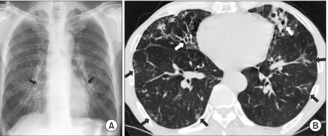
A 66-year-old male was referred to our hospital for manage- ment of probable NTM lung disease. He was an ex-smoker and had a history of previous treatment for pulmonary tuber- Copyright © 2014

The Korean Academy of Tuberculosis and Respiratory Diseases.

All rights reserved.

Introduction

Rapidly growing mycobacteria (RGM) are relatively com- mon pathogens of lung disease caused by nontuberculous

CASE REPORT

http://dx.doi.org/10.4046/trd.2014.76.1.30

ISSN: 1738-3536(Print)/2005-6184(Online) • Tuberc Respir Dis 2014;76:30-33

30

Address for correspondence: Won-Jung Koh, M.D.

Division of Pulmonary and Critical Care Medicine, Department of Medicine, Samsung Medical Center, Sungkyunkwan University School of Medicine, 81 Irwon-ro, Gangnam-gu, Seoul 135-710, Korea

Phone: 82-2-3410-3429, Fax: 82-2-3410-3849 E-mail: [email protected]

*Byeong-Ho Jeong and Su-Young Kim contributed equally to this work.

Received: Aug. 7, 2013 Revised: Sep. 4, 2013 Accepted: Sep. 16, 2013

cc

It is identical to the Creative Commons Attribution Non-Commercial

License (http://creativecommons.org/licenses/by-nc/3.0/).

(2)

M. bolletii lung disease

http://dx.doi.org/10.4046/trd.2014.76.1.30 31

www.e-trd.org

culosis 10 years prior. Six months before his visit to our hospi- tal, the patient reported chronic cough and sputum. He was initially diagnosed with acid-fast bacilli (AFB) smear-positive pulmonary tuberculosis and received isoniazid, rifampin,

ethambutol, and pyrazinamide for 6 months. However, AFB smears were persistently positive, and NTM were cultured from his sputum specimens. He was then referred to our hos- pital.

Figure 1. A 66-year-old male with bronchiectasis and nontuberculous mycobacterial lung disease caused by Mycobacterium abscessus sub- sp. bolletii. (A) Chest radiography revealed bilateral multifocal tram-track signs (black arrows), suggesting bronchiectasis. (B) A transverse chest computed tomography scan (3.0-mm-section thickness) revealed bilateral bronchiectasis (white arrows) in the middle right lobe and the lingular segment of the upper left lobe as well as multiple tree-in-bud appearances (black arrows), suggesting bronchiolitis in both lungs.

Figure 2. The phylogenetic position of isolate SMC-bol-002 and other species belong to the rapidly growing mycobacteria are based on rpoB

sequences. This tree was constructed by using the neighbor-joining method and visualized with MEGA 5.0. The percentages at the nodes

represent bootstrap levels which are supported by 1,000 re-sampled datasets. Scale bars indicate evolutionary distances in base substitutions

per site. M.: Myobacterium.

(3)

BH Jeong et al.

32 Tuberc Respir Dis 2014;76:30-33 www.e-trd.org

Physical examination showed that the patient was 172.3 cm tall and weighed 56.1 kg. Laboratory tests results were unremarkable. A human immunodeficiency virus antibody test was negative. The chest radiograph and computed to- mography (CT) scan revealed multifocal bronchiectasis and bronchiolitis in both lungs that suggested the nodular bron- chiectatic form of NTM lung disease (Figure 1).

The patient’s sputum was positive for AFB staining, and NTM were isolated more than three times in a liquid culture system (Bactec MGIT 960 system; BD Diagnostics, Sparks, MD, USA). To identify the etiological agent, bacteria were initially propagated in 7H9 broth (Difco Laboratories, Detroit, MI, USA) supplemented with 10% (vol/vol) oleic acid-albu- min-dextrose-catalase (BD Diagnostics) for 7 days at 37

o

C, sub-cultured in egg-based 3% Ogawa solid media (Shinyang, Seoul, Korea), and genomic DNA was extracted from the cul- tured bacteria. Initial species identification using a polymerase chain reaction-restriction fragment length polymorphism (PCR-RFLP) method based on the rpoB gene (Myco-ID; M&D Inc., Wonju, Korea) was M. abscessus complex

5

.

To speciate the isolates, sequencing analysis of the nearly complete 16S rRNA gene, partial hsp65, and rpoB sequences were performed using GenBank (http://blast.ncbi.nlm.nih.

gov/) with the BLAST algorithm

6

. The 16S rRNA sequences showed 100% identity with M. abscessus subsp. abscessus (GenBank accession no. NR074427), M. abscessus subsp.

massiliense (GenBank accession no. NR074421), M. chelonae (GenBank accession no. AY457082), and M. abscessus subsp.

bolletii (GenBank accession no. NR043236). The hsp65 and rpoB sequences were 100% identical to those of the M. ab- scessus subsp. bolletii type strain (GenBank accession nos.

FJ607778 and AY859692). Phylogenetic analysis based on the rpoB sequences from the isolate SMC-bol-002 and from those of closely related species within the RGM showed that this strain belonged to M. abscessus subsp. bolletii (Figure 2).

Drug susceptibility testing was performed using a broth

microdilution method (Table 1)

7

. At the genetic level, the isolate was genotyped for erm(41) polymorphism and for rrl mutations; these are known as the two main mechanisms of macrolide resistance

8

. The isolate had the erm(41) gene with a T28 sequevar and no rrl mutation acquisition.

The patient received oral clarithromycin and moxifloxacin, along with an initial 4-week course of intravenous amikacin and cefoxitin. During 4 weeks of initial antibiotic treatment, his symptoms and CT lesions were improved and sputum culture results were negative after 2, 3, and 4 weeks after treat- ment. Although he took oral clarithromycin and moxifloxacin for a total duration of 24 months, follow-up sputum cultures were persistently positive after 2 months after antibiotic treat- ment.

Discussion

This is the first reported case of M. abscessus subsp. bolletii lung disease in Korea. Previous reports showed that patients with M. abscessus subsp. bolletii lung disease had mild com- promised respiratory function, as was seen in the current report

9,10

. The reported number of patients with M. abscessus subsp. bolletii lung disease was less than 10 before this case

9,10

. The limited number of reports makes it difficult to character- ize patients with M. abscessus subsp. bolletii lung disease.

The M. abscessus complex has undergone many taxonomic classification changes since its first description in 1953

11

. The dramatic change in mycobacterial taxonomy came with the availability and reliability of DNA sequencing, including 16S rRNA, rpoB, and hsp65. Adekambi et al.

12

suggested that the M. abscessus complex should be divided into three species, M. abscessus sensu stricto, M. massiliense, and M. bolletii, according to the results of rpoB sequencing. Recent evidence based on comparative analysis of whole genome sequences has suggested that M. abscessus can be divided into three sub- Table 1. Antimicrobial drugs and MIC breakpoints

Drugs MIC (µg/mL) for category

MIC of the isolate (µg/mL)

Susceptible Intermediate Resistant

Amikacin ≤16 32 ≥64 8

Cefoxitin ≤16 32−64 ≥128 32

Imipenem ≤4 8−16 ≥32 32

Clarithromycin ≤2 4 ≥8 8

Ciprofloxacin ≤1 2 ≥4 16

Moxifloxacin ≤1 2 ≥4 8

Doxycycline ≤1 2−4 ≥8 >32

Drug susceptibility testing was performed by using a broth microdilution method, according to the guidelines of the Clinical and Laboratory Standards Institute (CLSI)

7

.

MIC: minimum inhibitory concentration.

(4)

M. bolletii lung disease

http://dx.doi.org/10.4046/trd.2014.76.1.30 33

www.e-trd.org

species: M. abscessus subsp. abscessus, M. abscessus subsp.

massiliense, and M. abscessus subsp. bolletii

13

. It is important to distinguish the three subspecies of M. abscessus complex, because of their differences in susceptibility to clarithromy- cin

3

. Commonly, M. abscessus subsp. abscessus, M. abscessus subsp. massiliense, and M. abscessus subsp. bolletii are in- ducible resistant, susceptible, and resistant to clarithromycin, respectively

3

.

The combination of macrolide, cefoxitin (or imipenem), and amikacin has been recommended as the first line antimicro- bial drug treatment for M. abscessus lung disease

14

. However, the appropriate duration of intravenous antibiotic therapy or combination of oral antibiotics is not known. We used intrave- nous antibiotics for 4 weeks as previously reported cases of M.

abscessus lung disease

15

, because of the phenotypic similari- ties between M. abscessus subsp. abscessus and M. abscessus subsp. bolletii. Although drug susceptibility testing showed in vitro resistance, clarithromycin and moxifloxacin were prescribed during outpatient treatment following the previous report

3,15

. We finally achieved improvement in his respiratory symptoms and prevented progression of radiological findings, although we were unable to achieve negative conversion of sputum AFB culture.

In summary, we report the first case of M. abscessus subsp.

bolletii lung disease in Korea. M. abscessus subsp. bolletii lung disease should be considered when the pathogen is iden- tified as M. abscessus complex by a non-sequencing method, such as PCR-RFLP, and has resistance to clarithromycin. In such cases, further molecular techniques may be needed to accurately identify the specific subspecies.

Acknowledgements

This work was supported by the Mid-career Researcher Pro- gram through a National Research Foundation grant funded by the Ministry of Education, Science and Technology (2011- 0015546).

References

1. Koh WJ, Kwon OJ, Jeon K, Kim TS, Lee KS, Park YK, et al. Clin- ical significance of nontuberculous mycobacteria isolated from respiratory specimens in Korea. Chest 2006;129:341-8.

2. Macheras E, Roux AL, Bastian S, Leao SC, Palaci M, Sivadon- Tardy V, et al. Multilocus sequence analysis and rpoB se- quencing of Mycobacterium abscessus (sensu lato) strains. J Clin Microbiol 2011;49:491-9.

3. Koh WJ, Jeon K, Lee NY, Kim BJ, Kook YH, Lee SH, et al. Clini- cal significance of differentiation of Mycobacterium massil-

iense from Mycobacterium abscessus. Am J Respir Crit Care Med 2011;183:405-10.

4. Koh WJ, Kwon OJ, Lee NY, Kook YH, Lee HK, Kim BJ. First case of disseminated Mycobacterium bolletii infection in a young adult patient. J Clin Microbiol 2009;47:3362-6.

5. Lee H, Park HJ, Cho SN, Bai GH, Kim SJ. Species identification of mycobacteria by PCR-restriction fragment length polymor- phism of the rpoB gene. J Clin Microbiol 2000;38:2966-71.

6. Kim H, Kim SH, Shim TS, Kim MN, Bai GH, Park YG, et al.

Differentiation of Mycobacterium species by analysis of the heat-shock protein 65 gene (hsp65). Int J Syst Evol Microbiol 2005;55(Pt 4):1649-56.

7. Clinical and Laboratory Standards Institute. Susceptibility testing of mycobacteria, nocardiae, and other aerobic actino- mycetes: approved standard. 2nd ed. Document no. M24-A2.

Wayne: CLSI; 2011.

8. Bastian S, Veziris N, Roux AL, Brossier F, Gaillard JL, Jarlier V, et al. Assessment of clarithromycin susceptibility in strains belonging to the Mycobacterium abscessus group by erm(41) and rrl sequencing. Antimicrob Agents Chemother 2011;55:775-81.

9. Adekambi T, Drancourt M. Mycobacterium bolletii respira- tory infections. Emerg Infect Dis 2009;15:302-5.

10. van Ingen J, de Zwaan R, Dekhuijzen RP, Boeree MJ, van Soolingen D. Clinical relevance of Mycobacterium chelonae- abscessus group isolation in 95 patients. J Infect 2009;59:324- 31.

11. Moore M, Frerichs JB. An unusual acid-fast infection of the knee with subcutaneous, abscess-like lesions of the gluteal region; report of a case with a study of the organism, Myco- bacterium abscessus, n. sp. J Invest Dermatol 1953;20:133-69.

12. Adekambi T, Berger P, Raoult D, Drancourt M. rpoB gene se- quence-based characterization of emerging non-tuberculous mycobacteria with descriptions of Mycobacterium bolletii sp. nov., Mycobacterium phocaicum sp. nov. and Mycobacte- rium aubagnense sp. nov. Int J Syst Evol Microbiol 2006;56(Pt 1):133-43.

13. Bryant JM, Grogono DM, Greaves D, Foweraker J, Roddick I, Inns T, et al. Whole-genome sequencing to identify transmis- sion of Mycobacterium abscessus between patients with cystic fibrosis: a retrospective cohort study. Lancet 2013;381:

1551-60.

14. Griffith DE, Aksamit T, Brown-Elliott BA, Catanzaro A, Daley C, Gordin F, et al. An official ATS/IDSA statement: diagnosis, treatment, and prevention of nontuberculous mycobacterial diseases. Am J Respir Crit Care Med 2007;175:367-416.

15. Jeon K, Kwon OJ, Lee NY, Kim BJ, Kook YH, Lee SH, et al. An- tibiotic treatment of Mycobacterium abscessus lung disease:

a retrospective analysis of 65 patients. Am J Respir Crit Care

Med 2009;180:896-902.

수치

Figure 1. A 66-year-old male with bronchiectasis and nontuberculous mycobacterial lung disease caused by Mycobacterium abscessus sub- sub-sp

참조

관련 문서

The phylogenetic position of isolated SMC-shi-001 strain from the patient in this report and other species belonging to the slow growing mycobacteria, based on the rpoB

On chest radiography B, there is more increased size of cystic lesion with air fluid level in both upper lung field and newly developed cystic lesion in right lower lung

본 연구의 한계점으로는 NTM 폐질환의 경우 원인균에 따라 임상 양상이 다르게 나타나며 치료 방법 또한 달라지기 때문에 1 원인균 동정이 필수적 이나 본 환자의 경우 타