Piperine에 의한 위암세포 AGS 증식 억제와 Apoptosis 유도
신성아1․이해님1․추강식1․김소정2․김형진1․박영석1 박병권1․김병수1․김상기1․이후장3․정지윤1
1공주대학교 특수동물학과
2충북대학교 의과대학 의학과 생화학교실
3경상대학교 수의과대학 동물의학연구소
Induction of Apoptosis and Inhibition of Growth in Human Gastric Cancer by Piperine
Seong-Ah Shin1, Hae-Nim Lee1, Gang-Sik Choo1, So-Jung Kim2, Hyeong-Jin Kim1, Young-Seok Park1, Byung-Kwon Park1, Byeong-Soo Kim1,
Sang-Ki Kim1, Hu-Jang Lee3, and Ji-Youn Jung1
1Department of Companion and Laboratory Animal Science, Kongju National University
2Department of Program in Cell Biology and Genetics, College of Medicine, Chungbuk National University
3Institute of Animal Medicine, College of Veterinary Medicine, Gyeongsang National University
ABSTRACT Piperine [(E,E)-5-(3,4-methtylenedioxyphenyl)-2,4-pentadienolypiperidide] is a principal of Piperaceae, including Piper nigrum L. and Piper longum Linn., which has been used as a spice and traditional medicine. In this study, we investigated whether or not piperine has anti-cancer effects on AGS human gastric cancer cells. The results demonstrated that piperine not only inhibited proliferation using MTT assay but also induced apoptotic bodies using DAPI assay in a dose-dependent manner in response to piperine. Expression levels of p53, Bax (pro-apoptotic), cleaved caspase-9, and cleaved-PARP increased, whereas expression levels of Bcl-2, XIAP (anti-apoptotic), and Akt decreased in a dose-dependent manner compared with the control by western blotting analysis. To identify the connection between phospo-Akt and Bcl-2 family in response to piperine, LY249002 (Akt inhibitor) was treated with piperine (150 μM).
The results were shown that expression of phospo-Akt was reduced whereas expression of Bax and cleaved-PARP increased in a dose-dependent manner. These results indicate that piperine induced apoptosis in AGS cells and may serve as a chemopreventive or therapeutic agent for human gastric cancer.
Key words: cancer cell, apoptosis, anticancer, piperine, Akt signaling
Received 18 July 2016; Accepted 24 August 2016
Corresponding author: Ji-Youn Jung, Department of Companion and Laboratory Animal Science, Kongju National University, Yesan, Chungnam 32439, Korea
E-mail: [email protected], Phone: +82-41-330-1526
서 론
의학의 발달과 생활환경의 개선으로 삶의 질은 높아졌지 만 여전히 암은 가장 치명적인 질병으로 여겨진다(1). 전 세 계에서 폐암 다음으로 가장 일반적으로 발병하며 우리나라 에서도 전체 암 중에서 가장 많은 비중을 차지하고 있는 위 암은 여성보다는 특히 남성에서 높은 발병률을 보이고 있다.
위암을 치료하기 위해 외과적 절제, 화학요법 등이 이용되고 있고, 치료의 효과를 높이기 위해서 여러 치료법이 병행되기 도 한다. 하지만 위암은 조기에 발견하지 못하면 좋은 예후 를 기대하기 어렵고, 값비싼 치료비에 대한 부담과 기존 항
암제에 의한 부작용으로 인해 새로운 치료방법의 모색이 필 요한 실정이다(2,3). 따라서 이러한 경제적 부담과 부작용을 줄이기 위해 안전성이 입증된 천연물질에서 항암성분을 찾 는 연구가 지속해서 진행되고 있다(4).
후추과(Piperaceae)에 속하는 후추(Piper nigrum L.)와 필발(Piper longum Linn.)은 아시아와 인도에서 향신료로 널리 사용되고 있으며, 소화불량, 통증 완화 등에 효능이 있 는 것으로 알려져 전통의학에서 사용되어 왔다. 이들은 다양 한 phytochemical들이 존재하고 있어 여러 생리활성을 나 타내는 것으로 알려져 있으며, 그중에서 가장 대표적인 al- kaloid-amid 성분인 piperine으로 인해 후추에서 특유의 향 과 맛이 나는 것으로 알려져 있다(5,6). 또한, piperine은 항 산화(7), 항염증(8), 항관절염(9), 항균(10), 항암(11) 작용 등의 여러 가지 생리활성을 가지는 것으로 보고되어 있다.
세포사멸은 세포자살이라고 할 수 있는 세포의 내적 활동 인 apoptosis와 세포 내 물질을 밖으로 누출시켜 조직의 염
Fig. 1. The structure of piperine (C17H19NO3).
증을 유발하는 necrosis로 구분된다. 특히 apoptosis는 생 체 내에서 손상된 세포의 복구가 어렵거나 불필요한 세포를 제거하는 등 항상성 유지에 있어 중요한 역할을 하고 있다 (12). Apoptosis는 물리적이나 화학적인 자극에 의해 DNA 손상을 받게 되면 이와 연관된 Bcl-2 family, p53, Fas, Akt 등 다양한 단백질들의 발현 조절에 의해서 유도되는 것으로 알려져 있다(13). 이러한 apoptosis 기능이 원활히 이루어 지지 않으면 면역질환이나 암을 포함한 각종 질병을 유발할 수 있는 것으로 알려져 있다(14).
Protein kinase B(PKB)라고도 불리는 Akt는 serine/
threonine kinase로서, 상위 kinase인 PI3K 경로를 통해 활성화된다. 활성화된 Akt는 핵으로 이동하여 전사인자를 포함한 여러 기질을 인산화한다. Akt 신호경로는 여러 암종 에서 높은 활성상태로 존재하며, 전사, 번역, 성장과 같은 기본적인 세포의 기능을 조절한다. 또한, 암의 발생과 진행 에 있어서 세포의 증식과 apoptosis의 균형 유지에 관여하 는 것으로 알려져 있다. 이러한 Akt 신호 경로의 조절은 종 양의 성장뿐만 아니라 암의 치료에 있어 중요한 역할을 하는 것으로 알려져 왔다(15,16).
p53은 종양 억제 유전자로 세포에 가해지는 스트레스, 비정상적 성장신호와 DNA 손상 등의 여러 신호에 의해 활 성화 상태가 된다. 활성화된 p53의 하위기전을 통해 종양의 발생을 억제한다. p53의 주요 기능 중 하나는 p21을 통하여 세포주기를 정지시켜 손상된 DNA를 수리하여 이상 DNA가 퍼지는 것을 방지하고, 또 다른 기능은 DNA의 손상이 심해 세포가 본래의 상태로 회복할 수 없는 경우 Bcl-2 family 단백질의 발현을 조절하여 apoptosis를 유도하는 것으로 알 려져 있다. 따라서 새로운 항암제 개발에 있어 종양 억제유 전자인 p53의 중요성은 강조되고 있다(17).
따라서 본 연구에서는 다양한 생리활성을 가지는 것으로 보고되어 있는 piperine이 인간 위암세포 AGS의 증식과 세 포사멸에 미치는 영향을 확인하고, 이러한 효과에 있어서 Akt 신호전달경로와 Bcl-2 family의 역할에 관해서 확인하 고자 하였다.
재료 및 방법
세포 및 재료
본 연구에 사용된 위암세포 AGS는 한국세포주은행(KCLB, Korea Cell Line Bank, Seoul, Korea)에서 구입하였다. 세 포배양에 사용된 RPMI-1640은 Welgene(Gyeonsan, Korea) 에서 구입하였고, fetal bovine serum과 streptomycin/
penicillin은 Gibco BRL(Grand Island, NY, USA)에서 구 입하였다. 본 연구에 사용한 piperine(Fig. 1)과 일반적인 시약은 Sigma-Aldrich Co.(St. Louis, MO, USA)에서 구입 하였다. 1차 항체 anti-Bax, anti-Bcl-2, anti-PARP, an- ti-p53, anti-XIAP, anti-Akt, anti-p-Akt, anti-caspase- 9, anti-β-actin과 2차 항체 anti-rabbit IgG는 Cell Signal-
ing Technology(Danvers, MA, USA)에서 구입하였다.
세포 배양
위암세포 AGS는 5% fetal bovine serum, 1% strepto- mycin/penicillin을 첨가한 RPMI-1640 배지를 사용하여 37°C, 5% CO2가 유지되는 incubator에서 배양하였다. 175 T flask에 세포 밀도가 80% 정도가 되면, PBS(pH 7.4)로 washing 한 후 trypsin-EDTA를 처치하여 계대배양 하였 고, 배지는 2~3일마다 교환하였다.
MTT assay
위암세포 AGS를 96 well plate에 2×10⁴ cells/mL로 분 주한 후 세포가 plate에 붙을 수 있도록 24시간 동안 배양시 킨 다음, piperine을 0, 50, 100, 150, 200 μM의 농도로 처치하였다. 24시간 후 AGS 세포가 배양된 96 well plate에 MTT[3-(4,5-dimethylthiazol-2-yl)-2,5-diphenylte- trazolium bromide] 용액(1mg/mL)을 40 μL/well씩 처치 한 다음 1시간 30분 동안 37oC, 5% CO2가 유지되는 in- cubator에서 배양하였다. MTT 용액을 제거한 후 dime- thylsulfoxide(DMSO)를 100 μL/well로 첨가하여 well에 형성된 formazan을 모두 녹인 후 ELISA-reader(Bio-Rad Laboratories Inc., Hercules, CA, USA)로 595 nm에서 흡 광도를 측정하였다.
DAPI staining
AGS 세포를 60 mm dish에 1×10⁵ cells/mL로 분주한 후 24시간 동안 안정화하였다. Piperine을 0, 100, 150 μM 의 농도로 처치하여 24시간 동안 배양한 다음 PBS로 두 번 washing 한 후 4% paraformaldehyde 용액으로 15분간 고 정하였다. 다시 한 번 washing 한 후 PBS에 10배로 희석한 DAPI 시약을 2 mL씩 처치하여 형광현미경(Zeiss Fluo- rescence Microscope, Thornwood, NY, USA)으로 200 배 시야에서 관찰하였다.
Western blotting analysis
175T flask에 AGS 세포를 37°C, 5% CO2가 유지되는 incubator에서 24시간 동안 배양하였다. Piperine에 의한 apoptosis와 Akt signaling pathway 단백질 발현을 확인하 기 위해 piperine을 0, 100, 150 μM의 농도로 처치하였다.
24시간 동안 배양하여 trypsin-EDTA를 첨가한 후 세포를 부유시켜 원심분리(1,200 rpm, 5 min, 4°C) 하였다. PBS로 세포를 washing 하고 원심분리 하여 cell pellet에 cell ly-
0 20 40 60 80 100 120
0 50 100 150 200
Piperine (μM)
Cell viability (% of control) .
*
*
*
*
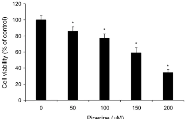
Fig. 2. Effect of piperine on the cell viability in AGS cells. AGS cells were treated with absence or presence of piperine (50, 100, 150, 200 μM) for 24 h by MTT assay. Datas are shown as mean
±SD of three independent experiments performed in triplicate.
Significance was determined by Dunnett's t-test with *P<0.05 considered as statistically significant compared with non-treated controls.
sis buffer(Invitrogen, Carlsbad, CA, USA)를 첨가한 후 4°C에서 20분간 반응시켰다. Lysate는 13,000 rpm에서 5 분 동안 원심분리 하여 상등액을 취해 cell lysate로 사용하 였다. 추출한 단백질의 농도는 Bradford protein assay를 이용해 측정하였다. 단백질(40 μg)을 12% sodium dodecyl sulfate polyacrylamide gel electrophoresis(SDS-PAGE) 로 크기별로 분리한 후 nitrocellulose membrane(Bio- Rad)에 이동시켰다. Membrane은 5% skim milk로 1시간 30분간 blocking 한 후 anti-p53, anti-Bax, anti-Bcl-2, anti-XIAP, anti-PARP, anti-caspase-9, anti-Akt, anti- p-Akt, anti-β-actin의 1차 항체를 각각 첨가하여 4°C에서 overnight 하였다. 그 후 2차 항체 anti-rabbit IgG를 첨가 하여 2시간 반응시켰다. 각 protein band는 ECL detection reagents(Pierce, Rockford, IL, USA)를 이용하여 실험 결 과를 확인하였다.
Akt와 apoptosis 관련 단백질의 역할을 확인하기 위해 LY294002(Akt inhibitor) 20 μM과 piperine 0, 150 μM의 농도로 병행 또는 단독으로 처치한 후 24시간 동안 배양하 여 단백질을 추출하였다. 추출한 단백질을 전기영동 과정을 거친 후 skim milk로 1시간 30분간 blocking 하고 anti- p-Akt, anti-Bax, anti-Bcl-2, anti-PARP, anti-β-actin 의 1차 항체와 2차 항체 anti-rabbit IgG를 첨가하여 반응시 킨 다음, 각 protein band는 ECL detection reagents를 이 용하여 실험 결과를 확인하였다.
통계 분석
모든 실험 결과는 평균치와 표준편차를 사용하여 나타내 고 각 군간 비교는 one-way ANOVA에 이은 t-test 분석을 시행하였다. 대조군과 비교하여 P값이 0.05 미만일 때를 통 계학적으로 유의성이 있다고 판정하였다
결과 및 고찰
Piperine이 위암세포의 생존에 미치는 영향
Piperine이 위암세포 AGS 생존에 미치는 영향을 확인하 기 위해 piperine을 0, 50, 100, 150, 200 μM의 농도로 처치 하여 MTT assay를 수행하였다. Piperine(0, 50, 100, 150, 200 μM)을 24시간 처치하였을 때 위암세포 AGS의 생존율 은 85.9, 77.3, 59.1, 34.3%로 대조군과 비교해 농도 의존적 으로 유의적인 감소를 확인할 수 있었다(Fig. 2). Do 등(18) 의 연구에 의하면 piperine을 유방암세포 SKBR3와 MCF- 7에 10, 25, 50, 100, 200 μM 농도로 처치하였을 때 SKBR3에서는 25 μM, MCF-7에서는 100 μM 농도에서부 터 농도 의존적인 감소를 보여주었다. 또한, Lin 등(19)의 연구에 의하면 piperine을 폐암세포 A549에 0, 25, 50, 100, 200, 400 μM 농도로 48시간 처치하였을 때 25 μM에 서부터 농도 의존적인 감소를 보여주었다. 이를 통해 본 연 구에서도 piperine이 위암세포 AGS에서 농도 의존적으로
세포생존율의 억제에 영향을 준 것으로 생각한다.
Piperine이 위암세포의 형태적인 변화에 미치는 영향 MTT assay를 통해 확인한 위암세포 AGS의 생존율 억제 가 apoptosis에 의한 결과인지를 확인하기 위해 piperine을 위암세포 AGS에 0, 100, 150 μM 농도로 24시간 처치한 후 DAPI 염색을 하여 형광현미경으로 관찰하였다(Fig. 3A).
그 결과 MTT assay에서 확인한 결과와 동일하게 농도 의존 적으로 세포의 수가 감소한 것을 확인할 수 있었다. 또한, 대조군과 비교하였을 때 piperine을 처치한 위암세포에서 더 많은 apoptosis의 특징인 염색질 응축과 apoptotic body 를 확인할 수 있었다. Apoptosis가 유도된 세포를 정량화하 여 분석하기 위해 DAPI 형광에 반응한 apoptotic 세포를 counting 한 결과 대조군과 비교하였을 때 piperine을 처치 한 군에서 apoptotic 세포가 유의적으로 증가한 것을 확인 할 수 있었다(Fig. 3B). Kim 등(20)의 연구에서 piperine을 대장암세포 HT-29에 40 μM 농도로 72시간 처치하였을 때 세포사멸의 특징인 apoptotic body가 관찰되었다. 이러 한 결과를 종합해볼 때 piperine의 위암세포 AGS에 대한 생존율 억제는 apoptosis 유도에 의해 이루어진 것으로 생 각한다.
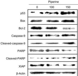
Piperine이 apoptosis 경로 단백질 발현에 미치는 영향 이전 실험 결과를 통해 위암세포 AGS에 piperine을 처치 한 투여군에서 암세포의 생존율 억제와 apoptosis 양성세포 의 유의적인 증가를 관찰하였으므로, western blotting을 통해 apoptosis 조절에 관여하는 단백질들의 발현 정도를 확인하였다. Piperine을 0, 100, 150 μM의 농도로 24시간 처치하여 apoptosis 경로에 관여하는 단백질의 발현을 대조 군과 비교한 결과 p53, Bax, cleaved-PARP, cleaved- caspase-9의 발현은 증가하였고, Bcl-2, XIAP의 발현은
A 0 μM 100 μM 150 μM
0 5 10 15 20 25 30 35 40
0 100 150
Piperine (μM)
Apoptotic cell (%) .
*
*
B
Fig. 3. The effect of piperine-induced apoptosis on AGS cells.
(A) AGS cells were treated with absence or presence of piperine (100 and 150 μM) for 24 h and apoptotic cells were stained with DAPI. Chromatin condensation, representing apoptotic cell death (arrow), was examined using a fluorescence microscope (×200). Indicated bar is 10 μm. (B) The graphs show quantifi- cation of DNA fragmentation and nuclear condensation in AGS cell. Each bar represents the mean±SD calculated from three independent experiments in triplicate. Significance was deter- mined by Dunnett's t-test with *P<0.05 considered as statistically significant compared with non-treated controls.
Piperine 0 100 150 p53
Bax Bcl-2
Caspase-9
Cleaved-caspase-9
PARP
Cleaved-PARP
XIAP β-Actin
Fig. 4. Effect of piperine-induced apoptosis proteins on AGS cell. AGS cells were treated with absence or presence of piperine (100 and 150 μM) for 24 h and cells were harvested to measure protein levels of p53 and Bcl-2 family members, caspase-9, PARP, XIAP by western blotting. We used the housekeeping protein β-actin as a positive loading control in all experiments.
Three independent repetitions of the experiment were performed.
감소하였다(Fig. 4).
P53은 가장 대표적인 종양 억제유전자로서 외부의 자극 을 받아 세포주기를 방해 또는 apoptosis를 유도하는 기능 을 가지고 있으며, DNA 손상이 심하여 손상 부위를 수리할 수 없을 때 Bcl-2 family의 발현을 조절하여 apoptosis를 유도한다. Bcl-2 family는 apoptosis 신호에 의해 미토콘드 리아막에 통로를 만들고, apoptosis를 매개하는 물질의 출 입을 조절한다. 이 단백질에는 apoptosis를 유도하는 활성 인자인 Bax, Mtd/Bok, Bak 등이 있고, apoptosis를 억제하 는 인자인 Bcl-2, Bcl-w, Bcl-xL 등이 존재하고 있다. 이들 Bcl-2 단백질의 조절로 여러 세부적인 과정을 거쳐 궁극적 으로 caspase를 활성화하여 apoptosis를 조절한다. 이들 외에도 apoptosis 과정을 조절하는 인자들로 poly-ADP ri- bose polymerase(PARP)는 세포핵에 존재하고 DNA를 수 선하는 데 있어 중요한 역할을 하며, inhibitor of apoptosis protein(IAP)의 일종으로 X-염색체 연관인자인 XIAP는 caspase-9에 붙어 비정상적인 형태로 활성 영역을 차단하 는 것으로 알려져 있다(17).
Do 등(18)의 연구에서 piperine을 유방암세포 SKBR3와 MCF-7에 10, 25, 50, 100, 200 μM 농도로 처치하였을 때 cleaved-PARP와 cleaved-caspase-3의 발현이 농도 의존적으로 증가하였고, Lin 등(19)의 연구에 의하면 pi-
perine을 폐암세포 A549에 0, 25, 50, 100, 200, 400 μM 농도로 처치하였을 때 Bax와 p53의 발현은 농도 의존적으 로 증가했지만 Bcl-2의 발현은 감소하였다. Kim 등(20)의 연구에서는 piperine을 대장암세포 HT-29에 40 μM 농도 로 처치하였을 때 cleaved-caspase-9의 발현이 증가하였 고, Lai 등(21)의 연구에 의하면 piperine을 마우스 유방암 세포 4T1 세포에 0, 70, 140, 280 μM/L로 처치하였을 때 Bcl-2의 발현은 감소했지만, Bax와 cleaved-caspase-3 의 발현이 농도 의존적으로 증가하였다. 또한, Fofaria 등 (22)의 연구에서는 piperine을 흑색종세포 SK-MEL-28에 0, 100, 150, 200 μM로 처치했을 때 p53의 발현은 농도 의존적으로 증가하였지만, XIAP 발현은 농도 의존적으로 감소하였다. 이러한 결과를 종합해 볼 때 piperine이 위암세 포 AGS에서 p53, Bax, Bcl-2, XIAP의 발현을 조절하고, PARP와 caspase-9의 분절을 유도하여 apoptosis를 유도 하는 것으로 여겨진다.
Piperine이 Akt 신호전달에 미치는 영향
Piperine이 Akt의 인산화에 미치는 영향을 확인하기 위 해 western blotting을 실시하였다. Piperine을 0, 100, 150 μM의 농도로 24시간 처치하였을 때 p-Akt의 발현이 농도 의존적으로 감소한 것을 확인할 수 있었다(Fig. 5).
Greenshields 등(23)의 연구에서 piperine을 유방암세포 MDA-MB-468에 150 μM 농도로 처치하였을 때와 Douc- ette 등(24)의 연구에서 piperine을 T 림프구에 100 μM 농 도로 처치하였을 때 Akt의 인산화가 억제되었다.
Piperine
0 100 150
p-Akt
Akt
β-Actin
Fig. 5. Effect of piperine on Akt phosphorylation in AGS cell.
AGS cells were treated with absence or presence of piperine (100 and 150 μM) for 24 h and cells were harvested to measure protein levels of Akt, p-Akt by western blotting. We used the housekeeping protein β-actin as a positive loading control in all experiments. Three independent repetitions of the experiment were performed.
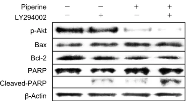
Piperine LY294002 p-Akt Bax Bcl-2 PARP Cleaved-PARP β-Actin
- - + + - + - +
Fig. 6. Effect of Akt inhibitor (LY294002) on Bcl-2 family in AGS cell. AGS cells were treated with absence or presence of piperine (150 μM) and LY294002 (20 μM) for 24 h and cells were harvested to measure protein levels of p-Akt, Bcl-2 family by western blotting. We used the housekeeping protein β-actin as a positive loading control in all experiments. Three independent repetitions of the experiment were performed.
Serine/threonine kinase인 Akt는 여러 암종에서 과 발 현된 상태로 발견되었으며, 세포의 증식, 분화, apoptosis와 같은 세포의 생존에 관여하고 있다(25). Park 등(26)의 연구 에 의하면 curcumin을 대장암세포 HT-29에 6시간 처치하 였을 때 Akt의 활성화 상태인 p-Akt의 발현이 농도 의존적 으로 증가하는 데 반해 p53의 활성이 증가하였다. 또한, Song 등(27)에 의하면 p53은 ubiquitin ligase인 Mdm2의 활성을 통해 조절되며, Mdm2는 Akt에 의해서 인산화가 이 루어지게 되는데, 이러한 Akt는 p53과 매개된 pro-apop- tosis 전사 반응을 방해하거나 p53의 분해나 비활성을 촉진 한다고 보고하고 있다. 따라서 본 실험 결과 piperine이 위 암세포 AGS에서 Akt의 인산화를 억제하고 p53의 발현을 증가시키므로 piperine에 의한 apoptosis 유도는 Akt와 p53의 발현과 연관이 있는 것으로 생각한다.
Akt 억제제가 Akt와 apoptosis 경로 신호전달에 미치는 영향
Akt 억제제인 LY294002와 piperine을 병행 또는 단독으 로 처치했을 때 Akt의 억제에 따른 p-Akt와 apoptosis 관련 단백질의 발현 양상을 확인하기 위해 western blotting을 실시하였다. 그 결과 대조군과 비교하여 LY294002 또는 piperine을 단독으로 처치했을 때 p-Akt와 Bcl-2의 발현은 감소하였고, Bax와 cleaved-PARP의 발현은 증가하였다.
LY294002와 piperine을 병행하여 처치했을 때 Bcl-2의 발 현은 piperine을 단독으로 처치했을 때와 유사한 감소 경향 을 보였지만, p-Akt의 발현은 더욱 감소하는 경향을 보였고 Bax와 cleaved-PARP의 발현은 강하게 증가하는 경향을 확인하였다(Fig. 6). Matsuzaki 등(28)에 의하면 해마신경 세포 rat primary hippocampal neurons에 LY294002를 처치했을 때 대조군과 비교해 Bax의 발현은 증가하였고, Bcl-2의 발현은 감소하였다. 또한, Kunnimalaiyaan 등(29) 에 의하면 인간 갑상선수질 암세포 TT cell에 LY294002를 처치했을 때 농도 의존적으로 cleaved-PARP의 발현이 증가
하였다. 이러한 결과를 종합해볼 때 piperine에 의한 apop- tosis 유도는 Akt 경로와 연관이 있을 것으로 생각한다.
요 약
본 연구는 후추과 식물의 주성분인 piperine이 위암세포 AGS의 항암 효과에 미치는 영향을 확인하기 위해 수행되었 다. Piperine에 의한 위암세포 AGS의 생존율을 확인하기 위해 MTT assay를 수행한 결과 농도 의존적으로 암세포의 생존율이 감소하는 것을 확인하였다. 이러한 암세포의 생존 율 억제가 apoptosis에 의한 효과인지를 확인하기 위해 DAPI staining을 실시한 결과 piperine을 처치한 군에서 apop- totic body와 염색질 응축이 증가하는 것을 확인하였다.
MTT assay와 DAPI staining의 결과를 바탕으로 piperine 이 위암세포 AGS에서 apoptosis와 관련한 단백질 발현 양 상에 미치는 영향을 확인하기 위해 western blotting을 실 시하였다. 그 결과 piperine에 의해 AGS 세포에서 apopto- sis를 유도하는 p53, Bax의 발현은 농도 의존적으로 증가하 였고, apoptosis를 억제하는 Bcl-2, XIAP의 발현은 농도 의존적으로 감소하였다. Apoptosis의 궁극적 단계인 cas- pase-9와 손상된 DNA를 복구하는 PARP의 분절은 증가하 였으며, apoptosis를 방해하는 인자인 Akt의 인산화는 농도 의존적으로 감소하였다. Akt 억제제인 LY294002와 pi- perine을 병행 또는 단독으로 처치하여 western blotting을 진행한 결과 Bcl-2의 발현은 piperine을 단독으로 처치했 을 때와 유사한 감소 경향을 보였지만, p-Akt의 발현은 대 조군과 비교해 LY294002와 piperine을 단독으로 처치했을 때보다 병행했을 때 더욱 감소하였다. 이와 반대로 Bax와 cleaved-PARP의 발현은 강하게 증가하였다. 본 연구의 결 과를 종합해볼 때 piperine이 위암세포 AGS에서 Akt 경로 를 통해 apoptosis를 유도하는 것으로 여겨지며, 향후 위암 예방제나 치료제로의 개발 가능성이 있을 것으로 생각한다.
REFERENCES
1. Song M, Shin GY. 1987. Efficaey of progressive muscle relaxation training in reducing nausea and vomiting, anxiety and symptom distress of cancer chemotherapy. J Nurs Acad Soc 17: 195-203.
2. Lee HJ, Yang HK, Ahn YO. 2002. Gastric cancer in Korea.
Gastric Cancer 5: 177-182.
3. Maehara Y, Hasuda S, Koga T, Tokunaga E, Kakeji Y, Sugimachi K. 2000. Postoperative outcome and sites of re- currence in patients following curative resection of gastric cancer. Br J Surg 87: 353-357.
4. Kim EJ, Part H, Lim SS, Kim JS, Shin HK, Yoon Park JH.
2008. Effect of the hexane extract of Saussurea lappa on the growth of HT-29 human colon cancer cells. Korean J Food Sci Technol 40: 207-214.
5. Sunila ES, Kuttan G. 2004. Immunomodulatory and anti- tumor activity of Piper longum Linn. and piperine. J Ethno- pharmacol 90: 339-346.
6. Srinivasan K. 2007. Black pepper and its pungent princi- ple-piperine: a review of diverse physiological effects. Crit Rev Food Sci Nutr 47: 735-748.
7. Vijayakumar RS, Surya D, Nalini N. 2004. Antioxidant effi- cacy of black pepper (Piper nigrum L.) and piperine in rats with high fat diet induced oxidative stress. Redox Rep 9:
105-110.
8. Mujumdar AM, Dhuley JN, Deshmukh VK, Raman PH, Naik SR. 1990. Anti-inflammatory activity of piperine. Jpn J Med Sci Biol 43: 95-100.
9. Bang JS, Oh DH, Choi HM, Sur BJ, Lim SJ, Kim JY, Yang HI, Yoo MC, Hahm DH, Kim KS. 2009. Anti-inflammatory and antiarthritic effects of piperine in human interleukin 1β- stimulated fibroblast-like synoviocytes and in rat arthritis models. Arthritis Res Ther 11: R49.
10. Karsha PV, Lakshmi OB. 2010. Antibacterial activity of black pepper (Piper nigrum Linn.) with special reference to its mode of action on bacteria. Indian J Nat Prod Resour 1: 213-215.
11. Bezerra DP, de Castro FO, Alves AP, Pessoa C, de Moraes MO, Silveira ER, Lima MA, Elmiro FJ, de Alencar NM, Mesquita RO, Lima MW, Costa-Lotufo LV. 2008. In vitro and in vivo antitumor effect of 5-FU combined with pi- plartine and piperine. J Appl Toxicol 28: 156-163.
12. Hsu HF, Houng JY, Kuo CF, Tsao N, Wu YC. 2008. Glos- sogin, a novel phenylpropanoid from Glossogyne tenuifolia, induced apoptosis in A549 lung cancer cells. Food Chem Toxicol 46: 3785-3791.
13. Oda E, Ohki R, Murasawa H, Nemoto J, Shibue T, Yamashita T, Tokino T, Taniguchi T, Tanaka N. 2000. Noxa, a BH3- only member of the Bcl-2 family and candidate mediator of p53-induced apoptosis. Science 288: 1053-1058.
14. Song Q, Kuang Y, Dixit VM, Vincenz C. 1999. Boo, a novel negative regulator of cell death, interacts with Apaf-1.
EMBO J 18: 167-178.
15. Osaki M, Oshimura M, Ito H. 2004. PI3K-Akt pathway: its functions and alterations in human cancer. Apoptosis 9: 667- 676.
16. Gottlieb TM, Leal JF, Seger R, Taya Y, Oren M. 2002. Cross- talk between Akt, p53 and Mdm2: possible implications for the regulation of apoptosis. Oncogene 21: 1299-1303.
17. Pecorino L. 2012. Molecular biology of cancer: mechanisms, targets, and therapeutics. Oxford University Press, Oxford, UK. p 157-182.
18. Do MT, Kim HG, Choi JH, Khanal T, Park BH, Tran TP, Jeong TC, Jeong HG. 2013. Antitumor efficacy of piperine in the treatment of human HER2-overexpressing breast can- cer cells. Food Chem 141: 2591-2599.
19. Lin Y, Xu J, Liao H, Li L, Pan L. 2014. Piperine induces apoptosis of lung cancer A549 cells via p53-dependent mi- tochondrial signaling pathway. Tumour Biol 35: 3305-3310.
20. Kim EJ, Park HS, Shin MJ, Shin HK, Yoon Park JH. 2009.
Induction of apoptosis in HT-29 human colon cancer cells by the pepper component piperine. J Korean Soc Food Sci Nutr 38: 442-450.
21. Lai LH, Fu QH, Liu Y, Jiang K, Guo QM, Chen QY, Yan B, Wang QQ, Shen JG. 2012. Piperine suppresses tumor growth and metastasis in vitro and in vivo in a 4T1 murine breast cancer model. Acta Pharmacol Sin 33: 523-530.
22. Fofaria NM, Kim SH, Srivastava SK. 2014. Piperine causes G1 phase cell cycle arrest and apoptosis in melanoma cells through checkpoint kinase-1 activation. PLoS One 9: e94298.
23. Greenshields AL, Doucette CD, Sutton KM, Madera L, Annan H, Yaffe PB, Knickle AF, Dong Z, Hoskin DW.
2015. Piperine inhibits the growth and motility of triple-neg- ative breast cancer cells. Cancer Lett 357: 129-140.
24. Doucette CD, Rodgers G, Liwski RS, Hoskin DW. 2015.
Piperine from black pepper inhibits activation-induced pro- liferation and effector function of T lymphocytes. J Cell Biochem 116: 2577-2588.
25. Kada F, Saji M, Ringel MD. 2004. Akt: a potential target for thyroid cancer therapy. Curr Drug Targets Immune Endocr Metabol Disord 4: 181-185.
26. Park SY, Lee SH, Park OJ, Kim YM. 2011. Apoptotic effects of curcumin and EGCG via Akt-p53 signaling pathway in HCT116 colon cancer cells. J Life Sci 21: 89-95.
27. Song G, Ouyang G, Bao S. 2005. The activation of Akt/PKB signaling pathway and cell survival. J Cell Mol Med 9: 59- 71.
28. Matsuzaki H, Tamatani M, Mitsuda N, Namikawa K, Kiyama H, Miyake S, Tohyama M. 1999. Activation of Akt kinase inhibits apoptosis and changes in Bcl-2 and Bax expression induced by nitric oxide in primary hippocampal neurons.
J Neurochem 73: 2037-2046.
29. Kunnimalaiyaan M, Ndiaye M, Chen H. 2006. Apoptosis- mediated medullary thyroid cancer growth suppression by the PI3K inhibitor LY294002. Surgery 140: 1009-1015.