가금 콕시듐증을 일으키는
Eimeria
spp.의 포자충 항원에 결합하는 닭의 재조합 항체(ScFv)의 개발박동운1․김언동1․김성헌1․한재용2․김진규1,†
1창원대학교 자연과학대학 미생물학과,2서울대학교 농업생명과학대학 농생명공학부 WCU 바이오모듈레이션 전공
The Development of Chicken Recombinant Single-chain Fv (ScFv) Antibody Reactive with Sporozoite Antigen of Eimeria spp. which Causes Avian Coccidiosis
Dong Woon Park1, Eon Dong Kim1, Sung Heon Kim1, Jae Yong Han2 and Jin-Kyoo Kim1,†
1Department of Microbiology, College of Natural Sciences, Changwon National University, Changwon 641-773, Korea
2WCU Biomodulation Major, Department of Agricultural Biotechnology, and Research Institute for Agriculture and Life Sciences, Seoul National University, Seoul 151-921, Korea
ABSTRACT The chicken monoclonal antibody (mAb), 13C8, reacts with sporozoite antigens of Eimeria spp. which causes avian coccidiosis. Since this mAb was produced at low amount due to genetic instability of chicken hybridoma, a recombinant 13C8 single-chain Fv (ScFv) antibody was constructed by amplification of the variable domain of heavy (VH) and light chain (VL) genes of antibody derived from chicken hybridoma. The constructed 13C8 ScFv was successfully expressed in E. coli and purified as a soluble form. In ELISA analysis, this recombinant 13C8 ScFv antibody showed antigen binding activity as the original mAb. In addition, nucleotide sequence comparison of 13C8 gene to the germline chicken VL and VH genes suggested that the gene conversion with Vλ and VH pseudogenes might contribute to the diversification of VL and VH genes in chickens.
(Key words : chicken monoclonal antibody, avian coccidiosis, single-chain Fv (ScFv), hybridoma, gene conversion)
†To whom correspondence should be addressed : [email protected]
서 론
조류의 coccidiosis는 장에 기생하는 기생충 속(genus)에 속하는 원생기생충(protozoan parasite)인 Eimeria 종에 의해 야기되는 장의 감염증이다. 감염되어진 조류는 체중 증가가 느려지고, 심한 경우 출혈성 설사와 빈혈 증상을 나타낸다 (Lillehoj and Lillehoj, 2000). 특히 대량 사육을 하는 양계 농가 에서 coccidiosis에 의한 경제적인 손실은 막대하다고 할 수 있다. 그러므로 효율적인 coccidiosis 치료 및 예방을 위해 화학적인 항콕시듐약제가 사용되었으나 높은 비용과 장기간 반복적으로 사용하게 되면 내성을 갖는 종 출현 등의 단점 으로 그 사용이 제한되어(Chapman, 1993), 이를 대체할 수 있 는 두 가지의 면역학적 방법이 보고되어졌다. 우선, 체내 항체 생성을 촉진시킬 수 있는 재조합 단백질 백신을 개발하는
방법(Lawn and Rose, 1982)이 보고되어졌다. 즉, coccidial 항 원에 대해 특이성을 나타내는 여러 종류의 생쥐 단일 클론 항체를 개발하여 그 항체가 인지하는 항원을 subunit va- ccine으로 이용하려는 방법이나, 생쥐의 항체에 의해 인식되 어지는 coccidial 항원은 닭의 면역체계에서 인식되어지는 항원과 차이점이 있어 닭에서 효과적인 면역성을 유도하지 못하여 생쥐의 단일 클론 항체를 이용한 subunit vaccine 개 발 방법은 큰 효과를 보지 못하고 있는 실정이다. 이러한 문 제점을 극복할 수 있는 또 다른 면역학적 방법으로 Eimeria 가 숙주세포로 침입하는 것을 직접적으로 막아주는 단일 클 론 중화 항체를 개발하여 수동 면역(passive immunity)을 획 득하게 하는 방법이 보고되었다(Sasai et al., 1996). 즉, 보고 되어진 항콕시듐증 중화 단일 클론 항체는 Eimeria acervulina 의 sporozoite antigen으로 닭을 immunization시켜서 얻은 비장
세포를 닭의 myeloma cells와 융합시켜 개발하였으며, 이 항 체는 20~21 kDa의 분자량를 갖는 E. acervulina의 sporozoite 항원과 결합하는 것으로 알려져 있다(Sasai et al., 1996). 또 한 인식되어지는 항원은 sporozoite가 닭의 CD8+ T cells로의 감염에 관여하는 것으로 알려져 있으며, 이는 in vitro에서 이 항체가 Eimeria sporozoites의 CD8+ T cells로의 감염을 차 단시키는 중화 효과(neutralization effect)를 나타냄으로써 증 명되었다(Sasai et al., 1996). 그러나 이러한 치료 목적의 닭 항체개발은 닭 하이브리도마 세포의 유전적 불안정성과 발 현되는 항체의 낮은 생산성 때문에 극히 제한되어져 있으므 로(Nishinaka et al., 1996), 이를 극복하기 위해 항체 공학 (Winter et al., 1994)을 이용한 재조합 닭 항체를 제조하는 방법이 보고되어졌다(Yamanaka et al., 1996).
항체 공학을 이용해 제조되는 재조합항체는 특이적 항원 결합력을 갖는 Fv 항체와 Single-chain Fv(ScFv) 항체(Hayden et al., 1997; Verma et al., 1998) 형태이다. Fv 구조는 항체의 중사슬 가변 부위(VH)와 경사슬 가변부 위(VL)가 비공유 결합으로 형성된 이형이량체로서 Y-shape의 모 항체와 같은 항원 결합 부위와 항원 특이성을 갖게된다. 그러나 단백질 발현시 불안정한 비공유결합으로 인해 VH와 VL이 분리되 기 때문에 이러한 단점을 극복하기 위해 VH유전자와 VL유 전자 사이에 linker peptide를 연결한 single chain Fv (ScFv) 형태가 제조되었다. 그 결과 발현되는 ScFv 항체는 공유 결 합인 펩티드 결합에 의해 VH와 VL을 연결하므로 비공유적 으로 결합하는 Fv보다 안정한 상태로 발현되어 Fv의 불안 정한 구조로 인한 항원 결합력의 감소를 최소화시킬 수 있게 된다(Bird et al., 1988; Huston et al., 1991). 이 방법에 의하면 hybridoma 배양에 의한 단일 클론 항체의 제조보다 짧은 시 간에 대량의 단일 클론 항체 제조가 가능하며, 또한 유전자 조작이 용이해서 무작위 돌연변이(random mutation)을 이용 하여 인위적으로 재조합 항체의 항원에 대한 결합친화력을 증가시킬수 있는 잇점이 있다. 또한 높은 친화력을 갖는 닭 의 재조합 ScFv를 이용한 affinity chromatography 또는 im- munoprecipitation 등을 통해, 인지되는 항원의 탐지 및 정제가 용이해져서 정제된 항원을 이용하여 Coccidiosis를 예방할 수 있는 subunit vaccine 개발 가능성 또한 제시해 줄 것이다.
한편, phage-displayed antibody technology를 이용하여 닭의 비장 조직을 항체유전자의 naive repertoire source로 이용 가 능하기 때문에 닭항체의 유전자도서관(gene library)을 제조, hybridoma technology에 의한 단일 클론 항체 제조 방법에 의해서는 거의 불가능한 다양한 항원에 대해 특이성을 갖는 재조합 단일 클론 항체의 개발 또한 가능하게 될 것이다.
본 연구에서는 닭 hybridoma의 유전적 불안정성과 항체 발 현량의 제한성(Nishinaka, et al., 1996)을 극복하기 위해 같은 Eimeria에 속하는 Eimeria acervulina, Eimeria maxima, Eime-
ria tenella 포자충(sporozoites) 항원에 특이적으로 결합하는
닭 단일 클론 항체 13C8 hybridoma로부터 ScFv 항체 유전자 를 구축하였다. 구축된 닭 13C8 ScFv 항체는 E.coli를 숙주 (host)로 하여 성공적으로 발현 및 정제되어 졌다. ELISA를 통해 제조된 재조합 13C8 ScFv 항체의 항원 결합력을 측정 하였고, 13C8 ScFv 항체 유전자의 염기 및 아미노산 배열 분 석을 통해 13C8 ScFv 항체 단백질의 3차 구조를 예측해 보 았으며, 이미 알려진 닭의 germline 항체 유전자와 비교분석 하여 13C8 항체의 VH 및 VL유전자는 gene conversion 기작 에 의해 pseudogenes들로부터 유래되어짐을 확인하였다.
재료 및 방법
1. Variable Region Gene의 유전자 증폭 및 분석
Eimeria spp. sporozoite 항원과 결합하는 단일 클론 항체
를 분비하는 닭의 13C8 hybridoma 세포주로부터 TRIzol(In- vitrogen, U.S.A.)을 이용해 Total RNA를 추출하였다. 5μg의 total RNA와 Oligo d(T)18 primer 그리고 SuperScript Ⅱ re- verse transcriptase(Invitrogen, U.S.A.)를 이용하여 First strand cDNA를 합성하였고, 제조된 cDNA를 주형으로 하여 각각 의 primer set에 의해 VH와 VL 유전자가 증폭되었다. 즉, CK VLB(VL reverse primer)와 CKVLF(VL forward primer) 를 이용하여VL유전자를 증폭하였고, CK VHB(VH reverse primer)와 CKVHF(VH forward primer)는 VH 유전자의 증 폭을 위해 사용되었다(Table 1). 특히 Splicing by Overlap Extension(SOEing) PCR을 위해 상보적인 linker sequence (italics: GSTSGKPSEGKG)를 CKVLF와 CK VHB primer에 포함시켰다(Table 1). PCR products를 1% agarose gel에서 전개하여 증폭된 DNA fragment를 QIAEX Ⅱ gel extraction kit(QIAGEN, Germany)를 이용해 gel로부터 회수하였다. 회 수되어진 PCR products는 pGEM-T vector(Promega, U.S.A.) 로 클로닝하여 유전자 염기 배열을 분석하였다. 얻어진 se- quencing 결과를 이미 알려진 닭의 heavy chain 및 light chain 의 germline sequence(Reynaud et al., 1987)와 배열하여 비교 분석하였다.
2. 13C8 ScFv 항체유전자의 구축
1차 PCR에 의해 얻어진 VH와 VL 유전자는 증폭 시 첨가 된 linker의 각각 상보적인 sequence들을 포함하고 있으므로
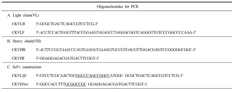
Table 1. Primers used for PCR amplification for chicken ScFv construction
Oligonucleotides for PCR A. Light chain(VL)
CKVLB 5'-GCGCTGACTCAGCCGTCCTCG-3'
CKVLF 5'-ACCTCCACTGGGTTTACCGGAAGTAGAGCCTAGGACGGTCAGGGTTGTCCCGGCCCCAAA-3' B. Heavy chain(VH)
CKVHB 5'-ACTTCCGGTAAACCCAGTGAAGGTAAAGGTGCCGTGACGTTGGACGAGTCCGGGGGCGGC-3'
CKVHF 5'-GGAGGAGACGATGACTTCGGT-3'
C. ScFv construction
CKVLSfi 5'-GTCCTCGCAACTGCGGCCCAGCCGGCCATGGC GCGCTGACTCAGCCGTCCTCG-3' CKVHNot 5'-GGCCACCTTTGCGGCCGC GGAGGAGACGATGACTTCGGT-3
CKVLB, reverse primer for light chain variable region. CKVLF, forward primer for light chain.
CKVHB, reverse primer for heavy chain variable region. CKVHF, forward primer for heavy chain.
Italic represents linker sequences for gene assembly of final ScFv constructio.
Sfil and Notl restriction sites are underlined.
VLSfi(SfiI restrictionsite is underlined):와 VHNot(NotI restric- tion site is underlined)의 primer를 이용한 2차적인 Splicing by Overlap Extension PCR(SOEing PCR)에 의해 ScFv가 최 종 완성되어졌다. 증폭된 13C8 ScFv DNA는 DNA 제한 효소 SfiI과 NotI에 의해 5'-pelB leader sequence와 3'-hexahistidine tag을 가진 pUC119 벡터로 클로닝하였다.
3. 13C8 ScFv 항체의 발현, 정제 및 Westernblot ScFv 유전자를 포함한 발현용 pUC119 plasmids로 E. coli BMH 71-18을 형질전환하고, 형질 전환된 E. coli BMH 71- 18을 100μg의 ampicillin과 1%(w/v) glucose를 포함한 2XTY 액체배지에 접종하여 30℃, 180 rpm에서 밤새 진탕 배양하 고. 2,500 g에서 10분 동안 원심분리하여 상등액을 제거한 다. 2XTY 액체 배지를 이용해 한번 세척해 주고, 100μg의 ampicillin과 0.25 mM isopropyl-β-D-thiogalactopyranoside를 포함한 2XTY 액체배지에 bacteria pellet을 재현탁하여 25℃, 5.5시간 동안 배양하여 단백질 발현을 유도하였다. 배양후 세포 배양액을 원심분리하여 상등액을 제거하고 bacteria cell pellet을 차가운 TES buffer(0.5M sucrose, 0.1mM EDTA 그 리고 200mm Tris-Cl, pH 7.5)를 이용하여 현탁하고 4℃에서 1시간 동안 배양하였다. 다시 원심분리하여 수거된 상등액을 PBS를 이용해 밤새 투석한 다음 단백질 샘플 용액을 Ni-NTA agarose에 결합시키고, TN buffer I(0.1M Tris-Cl, 0.5M NaCl, pH 7)과 Ⅱ(0.1M Tris-Cl, 0.5M NaCl, pH 8.0)를 이용해 세척
해 준다. 세척이 끝난 Ni-NTA agarose에 250 mM immida- zole을 처리해 단백질을 용출해 낸다. TES buffer 처리 후 남은 bacteria cell pellet을 Sonication buffer(0.1M Tris-Cl, 0.5 M NaCl, pH 7.5)에 재현탁 후 초음파 파쇄하고, 원심분리 후 얻 어진 상등액을 다시 위와 같은 방법으로 정제에 이용한다. 정 제된 단백질들은 2XSDS-PAGE buffer와 섞어 100℃에서 5분 간 끓이고 15% polyacrylamide gel에 전기영동한 후 0.25%
coomassie brilliant blue R-25 용액을 이용해 염색하였다.
Western blot 분석을 위해 정제된 13C8 ScFv 단백질은 전기 영동방법으로 SDS-PAGE하고 PVDF membrane(Millipore, U.S.A.)으로 transfer 하였다. 그리고 transfer되어진 membrane 을 2%(w/v) skim milk를 포함한 PBS-T buffer를 이용하여 실온에서 1시간동안 blocking한 다음, PBS-T로 2회 세척하 였다. 발색을 위해 2차 항체인 peroxidase-conjugated polyhis- tidine monoclonal antibody(Sigma-Aldrich, U.S.A.)를 1:2,000 (v/v)으로 희석해서 membrane에 처리하여 실온에서 1시간 동 안 incubation하였다. PBS-T washing buffer로 5회 세척한 다 음, 3,3-diaminobenzidine(Sigma-Aldrich, U.S.A.)를 이용해 발색시켜 13C8 ScFv 항체의 발현을 재확인하였다.
4. 13C8 ScFv의 3차 구조 모델링
분석된 13C8 ScFv 항체 유전자로부터 아미노산 배열 정보 를 결정한 다음 WAM(http://antibody.bath.ac.kr/) 프로그램을 이용하여 3차 구조 모델링을 실시하였다. 결정된 13C8의 VH
및 VL의 아미노산 서열을 입력하고, 이미 3차 구조가 밝혀 진 항체들 중에서 가장 유사한 아미노산 서열을 갖고 있는 항체를 RCSB protein data bank(http://www.rcsb.org/pdb/)에 서 찾아내어 이를 주형으로 항원 결합부위인 complementa- rity determining region(CDR)의 loop와 항체의 구조적인 안 정화 역할을 하는 framework region(FR)의 β-sheet의 3차 구 조를 결정하였다. 추정된 3차 구조는 PyMol(Delano Scien- tific, U.S.A.) 프로그램을 통해 형상화 하여 분석하였다.
5. Eimeria항원 준비
Eimeria acervulina sporozoites 포자충(sporozoite) 항원을 0.125%(w/v)의 trypsin과 1%(w/v)의 taurodeoxycholic acid를 포함한 Hanks’ balanced salt solution pH 7.6(Sigma-Aldrich, U.S.A.)에 현탁해 41℃의 humidified incubator에서 10분 동안 배양하였고, 원심분리에 의해 sporozoites가 가라앉으면, 상 등액을 제거하고 Dulbecco’s phosphate buffered saline(Sigma- Aldrich, U.S.A.)에 현탁해 6번의 냉동과 해동을 반복하고, 초음파 파쇄하여 사용하였다. 같은 Eimeria(genus)속에 속하 는 Eimeria maxima 그리고 Eimeria tenella 포자충 항원도 동일한 방법으로 준비되었다.
6. Enzyme Linked Immunosorbent Assay(ELISA) 13C8 ScFv 항체의 Eimeria sporozoites antigen과의 결합 력을 확인하기 위해 2μg의 항원을 4℃에서 밤새 coating시 킨 96-well maxisorp plate(Nunc, Denmark)를 이용한 ELISA 를 수행하였다. 항원이 coating된 plate를 0.1%의 tween 20을 포함한 PBS(PBS-T)를 이용하여 3회 세척하고, 2% skim milk 를 포함한 PBS-T를 이용하여 blocking해 주었다. 2μg의 13C8 ScFv 항체를 포함한 blocking buffer를 처리하여 실온 에서 1시간 동안 항체가 결합할 수 있도록 한 후, PBS-T를 이용한 3번의 세척 후 horseradish peroxidase가 conjugated된 polyhistidine monoclonal antibody(Sigma-Aldrich, U.S.A.)를 처리하여 ABTS로 발색하여 Microplate reader(Expert 96, Asys, Austria)로 492 nm 파장에서 흡광도를 측정하였다.
결과 및 고찰
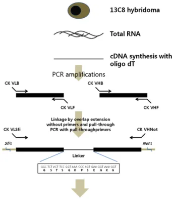
닭의 VH와 VL 유전자는 닭 hybridoma cell line 13C8으 로부터 RT-PCR에 의해 증폭되어졌고, SOEing PCR에 의해 성공적으로 ScFv 항체유전자가 구축되었다(Fig. 1 and Fig.
2B). 닭의 VH와 VL 유전자의 크기는 각각 350 bp와 325 bp 정도이지만, SOEing PCR에 의한 최종 ScFv PCR product는
Fig. 1. Schematic outline for the construction of chicken 13C8 ScFv antibody. The total RNA was extracted from 13C8 hybridoma. cDNA was synthesized by using oligo d(T)18. The locations of all PCR primers on the two variable region genes are shown. Two step PCR reactions were performed. The first and second PCRs link and amplify full-length 13C8 ScFv gene, which were cloned into pUC119 plasmid. The DNA sequence encoding the fle- xible linker is depicted in detail.
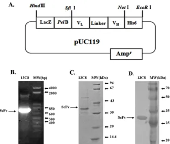
VH와 VL 유전자 사이에 linker peptides 유전자가 포함되어 져 750 bp의 유전자로 증폭되어졌다(Fig. 1 and Fig. 2B). 증 폭된 ScFv 유전자는 Sfi I 및 Not I의 두 제한 효소를 이용하 여 박테리아 발현 벡터로 클로닝 되어졌다. 본 연구에서 이용 된 발현 벡터는 IPTG에 의한 전사(transcription)를 활성화시 키기 위해 lac Z promoter를 사용하였고, N-terminus에 pel B leader peptide(Kim et al., 1994)를 첨가하여 발현된 단백질 을 E. coli의 periplasmic space로 이동시켜 분비를 촉진시켰 다. 또한 C-terminus에는 6개의 histidine을 첨가시켜 Ni-NTA- Agarose affinity column를 사용하여 발현된 단백질의 정제 를 가능케 하였다(Fig. 2A).
재조합 13C8 ScFv 발현 벡터는 항체 단백질의 발현을 위 해 E. coli를 형질 전환시켜 IPTG에 의해 단백질 발현을 유 도하였다. 발현된 단백질은 Ni-NTA affinity column에 의해 정제되어지고 SDS-PAGE를 수행한 결과 약 32 kDa의 13C8
Fig. 2. (A) Construction of recombinant ScFv plasmid. Lac Z promoter is activated by IPTG for overexpression of ScFv antibody. Pel B leader transports expressed pro- teins into periplasmic space for secretion. His6 is hexa- polyhistidine tag for detection and purification of ex- pressed ScFv proteins. (B) The recombinant ScFv(750 bp) was amplified with chicken VLBsfi and VHFNot oli- gonucleotides and resolved on 1% agarose gel and stained with ethidium bromide. DNA size markers are shown on the right. (C) The ScFv proteins purified from E.
coli was resolved by SDS-PAGE and stained with Coo- massie Blue. The protein size markers are shown on the right. (D) The ScFv protein was blotted on to PVDF membrane and stained with horseradish peroxidase- conjugated anti-polyhistidne secondary antibody. The pro- tein size markers are shown on the right.
ScFv 항체 발현을 확인할 수 있었다(Fig. 2C). 또한 재조합 13C8 ScFv는 C-terminus에 6개의 histidine을 포함하므로 이 histidine에 결합하는 항체를 이용한 Western blot를 실시하 여 13C8 ScFv의 발현을 재확인하였다(Fig. 2D). 13C8의 VH와 VL 유전자의 염기배열은 CB strain의 닭으로부터 분석된 ger- mline sequence(Reynaud et al., 1987, 1989)와 비교 분석하였 고, Framework region(FR)과 complementarity determining re- gions(CDRs)는 Kabat sequence에 따라 결정되었다(Kabat et al., 1991). Germline sequence와 13C8 항체의 염기배열상의 차이는 주로 CDRs region에서 발견되었다(Fig. 3). 예를 들 면 VLCDR3의 경우 16개의 nucleotides(TGATAGTGATTA T GTT)가 삽입되었고, 3개의 nucleotides(ACA)가 삭제되어 진 변화를 보여주고 있다(Fig. 3).
한편 CB strain으로부터 알려진 다수의 pseudogenes 정보 (Reynaud et al., 1987, 1989)를 이용하여 13C8 항체의 VH 및
VL 유전자가 유래된 pseudogenes을 추정한 결과 13C8 항체의 VH 및 VL 유전자는 모두 고유한 특정pseudogene에서 유래되 었음을 확인할 수 있었다(Fig. 4). 따라서 무수히 많은 항원 에 대한 특이성을 나타내기 위한 닭 항체 유전자의 다양성 은 다수의 pseudogenes이 gene conversion이라고 하는 특정 recombination 기작에 의해 다양한 형태로 변화되어 닭 항체 유전자의 다양성을 유지한다는 것을 확인할 수 있었다(McCor- mack and Thompson, 1990).
3차 구조 모델링을 통해 예측되어진 13C8 ScFv 항체의 구조를 보면 VH와 VL 모두 각각 3개씩의 CDR 지역이 고유 한 loop를 형성하고, 이 총 6개의 CDR loop들이 모여 hydro- phobic pocket을 형성하여 항원과 특이적으로 결합하는 부 위를 형성하게 된다(Fig. 5).
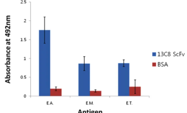
재조합 13C8 ScFv 항체의 주된 기능인 항원 결합 능력을 측정하기 위해 ELISA 분석을 시도하였다. 같은 Eimeria에 속하는 Eimeria acervulinar, Eimeria maxima 그리고 Eimeria tenella 포자충(sporozoite) 항원이 고정된 plate에 재조합 13C8 ScFv 항체를 처리하여 항원 결합력을 측정한 결과, 사용되 어진 세가지 Eimeria 종류의 포자 충항원을 모두 인지하는 것으로 확인되었다(Fig. 6).
이 결과는 13C8 ScFv 항체가 세 종류의 Eimeria strain에 공통적으로 존재하는 항원에 결합하는 가능성을 제시해 주 고 있으므로, 13C8 ScFv 항체는 언급된 세 종류 Eimeria가 일으키는 조류 콕시듐증에 공통적으로 사용할 수 있는 진단 및 치료목적의 항체로 개발되어질 수 있는 잇점을 가지게 될 것이다. 또한 13C8 항체가 인지하는 항원을 탐지해내는 연구를 통해 재조합 vaccine 개발 또한 가능할 것이다.
적 요
닭의 단일 클론 항체인 13C8 항체는 조류의 콕시듐증을 유발하는 것으로 알려진 Eimeria acervulina의 포자충(sporo- zoites) 항원에 결합하는 닭 항체이다. 그러나 닭 하이브리도 마 유전자의 불안정성 때문에 분비되는 항체의 생산량이 낮 은 단점이 있다. 이러한 단점을 보완하기 위해 hybridoma로 부터 항체의 중사슬 가변 부위(VH)유전자와 경사슬 가변 부 위(VL) 유전자를 증폭하여 linker peptide로 연결해준 재조 합 ScFv 항체 유전자를 구축하고, E. coli를 형질 전환시켜 재 조합 단백질로 발현 정제하였다. ELISA 분석 결과 재조합 13C8 ScFv 항체는 세 종류의 Eimeria spp.에 대해 모두 항 원 결합력을 나타내었으며, 염기서열 분석을 수행하여 ger- mline sequence와 비교한 결과 닭 항체유전자의 다양성(di-
A. Heavy chains B. Light chains Fig. 3. Nucleotide sequences of VH(a) andVL(b) genesof 13C8ScFv. Germline sequences fromthe CBstrainare shownatthe topandthosefromthe 13C8 hybridoma celllinesbelow. Nucleotide identitiestothe germline sequences are shownbydashes().Nucleotide deletions are indicatedby a colon(:). Complementarity-determiningregions (CDRs)andPCRprimers are underlinedinthe germline sequences.Regions tobe filledwithDsegment inthe germline heavychainare shownbyN.
Fig. 4. Donor pseudogenes used in variable regions of λ-light chain(VL) and heavy chain(VH). Boundaries between inserted pseudogenes and the germline sequences cannot be specified. If the number of candidate pseudogenes exceeded one, all are listed anddivided by slashes.
Fig. 5. Deduced amino acid sequences of 13C8 ScFv (A). The colored complementarity-determing regions (CDRs) are indicated with underlines in the ScFv sequences. Ho- mology modeling of chicken 13C8 ScFv (B). (1) Side view. (2) Top view. Colored regions represent VH CDR (HCDR1, 2, 3) and VL(LCDR1, 2, 3) (HCDR 1: red, HCDR2: green, HCDR3: blue, LCDR 1: yellow, LCDR 2: magenta and LCDR 3: Cyan).
versity)은 pseudogene들의 gene conversion 기작에 의해 이 루어짐을 제시해 주었다.
(색인어: 조류 단일클론 항체, 조류 콕시듐증, single-chain Fv (ScFv) 항체, 하이브리도마, 유전자 재조합)
사 사
본 연구는 2011년도 농촌진흥청 Biogreen 21 지원사업 (No. 2007040103401)에 의해서 수행되었으며, 이에 감사드 립니다.
Fig. 6. ELISA analysis of three Eimeria spp. (E. acervulina, E.
maxima and E. tenella) sporozoite antigens with 13C8 ScFv antibody. 13C8 ScFv antibody or BSA (negative control) were reacted with three soluble Eimeria spp.
sporozoite antigens in a 96-well microplate and the ab- sorbance at 492 nm was measured.
인용문헌
Bird RE, Hardman KD, Jacobson JW, Johnson S, Kaufman BM, Lee SM, Lee T, Pope SH, Riordan GS, Whitlow M 1988 Single-chain antigen-binding proteins. Science 242:
423-426.
Chapman HD 1993 Resistance to anticoccidial drugs in fowl.
Parasitol Today 9:159-162.
Hayden MS, Gilliland LK, Ledbetter JA 1997 Antibody engi- neering. Curr Opin Immunol 9:201-212.
Huston JS, Mudgett-Hunter M, Tai MS, McCartney J, Warren F, Haber E, Oppermann H 1991 Protein engineering of single- chain Fv analogs and fusion proteins. Methods Enzymol 203:46-88.
Kabat EA, Wu TT, Perry HM, Gottesman KS, Foeller C 1991 Sequences of proteins of immunological interest. U.S.
Dept Health and Human Services, NIH publication No. 91- 3242. 5th ed.
Kim JK, Tsen MF, Ghetie V, Ward ES 1994 Identifying amino acid residues that influence plasma clearance of murine IgG1 fragments by site-directed mutagenesis. Eur J Immunol 24:542-548.
Lawn AM, Rose ME 1982 Mucosal transport of Eimeria te- nella in the cecum of the chicken. J Parasitol 68:1117-1123.
Lillehoj HS, Lillehoj EP. 2000 Avian coccidiosis. A review of acquired intestinal immunity and vaccination strategies.
Avian Dis 44:408-425.
McCormack WT, Thompson CB 1990 Chicken IgL variable region gene conversions display pseudogene donor prefe- rence and 5' to 3' polarity. Genes Dev 4:548-558.
Nishinaka S, Akiba H, Nakamura M, Suzuki K, Suzuki T, Tsubokura K, Horiuchi H, Furusawa S, Matsuda H 1996 Two chicken B cell lines resistant to ouabain for the pro- duction of chicken monoclonal antibodies. J Vet Med Sci 58:1053-1056.
Reynaud CA, Anquez V, Grimal H, Weill JC 1987 A hyper- conversion mechanism generates the chicken light chain preimmune repertoire. Cell 48:379-388.
Reynaud CA, Dahan A, Anquez V, Weill JC 1989 Somatic hy- perconversion diversifies the single Vh gene of the chicken with a high incidence in the D region. Cell 59:171-183.
Sasai K, Lillehoj HS, Matsuda H, Wergin WP 1996 Cha-
racterization of a chicken monoclonal antibody that recog- nizes the apical complex of Eimeria acervulina sporo- zoites and partially inhibits sporozoite invasion of CD8+ T lymphocytes in vitro. J Parasitol 82:82-87.
Verma R, Boleti E, George AJ 1998 Antibody engineering:
comparison of bacterial, yeast, insect and mammalian ex- pression systems. J Immunol Methods 216:165-181.
Winter G, Griffiths AD, Hawkins RE, Hoogenboom HR 1994 Making antibodies by phage display technology. Annu Rev Immunol 12:433-455.
Yamanaka HI, Inoue T, Ikeda-Tanaka O 1996 Chicken mo- noclonal antibody isolated by a phage display system. J Immunol 157:1156-1162.
(접수: 2011. 10. 4, 수정: 2011. 11. 21, 채택: 2011. 11. 22)