Res. Plant Dis. 17(3) : 364−368 (2011) © The Korean Society of Plant Pathology
Gray Mold on Carrot Caused by Botrytis cinerea in Korea
Kyeong-Hun Park, Kyoung-Yul Ryu, Hye-Jeong Yun, Jeong-Chul Yun, Byeong-Seok Kim, Kyu-Sik Jeong
1, Young-Seok Kwon
2and Byeongjin Cha
3*Microbial Safety Division, National Academy of Agricultural Science, Rural Development Administratiom Suwon, 441-707, Korea
1Variety Testing Division, Korea Seed and Variety Service, Suwon, 443-400, Korea
2Highland Agriculture Research Center, Natiomal Institute of Crop Science, Rural Development Administration, Pyeongchang 232-955, Korea
3Department of Plant Medicine, Chungbuk National University, Cheongju 361-763, Korea (Received on September 26, 2011; Revised on December 6, 2011; Accepted on December 15, 2011)
Gray mold caused by Botrytis cinerea was found on a carrot seedling in a greenhouse and a field at Daegwallryeong, Gangwon Province in 2007–2009. Symptoms included irregular, brown, blight, or chlorotic halo on leaves and petioles of the carrots. Fungal conidia were globose to subglobose or ellipsoid, hyaline or pale brown, nonseptate, one celled, 7.2–18.2 × 4.5–11 µm (12.1
× 8.3 µm) in size, and were formed on botryose heads. B. cinerea colonies were hyaline on PDA, and then turned gray and later changed dark gray or brown when spores appeared. The fungal growth stopped at 35
oC, temperature range for proper growth was 15–25
oC on MEA and PDA.
Carrots inoculated with 1 × 10
5ml conidial suspension were incubated in a moist chamber at 25
± 1
oC for pathogenicity testing. Symptoms included irregular, brown, water-soaked rot on carrot roots and irregular, pale brown or dark brown, water-soaked rot on leaves. Symptoms were similar to the original symptoms under natural conditions. The pathogen was reisolated from diseased leaves, sliced roots, and whole roots after inoculation. As a result, this is the first report of carrot gray mold caused by B. cinerea in Korea.
Keywords : Botrytis cinerea, Daucus carota L. subsp. sativas (Hoffm), Leaf blight
Gray mold, caused by the important pathogen Botrytis cinerea, affects most vegetables and fruit crops as well as a large number of trees, flowers, and weeds (Jarvis, 1977). Carrot (Daucus carota L. subsp. sativas, Hoffm.), belongs to the family Umbelliferae. Carrot roots are consumed as an edible vegetable and contain many nutritive elements such as vitamins, minerals, and fiber (Rubatzky et al., 1999).
Gray mold symptoms were first found on leaves and petioles of carrots in the field. A fungus was isolated from infected tissues and re-isolated from inoculated samples using a conidial suspension.
However, gray mold caused by B. cinerea on carrot has not been reported in many countries. In many
crops including vegetables the mold causes brown spot on the leaves, petioles, and petals (Ellis, 1971;
Hennebert, 1973). The pathogenic fungus, B.
cinerea, causes a serious gray mold disease wherever umbelliferous crops are grown. This fungus is associated with significant postharvest carrot disease in the USA (Davis and Raid, 2002) but has not been reported on the leaves and petioles of carrots infected in the field in Korea. This fungus has low activity in storage at 0
oCbut can spread to adjacent tissue at 4
oC(Goodliffe and Heale, 1977). Additionally, B. cinerea is a problem on fruits and vegetables as well as carrots in cold storage and subsequent shipping, because the fungus is able to remain active at temperatures just above freezing (Hennebert, 1973). Gray mold originates on some parts of the carrot taproot, and the first symptoms are water-
*Corresponding author
Phone)+82-43-261-2557, Fax) +82-43-271-4414 Email)
[email protected]
http://dx.doi.org/10.5423/RPD.2011.17.3.364 Note Open Access
soaked, light brown to tan lesions. When the fungus expands, the lesions become covered with gray conidia that penetrate into inner tissues (Sharman and Heale, 1977).
This study was conducted to identify the casual agent related with blight lesions on carrot leaves collected from the field in Pyeongchang and to investigate the morphology, cultural characteristics, and â-tubulin analysis of the pathogen and pathogenicity on carrot.
Fungi were isolated from gray mold lesions on carrots grown in Pyeongchang. The surface of the diseased leaf tissue pieces was sterilized with 70%
ethanol for 1 minute, rinsed twice with sterilized distilled water, and then air dried on sterilized filter paper. Dried samples were placed on water agar (WA) and then incubated at 25 ± 1
oC.After 3 days of inoculation, growing mycelial tips were cultured on potato dextrose agar (PDA) plates for 2 weeks.
Single spores were isolated by the dilution method on WA and cultured on PDA for 7 days.
The shape, size, color, and conidia of the isolates were investigated under a microscope (Eclipse E600;
Nikon, Tokyo, Japan) after a 25 ± 1
oC incubation for 2 weeks on CMA, MEA, PDA, V8A, and WA.
Cultural characteristics of the isolates were observed on various culture media incubated for 2 weeks at 25
± 1
oC. Mycelial growth on the media was observed after a 7 day incubation at 25 ± 1
oC. Colonies were incubated on PDA from 5–35
oC at intervals of about 5
oC with three replications to investigate the temperature range of growth. Agar disks of appropriate size (1 mm thick) were cut from a B.
cinerea isolate on PDA for scanning electron microscopy (SEM). Samples were immersion-fixed in 2.5% glutaraldehyde in 0.1 M phosphate buffer (pH 7.2–7.4) for 4 h. The fixed samples were rinsed with 0.1 M phosphate buffer th
ree times for 20 min each.Samples were post-fixed in 1% osmium tet
roxide in 0.1 M phosphate buffer for 1 h. Samples were washed in 0.1 M phosphate buffered saline (three time for 20 min each), dehydrated in a graded ethanol series (30, 50, 60, 70, 80, 90, 95, and 100%) for 15 min per step, and then rinsed in absolute ethanol three times
for 30 min each. After critical point drying, the specimens were sputter-coated with gold using a sputter coater and then observed under SEM (Hitachi S-2460N;
Hitachi Instruments, Hazel- brook, Australia) at 5–20 kv.
Isolates were used to test carrot pathogenicity.
Mycelial agar blocks, made from an 8 mm cork borer, were subcultured on PDA for 14 days.
Conidia suspensions were made by adding 3 ml of sterile distilled water in PDA, scraping the colonies with a rubber spatula, and then harvesting them by filtering through four layers of cheesecloth. Leaves, sliced roots, and whole roots of carrot with or without wounds were inoculated with 30 µl drops of conidial suspension (1 × 10
5conidia per ml). Inoculated samples were laid in a plastic box (292 × 225 × 120 mm) under 100% humidity, and incubated at 25 ± 1
oC for 2 weeks.
To identify the isolates, the β-tubulin gene was amplified with the Bcin-366r and BT-2M-up primers (Spotts et al., 2008) and directly sequenced. The sequence was compared with data in GenBank (Accession No. FQ790278 and Z69263). A molecular phylogenetic analysis was carried out with MEGA4 using the neighbor-joining method and the Tajima- Nei distance model.
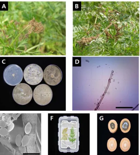
Gray mold was found on carrot leaves in September 2007. Gray mold originally appeared on the edge of the infected leaves with a small spot or chlorotic halo. Irregularly shaped and water-soaked lesions developed on infected leaves. Subsequently, infected petioles and stems became slender and were totally blighted on the above ground part of the carrot. The fungal colonies were hyaline and then turned gray, and the spores changed dark gray or brown (Fig. 1A, B). Colonies were hyaline or light gray to gray, scare, and weak on CMA and WA.
Sclerotia were black in color and irregular in shape
but were absent on CMA and WA. The size of the
sclerotia on MEA was larger than that on other
media (Fig. 1C). Hyphae were hyaline to gray, with
granules, and articulate-septated. Conidia that formed
on conidiophores in PDA were not in chains, hyaline to pale brown, one-celled, globose to subglobose or ellipsoidal, and measured 7.2–18.2 × 4.5–11 µm (12.1 × 8.3) in size (Fig. 1D). SEM revealed that the conidia of the isolate had
a smooth surface (Fig. 1E). All inoculated samples on the wounded carrot developed gray mold symptoms. No symptoms were observed on the control carrot or whole root without a wound. Typical symptoms on the wounded
Fig. 1. Symptoms and conidial structure of gray mold on carrot caused by Botrytis cinerea. Infected leaves A, petiole B, colony formed on media after 2 weeks of incubation; C, conidia and conidiophore; D, scanning electron micrograph of conidia; E, pathogenicity test of gray mold on the artificially inoculated leaves; F, and sliced roots; G, Bar = 100 µm for D and 10 µm for E.Table 1. Morphological characteristics of Botrytis cinerea cultured in various media
Characteristics Description of B. cinerea
Ellis(1971) CMA MEA PDA V8A WA
Colony color Pale brown Light gray Dark gray Dark brown Gray Gray
Sclerotia Present Absent Present Present Present Absent
Conidia Abundant Scarce Abundant Abundant Abundant Scarce
Color Pale brown Gray Pale brown Dark brown Pale brown Light gray
Shape Ellipsoidal or obovoid
Ellipsoidal or globose
Ellipsoidal or globose
Ellipsoidal or globose
Ellipsoidal or globose
Ellipsoidal or globose Size(µm) 6−18 × 4−11 9.7−18.9× 5.1−12.1 8.1−20.8× 5.1−13.2 7.2−18.2× 4.5−11 8−20.2× 5.4−11.9 6.4−19.1× 5.1−12.8
(mean) − 14.2 × 8.8 12.6 × 8.8 12.1 × 8.3 12.5 × 8.5 12.2 × 8.8
sample appeared 5 days after inoculation. Lesions seemed to expand readily on sliced carrot roots. Symptoms appeared in rot on carrot roots and blight on green leaves as well as petioles (Fig. 1F, G). Conidiophores on PDA were tall, fine, hyaline or pigmented, branched irregularly on the top, apical cells expanded or round-shaped, bearing clusters of conidia on short
denticles, and usually over 1 mm high (Table 1).
Fungal colonies reached 8 cm diameter on MEA, PDA, and V8A for 7 days at 25
oC. The fungal growth stopped at temperatures > 35
oC, and the temperature range for proper growth was 15–25
oC on MEA and PDA. However, mycelial growth did not stop at 5
oC (Fig. 2). Growth was similar to that of B. cinerea at low temperatures reported by Ellis (1971). After 7 days of inoculation, the diseased lesions enlarged, water-soaked, and coalesced in the mass. Common symptoms included a gray to brown discoloration, water soaking, and a pale gray to tan mold growing on the surface of diseased areas.
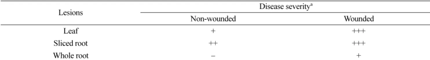
However, the pathogenicity of B. cinerea on non- wounded leaves and roots was very weak (Table 2).
Hennebert (1977) showed that symptoms of Botrytis diseases vary greatly depending on the host and plant part attacked. Gray mold symptoms, which were induced on plants after inoculation with the isolates, were very similar to those observed in the
Table 2. Pathogenicity of Botrytis cinerea isolates inoculated on carrot
Lesions Disease severitya
Non-wounded Wounded
Leaf + +++
Sliced root ++ +++
Whole root − +
a Length of lesions was measured 7 days after inoculation with 105 conidia/ml. +++, > 21 mm in length; ++, 11–20 mm in length; +, 1–10 mm in length; −, no lesion.
Fig. 2. Colony growth of Botrytis cinerea at different temperatures on five kinds of media.
Fig. 3. Phylogenic tree of the isolated Botrytis cinerea on Daucus carota L. subsp. sativas (Hoffm.) inferred by the neighbor-joining method with â-tubulin gene sequences. Bars show the number of nucleotide substitutions per site.