Res. Plant Dis. 17(1) : 52-58 (2011)
© The Korean Society of Plant Pathology
십자화과 작물 종자에서 종자전염 세균 및 바이러스 동시 검출을 위한 One-step Multiplex RT-PCR 방법
정규식·소은희*
국립종자원 재배시험과
One-step Multiplex RT-PCR Method for Simultaneous Detection of Seed Transmissible Bacterium and Virus Occurring on Brassicaceae Crop Seeds
Kyusik Jeong and Eunhee Soh*
Variety Testing Division, Korea Seed and Variety Service, Suwon 443-400, Korea (Received on February 9, 2011; Accepted on February 17, 2011)
The aim of this research was to develop specific and sensitive PCR-based procedures for simultaneous detection of economically important plant pathogenic bacteria and seed borne virus in commercial Brassicaceae crop seeds, Xanthomonns campestris pv. campestris (Xcc) and Lettuce Mosaic Virus (LMV).
Bacterial and virus diseases of Brassicaceae leaves are responsible for heavy losses. PCR with arbitral primers: selection of specific primers, performance of PCR with specific primers and determination of the threshold level for pathogens detection. To detect simultaneously the Xcc and LMV in commercial Brassicaceae crop seeds (lettuce, kohlrabi, radish, chinese cabbage and cabbage), two pairs of specific primer (LMV-F/R, Xcc-F/R) were synthesized by using primer-blast program (http://www.ncbi.nlm.nih.gov/tools/
primer-blast/). The multiplex PCR for the two pathogens in Brassicaceae crop seeds could detect specifically without interference among primers and/or cDNA of other plant pathogens. The pathogen detection limit was determined at 1 ng of RNA extracted from pathogens. In the total PCR results for pathogen detection using commercial kohlrabi (10 varieties), lettuce (50 varieties), radish (20 varieties), chinese cabbage (20 varieties) and cabbage (20 varieties), LMV and Xcc were detected from 39 and 2 varieties, respectively. In the PCR result of lettuce, LMV and Xcc were simultaneously detected in 8 varieties.
Keywords : Brassicaceae crop, Mutliplex RT-PCR, Seed pathogens
우리나라에서 주로 재배되는 십자화과 작물은 무, 배추, 상추, 양배추 그리고 순무 등이 있으며 십자화과작물의 주요 종자전염 병원균은 세균인 Xanthomonas campestris pv. campestris(Xcc)와 바이러스인 Lettuce Mosaic Virus (LMV)가 있다. 검은썩음병(black rot)의 원인인 Xanthomonas campestris pv. campestris는 십자화과 작물에 광범위하게 발생하여 매년 큰 피해를 주고 있는 세균병으로써 다습 한 조건에서 작물의 생육기 동안 발생하고 주로 작물의 지상부에 피해를 일으키는 반면 순무나 무에서는 다육질 의 뿌리에 침해하여 건부(dry rot) 증상을 나타내기도 한
다(Zacchardelli 등, 2007). 포장에서의 일반적인 병징은 큰 V자 모양의 퇴록 현상으로 잎 가장자리 부위에서 발 생하며 병이 진전됨에 따라 엽맥이 검은색으로 변하면서 나중에는 갈변하여 말라버리며 유묘의 경우에는 왜화하 여 시들어버린다(식물병리학 교재연구모임, 2006). Potyviridae 과에 속하는 사상형의 외가닥 RNA 바이러스인 LMV도 전세계적으로 십자화과 작물에 광범위하게 발병하여 잎 에 얼룩, 누렁증상 후에 뒤틀림 증상이 나타나고 마지막 엔 잎 주위가 괴사하여 상품적 가치를 떨어뜨려 Xcc와 마찬가지로 재배 농가에 경제적 피해를 입히는 병원균이 다(Lee, 1981). 두 병원균 모두 대부분 종자로 전염을 하 며 감염 후에는 세균의 경우 세포 간극에 존재하고 바이 러스는 세포의 핵안에 존재하므로 화학적 방법으로 방제
*Corresponding author
Phone) +82-31-8008-0230, Fax) +82-31-203-7431 Email) [email protected]
DOI: 10.5423/RPD.2011.17.1.052 보문 Open Access
하거나 치료가 매우 어렵다(식물병리학 교재연구모임, 2006). 따라서 종자 상태에서부터 병원균 검출을 통한 종 자의 병원균 감염 유ㆍ무를 확인하고 무균 종자를 보급 하는 방법만이 병 발생을 최소화 시킬 수 있는 대안책이 됨에 따라 이러한 병원균을 검출하기 위한 다양한 방법 들이 예전부터 시도되어 보고되어 왔다. 세균을 분리하여 검출하기 위해 보편적으로 사용되고 있는 선택배지 배양 법(Schaad, 1980)과 혈청학적 방법(Schaad 등, 1979;
Franken, 1992; Jin 등, 2001) 그리고 바이러스 검출을 위 한 ELISA 방법(Ghabrial 등, 1982; Ro 등, 2006)들이 사 용되었다. 하지만 이런 검출법은 검사 시간이 오래 걸리 고 검출 민감도가 낮기 때문에 신뢰할 수 있는 검사 결 과를 얻기가 매우 어려웠다. 따라서 이러한 방법의 대안 으로 병원균의 특정 유전자만을 증폭시켜 육안으로 병원 균의 검출 유ㆍ무를 확인할 수 있는 PCR 방법(Massomo 등, 2003; Park 등, 2004; 홍 등, 2007; Zaccardelli 등, 2007)이 개발되었고 PCR 방법에 기초하여 검출 민감도 를 높이기 위한 다양한 변형된 PCR 방법이 개발되어 병 원균 검출에 사용되고 있다.
지금까지 개발된 유전자 진단 기술은 PCR 방법을 기 본으로 하여 세균과 바이러스를 각각 따로 검출하는 방 법들이 보고되었고(De Leon 등, 2006; Park 등, 2006; Lee 등, 2009) 시간과 비용을 줄이기 위하여 종자를 직접 PCR 에 이용한 SDT/RT-PCR 방법(Suehiro 등, 2005)과 PCR 반 응을 저해시키는 요소를 제거하고 민감도를 높이기 위해 서 면역학적 방법을 결합한 IC/RT-PCR(Sharman 등, 2000;
Kim 등, 2006) 등이 개발되어 병원균 검출에 이용되어 왔 다. 다양한 병원균 검출에 이러한 방법을 적용하여 동시 에 여러 바이러스와 세균 병원균을 검출할 수 있는 multiplex- PCR 방법과 검출 민감도를 향상시킨 multiplex IC/RT-PCR 방법, multiplex real-time PCR 방법 등이 여러 작물을 대 상으로 개발되었다(Berg 등, 2006; Kim 등, 2006; Cho 등, 2007). 특히 박과작물 종자 바이러스(CGMMV, ZGMMV, KGMMV) 병원균 검출 방법은 직접 바이러스의 Viron을 포획하여 PCR에 이용하여 시간과 비용은 줄이고 검출 민 감도를 높힌 VC/RT-PCR 방법을 개발하였다(Cho 등, 2007).
그리고 버섯에서 바이러스를 검출하기 위한 버섯의 단백 질을 이용한 biosensor chip(Kim 등, 2007)이 개발되었다.
이와 같이 이미 개발된 방법들은 세균과 바이러스를 대 상으로 하여 여러가지 세균과 바이러스를 각각 따로 검 출함에 따른 오랜 시간 소요와 multiplex IC/RT-PCR 방 법, multiplex real-time PCR 방법 등은 검사 비용의 단점 들을 가지고 있다. 이에 따라 시간 및 비용을 절감하며 세균과 바이러스를 동시에 검출할 수 있는 One-step
multiplex RT-PCR 방법을 개발하여 유통중인 십자화과 작 물 종자에 적용하여 검출 효능을 검정하였다. 개발된 One- step multiplex RT-PCR 방법으로 민원 발생 종자의 분쟁 원인을 파악하고 신속한 민원 해결과 농업 현장의 지도 연구기관에서 보다 저렴하고 신속하며, 신뢰성 있는 진단 에 도움을 주기 위해 새로운 동시검출법을 국내 최초로 개발하여 보고하고자 한다.
재료 및 방법
바이러스와 세균 분리 균주. 세균인 Xcv, Ecc, Cmm, Xcc, Pst는 농촌진흥청 유전자원센터(RDA gene bank)에 서 분양받아 사용하였고 바이러스인 LMV, MNSV, SqMV, CGMMV, KGMMV, PMMoV, TMGMV는 농촌진흥청 농 업과학원(RDA NAAS)에서 바이러스 감염주를 분양받아 사용하였다(Table 1). 그리고 각각의 병원균은 −80oC 초 저온 냉동고에 보관하며 실험에 사용하였다.
병원균 검출 특이적 프라이머 제작. 십자화과 작물에 발생하는 종자 전염 바이러스(1종)과 세균(1종)의 검출을 위한 프라이머 2셋트를 제작하였다(Table 2). 프라이머 제 작 프로그램은 primer-blast(http://www.ncbi.nlm.nih.gov/
tools/primer-blast/)로 NCBI 홈페이지에서 이용 가능한 프 로그램을 사용하였다. 각각의 프라이머는 2종의 세균과 바이러스를 동시 진단할 경우에 프라이머간의 간섭 현상 이 없는 조합을 선발하여 One-step multiplex RT-PCR에 이용하였다.
Total RNA 추출. Nutrient agar 플레이트에서 배양된 세균과 분양받은 바이러스 감염주로부터 RNA 추출은 NucleoSpinR RNA II kit(Macherey-Nagel, Germany)를 이 용하여 추출하였다. 추출된 RNA는 QubitR fluorometer (Invitrogen, USA)를 이용하여 정량하였다.
PCR 민감도와 프라이머 특이성 확인. 추출된 RNA를 100 ng으로 정량한 후 10배 희석법으로 각각 100, 10, 1, 0.1 ng이 되도록 희석한 후에 PCR 민감도 검사에 이용하 였고 10 ng으로 정량한 여러 세균과 바이러스 추출 RNA 를 동량으로 혼합하여 Multiplex RT-PCR을 수행 후 프라 이머 특이성을 확인하였다.
종자 추출완충액으로부터 RNA 추출. 종자에서 병원 균을 검출하기 위한 추출 완충액은 Guanidine-hydrochloride 286.5 g, 4 M NaAC buffer(pH 5.2) 25 ml, EDTA 4.65 g, PVP-10 12.50 g을 혼합한 후 멸균증류수로 500 ml까지 mass up하였다. 1 g의 종자 시료를 지퍼백에 넣고 막자 로 으깬 시료를 2 ml 튜브에 넣고 추출완충액 1 ml을 첨 가한 후 1시간 동안 방치하였다. 1시간 후에 13,000 rpm
으로 원심 분리 후 상층액에서 0.3 ml을 취해서 RNA 추 출에 이용하였다. RNA 추출은 NucleoSpinR Plant kit (Macherey-Nagel, Germany) 사용하여 추출하였다.
One-step multiplex RT-PCR 조건. 특이적으로 합성 한 프라이머 세트(Table 2)를 이용하였다. One-step multiplex RT-PCR은 RobusTTM I RT-PCR kit(Finnzymes, Finland)의 조건을 약간 수정하여 수행하였으며 프라이머의 특이성 을 검정하기 위한 PCR 조건과 종자에서 병원균을 검출 하기 위한 PCR 조건 등 2가지로 나누어서 수행하였다.
특이성 검정을 위한 PCR 조건으로 추출 키트를 이용하 여 각각의 병원균에서 추출한 50 µl의 RNA중에 2 µl를 Templete(RNA)로 이용하였고, 종자에서 병원균 검출을 위 한 PCR 조건으로 50 µl의 추출 RNA중에 5 µl를 이용하 였다. 나머지 PCR 반응액 조건은 다음과 같이 동일한 조 건으로 수행하였다. 1 µl 5’,3’-primer (10 µM), 5 µl 10×
buffer, 1µl dNTP mixture (2.5 mM), 2.5 µl MgCl2 (50 mM), 1µl AMV RT Taq polymerase, 2 µl DNA Taq polymerase 와 DEPC D.W.를 첨가하여 전체 용량을 50 µl로 조절 하 였다. PCR 온도조건은 Tm을 48oC, 50oC, 53oC, 55oC, 58oC로 설정하고 42oC에서 60분간 Reverse Transcription 반응을 시키고 94oC 2분간 denaturation시킨 다음, 94oC에 서 30초, Tm에서 30초, 72oC에서 1분간의 과정으로 40회
반복 수행 후, 최종적으로 72oC에서 10분간 반응시켰다.
PCR 반응 후에 반응산물을 6× Lodding STAR(DYNE bio, Korea) 염색시약과 혼합하였고 이중 10 µl를 전기영동에 사용하였다.
결과 및 고찰
One-step multiplex RT-PCR의 프라이머 특이성과 검 출 민감도 확인. Xcc는 독특한 병원성 인자 발현유전자 인 hrpF의 exon 부분의 염기서열을, LMV 경우는 외피단 백질 유전자의 염기서열을 NCBI 홈페이지를 이용하여 탐 색하였고 증폭산물이 일정한 크기로 각각 구별될 수 있 도록 증폭산물의 크기를 고려하여 프라이머를 합성하였 다(Table 2). 합성된 프라이머를 이용하여 Table 1에 열거 된 병원균중에서 십자화과 작물 종자 전염병원균인 Xcc, LMV의 RNA를 추출하여 Table 2에서 열거된 프라이머 염기서열을 근거로 Tm을 48oC, 50oC, 53oC, 55oC, 58oC 로 각각 설정하고 바이러스와 세균에서 추출한 RNA를 동량으로 혼합한 시료에 합성된 프라이머 세트(Table 2) 를 넣고 RT-PCR을 수행한 결과, Tm 50oC의 경우 Xcc가 증폭되지 않았고 Tm 58oC의 경우 LMV가 증폭되지 않 았다. 그러나 Tm 48oC, 53oC의 경우 비특이적으로 결합 Table 1. Bacteria and viruses used in this study
Pathogens Source
Xanthomonas campestris pv. campestris KACC10377 (Xcc KACC10377) RDA Genebank Xanthomonas campestris pv. vesicatoria KACC11157 (Xcv KACC11157) RDA Genebank Clavibacter michiganensis subsp. michiganensis KACC20779 (Cmm KACC20779) RDA Genebank Erwinia carotovora subsp. carotovora KACC13966 (Ecc KACC13966) RDA Genebank Pseudomonas syringae pv. tomato KACC10560 (Pst KACC10560) RDA Genebank
Lettuce Mosaic Virus 53 (LMV 53) RDA NAAS
Pepper Mild Mottle Virus PAP1 (PMMoV PAP1) RDA NAAS
Tobacco Mild Green Mosaic Virus-Kr PV-0429 (TMGMV-Kr PV-0429) RDA NAAS Cucumber Green Mottle Mosaic Virus CO8004 (CGMMV CO8004) RDA NAAS Kyuri Green Mottle Mosaic Virus-C1 PV-0002 (KGMMV-C1 PV-0002) RDA NAAS
Melon Necrotic Spot Virus 60 (MNSV 60) RDA NAAS
Squash Mosaic Virus S23 (SqMV S23) RDA NAAS
Table 2. Primer sequences and amplified size for detection of the two pathogens infecting Brassicaceae seeds
Target pathogens Primer name Sequences (5'-3') Amplified size (bp)
Xcc Xcc-F CCGTAGCACTTAGTGCAATG
Xcc-R GCATTTCCATCGGTCACGATTG 617
LMV LMV-F ACAAGAAGAAACCGTATATGCC
LMV-R GCCAACACACGCCTTTAGTG 298
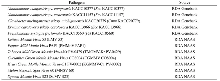
된 증폭산물이 확인되었다. 이는 낮은 Tm으로 인해 Annealing 과정에서 프라이머가 타겟 유전자 부분 이외 의 부분에 비특이적으로 결합되어 증폭되어진 것으로 추 측 되어진다. 반면 Tm 55oC로 설정하고 PCR 증폭 산물 을 NucleoSpinR Extract II kit(Macherey-Nagel, Germany) 을 이용하여 세척한 후에 전기영동한 결과 모든 병원균 이 특이적으로 증폭되는 것을 확인하였으며 각각의 프라 이머와 RT(Reverse Transcription) 단계에서 만들어진 병 원균들의 cDNA간에 상호간섭 없이 PCR이 수행되어 특 이적으로 각각의 산물이 증폭되는 결과를 얻었다(Fig. 1).
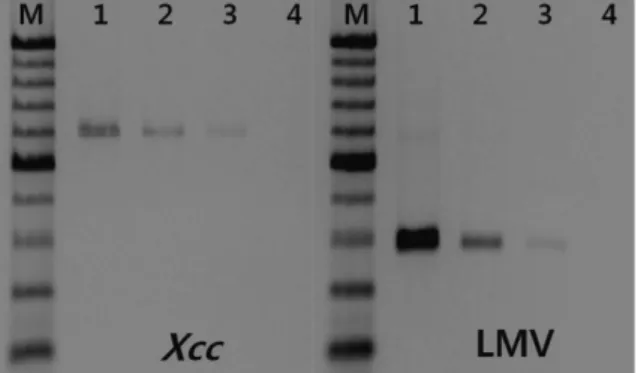
PCR 수행 결과는 agarose gel의 농도를 1.5%로 하여 110 volts로 90분간 수행하였을 때 증폭산물의 크기별로 뚜렷 이 구별되었다. 이러한 결과로 multiplex RT-PCR을 수행 하기 위한 최적의 Tm을 55oC로 결정하였다. Tm을 55oC 로 설정하고 각각의 병원균에서 추출한 RNA를 100 ng부 터 0.1 ng까지 10배 희석법을 이용하여 희석시킨 시료를 가지고 One-step RT-PCR을 수행한 결과, 1 ng까지 PCR 증폭 산물을 확인할 수 있었다(Fig. 2). 따라서 병원균 검 출 한계의 최소 농도를 1 ng으로 결정하였다.
유통 종자로부터 병원균 검출. 개발된 PCR 검출 방법 을 실제 유통 중인 십자화과 작물 종자인 순무(10품종), 상추(50품종), 무(20품종), 배추(20품종) 그리고 양배추(20 품종) 종자에 적용하여 전염병원균의 유ㆍ무를 조사하였 다. 종자 표면이 약품으로 코팅되어 있는 경우 코팅 처리 된 약품이 PCR 반응을 저해할 수도 있기 때문에 종자를 멸균된 증류수로 잘 씻어 내고 하루 동안 건조시킨 후에 RNA 추출 방법을 이용하여 추출하였다.
4품종의 콜라비 종자, 20품종의 상추 종자, 5품종의 무 종자, 6품종의 배추 종자 그리고 4품종의 양배추 종자 시 료에서 Positive control의 2개 증폭 단편 중 LMV의 증폭 단편과 같은 크기로 증폭된 DNA 단편을 확인하였다(Fig.
3, 4, 5). 8개의 상추 품종 시료에서는 두 가지 병원균의 Fig. 1. One-step RT-PCR (Lane 1 and 2) and One-step multiplex
RT-PCR (Lane 3) with mixed RNA samples which were extracted from pathogens at Tm 55oC. Primer specificity was conformed and amplified products were well separated on 1.5%
agarose gel. M, size marker (100 bp DNA ladder), Lane 1, Detection of LMV 53; Lane 2, Detection of Xcc KACC10377;
Lane 3, Simultaneous detection of LMV 53 and Xcc KACC10377.
Fig. 2. Detection sensitivity test (Xcc KACC10377 and LMV 53) with 10 fold dilution RNA samples which were extracted from each pathogen. M, size marker (100 bp DNA ladder), Lane 1, 100 ng RNA extracted from each pathogen; Lane 2, 10 ng RNA extracted from each pathogen ; Lane 3, 1 ng RNA extracted from each pathogen; Lane 4, 0.1 ng RNA extracted from each pathogen.
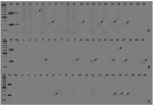
Fig. 3. Detection of pathogens using RNA extracted from commercial kholrabi seeds (‘commercial variety name’). M, size marker (100 bp DNA ladder); Pc, Positive control (RNA mixture of five pathogens); Nc, Negative control (Distilled water). (A) Lane 1, ‘Red olle’;
Lane 2, ‘Worldcol’; Lane 3, ‘Emaerald’; Lane 4, ‘Green daia’; Lane 5, ‘Red daia’; Lane 6, ‘Queen ball’; Lane 7, ‘King ball’; Lane 8, ‘Red duke’; Lane 9, ‘UFO’; Lane 10, ‘Vinus red’ Black arrows ( ) are indicated amplified pathogen product. ←
증폭 단편과 같은 크기의 증폭된 DNA 단편을 확인하여 두 병원균이 동시 검출된 것을 확인하였다(Fig. 4). 증폭 된 DNA가 LMV의 유전자 염기서열과 동일 여부를 검증 하기 위해 NucleoSpinR ExtractII kit(Macherey-Nagel, Germany)를 이용하여 증폭된 단편을 추출하여 염기서열 을 분석하였다. NCBI Blast 프로그램을 이용하여 분석된 염기서열의 homology를 조사한 결과 LMV의 유전자 염 기서열과 100%의 homology를 보여 검출된 병원균이 LMV 임을 확인하였다. 따라서 개발된 One-step multiplex RT- PCR 방법이 종자의 cDNA와 간섭없이 성공적으로 RT- PCR이 수행되어 특이적으로 LMV를 검출하였다는 것을 확인할 수 있었다. 콜라비, 상추, 무, 양배추 그리고 배추 품종 종자에서 LMV의 감염율은 각각 40%, 40%, 25%, 30% 그리고 20%를 나타냈다.
각각 1품종의 무 종자와 배추 종자 시료에서 Xcc의 증
폭 단편과 같은 크기로 증폭된 DNA 단편을 확인하였으 며(Fig. 5) 증폭된 단편의 유전자를 분석하여 homology를 조사한 결과 Xcc의 염기서열과 100%의 homology를 보 여 특이적으로 Xcc를 검출하였다는 것을 확인할 수 있었 다. 무와 배추 품종 종자에서 Xcc의 감염율은 각각 5%씩 나타냈다. LMV에 비해 상대적으로 감염율이 떨어지는 것은 세균보다는 바이러스에 의한 종자 감염이 심각하다 는 것을 보여준다고 볼 수 있겠다.
특히 50개의 상추 품종 종자 시료 중에 8개의 품종 시 료에서 LMV와 Xcc의 증폭 단편과 같은 크기로 증폭된 DNA 단편을 확인하였으며(Fig. 4) 증폭된 단편의 유전자 를 분석하여 homology를 조사한 결과 LMV와 Xcc의 염 기서열과 100%의 homology를 보여 특이적으로 LMV와 Xcc를 검출하였다는 것을 확인할 수 있었다. 동시 검출 되었다는 것으로 개발된 PCR 방법이 복합 감염된 종자 Fig. 4. Detection of pathogens using RNA extracted from commercial lettuce seeds (‘commercial variety name’). M, size marker (100 bp DNA ladder); Pc, Positive control(RNA mixture of five pathogens); Nc, Negative control (Distilled water). Lane 1, ‘Delicious flat leaf lettuce’; Lane 2, ‘Sunmang red rough leaf lettuce’; Lane 3, ‘Dukseom red rough leaf lettuce’; Lane 4, ‘Gohyang lettuce’; Lane 5, ‘Ohyang red flat leaf lettuce’; Lane 6, ‘Bobae red flat leaf lettuce’; Lane 7, ‘Cheonga blue flat leaf lettuce’; Lane 8, ‘Honga red rough leaf lettuce’;
Lane 9, ‘Asiaoraedda red rough leaf lettuce’; Lane 10, ‘Black flat leaf lettuce’; Lane 11, ‘Blue flat leaf lettuce’; Lane 12, ‘Red flat leaf lettuce’; Lane 13, ‘Jinaloe’; Lane 14, ‘Hongiljeom’; Lane 15, ‘Summer blue rough leaf lettuce’; Lane 16, ‘Asakee fresh lettuce’; Lane 17,
‘Dukseom rough leaf lettuce 1’; Lane 18, ‘New tabacco’; Lane 19, ‘Red flat leaf lettuce’; Lane 20, ‘Blue flat leaf lettuce’; Lane 21,
‘Summer red flat leaf lettuce’; Lane 22, ‘Redsun red rough leaf lettuce’; Lane 23, ‘Blacksunj violet flat leaf lettuce’; Lane 24, ‘Blue flat leaf lettuce 2’; Lane 25, ‘Red flat leaf lettuce 2’; Lane 26, ‘Ganghan blue flat leaf lettuce’; Lane 27, ‘Jinjeok red rough leaf lettuce’; Lane 28, ‘Hongmiinpochab’; Lane 29, ‘Dukseom red rough leaf lettuce’; Lane 30, ‘Blue flat leaf lettuce 3’; Lane 31, ‘Summer dukseom red rough leaf lettuce’; Lane 32, ‘Red flat leaf lettuce 3’; Lane 33, ‘Meok flat leaf lettuce’; Lane 34, ‘Summer blue flat leaf lettuce’; Lane 35,
‘Blue flat leaf lettuce 4’; Lane 36, ‘Dukseom red rough leaf lettuce’; Lane 37, ‘Red flat leaf lettuce 4’; Lane 38, ‘Nakdongmeok flat leaf lettuce’; Lane 39, ‘Goyangduk red rough leaf lettuce’; Lane 40, ‘Tojong red rough leaf lettuce’ Lane 41, ‘Blue flat leaf lettuce’; Lane 42,
‘Red flat leaf lettuce’; Lane 43,’Taifoon’; Lane 44, ‘Summer red flat leaf lettuce’; Lane 45, ‘Dukseom rough leaf lettuce’; Lane 46, ‘Red flat leaf’; Lane 47, ‘Blue flat leaf’; Lane 48, ‘Hyundae blue oke’; Lane 49, ‘Hyundae red oke’; Lane 50, ‘Hyundae blue romein’ Black arrows ( ) are indicated amplified pathogen product and red arrows ( ) are indicated simultaneously amplified pathogen products.← ←
시료에서 두 가지의 병원균을 성공적으로 동시 검출할 수 있다는 것을 의미하겠다.
기존의 multiplex PCR 방법은 세균과 바이러스를 각각 단독으로 개발하여 세균과 바이러스를 검출하기 위해서 개별적인 PCR을 수행하였기 때문에 시간과 비용이 이중 으로 소비되었지만 본 실험에서 개발된 One-step multiplex RT-PCR 방법은 세균과 바이러스를 동시에 검출할 수 있 으므로 시간과 비용을 절감하면서 효율적으로 종자 감염 병원균을 검출할 수 있었다.
요 약
우리나라에서 주로 재배되는 십자화과 작물(상추, 콜라 비, 무, 배추, 양배추)의 종자 전염 병원균 중에서 세균 성 병원균 Xanthomonns campestris pv. campestris(Xcc)와
바이러스 병원균 Lettuce Mosaic Virus(LMV)를 종자에 서 동시검출하기 위한 One-step multiplex RT-PCR을 개 발하였다. 각각의 병원균을 특이적으로 증폭시키는 병원 균 검출용 프라이머 2종(Xcc-F/R, LMV-F/R)을 primer- blast 프로그램을 이용하여 제작하였고 이들 프라이머 세 트는 프라이머간 또는 병원균 cDNA간의 간섭없이 특이 적으로 타겟 병원균만을 검출하였다. PCR을 이용한 병 원균의 검출 최소 민감도는 1 ng이었다. 십자화과 작물 중에서 유통 중인 콜라비 10품종, 상추 50품종, 무 20품 종, 배추 20품종 그리고 양배추 20품종에 대한 종자 감 염 병원균 검출을 위한 One-step multiplex RT-PCR 수 행 결과, LMV는 전체 120품종 중에서 39품종에서 검출 되었고, Xcc는 2개 품종에서 검출되었다. 그리고 50품종 의 상추 종자 시료 중에 8품종의 시료에서 LMV와 Xcc 가 동시 검출되었다.
Fig. 5. Detection of pathogens using RNA extracted from commercial radish (A), chinese cabbage (B) and cabbage (C) seeds(‘commercial variety name’). M, size marker (100bp DNA ladder); Pc, Positive control(RNA mixture of five pathogens); Nc, Negative control (Distilled water). (A) Lane 1, ‘Meein altari’; Lane 2, ‘Meotjinmatcal’; Lane 3, ‘Onsesangyeul’; Lane 4, ‘Miyeong’;
Lane 5, ‘Sinnamwonyeol’; Lane 6, ‘Keien888’; Lane 7, ‘Sanggam’; Lane 8, ‘Nokbongbom’; Lane 9, ‘Kwonnong summer altari’; Lane 10, ‘Kwonnong siraegi’; Lane 11, ‘Superhongbityeol’; Lane 12, ‘Daebong’; Lane 13, ‘Hajicheongjeong’; Lane 14, ‘Myeongjak altari’;
Lane 15, ‘Sindongsanyeol’; Lane 16, ‘Poonggang altari’; Lane 17, ‘Keumjang siraegi’; Lane 18, ‘Mansahyeongtong’; Lane 19,
‘Manchookwandong’; Lane 20, ‘Sinnamchonyeol’ (B) Lane 1, ‘Saelreodeu’; Lane 2, ‘Sweet ball’; Lane 3, ‘Nokdongja’; Lane 4,
‘Waialgaelreoksi’; Lane 5, ‘Waialekopeulreoseu’; Lane 6, ‘Royalston’; Lane 7, ‘Joongsaengakkaroobi’; Lane 8, ‘Waialgaelreoksi 1’;
Lane 9, ‘Smat’; Lane 10, ‘YR jeongwol’; Lane 11, ‘YR onnoori’; Lane 12, ‘YR hoyeong’; Lane 13,’Crown red’; Lane 14, ‘Keumywol’;
Lane 15, ‘Booltina’; Lane 16, ‘Onseosang’; Lane 17, ‘Dongywol’; Lane 18, ‘Crown red 1’; Lane 19, ‘Whoyoukki’; Lane 20, ‘Akkaroobi’
(C) Lane 1, ‘Kosohan’; Lane 2, ‘Ssialchoegobong’; Lane 3, ‘Ssialharooyosi’; Lane 4, ‘Saesimbong’; Lane 5, ‘Samyuko’; Lane 6,
‘Aramchansin’; Lane 7, ‘Chunyeongbom’; Lane 8, ‘Ssialwatdajinnorang’; Lane 9, ‘Gihoeotgali’; Lane 10, ‘Choowang’; Lane 11,
‘Dongbangsingi’; Lane 12, ‘Ssialmaeriteu’; Lane 13, ‘Danbieotgali’; Lane 14, ‘Miraeweilbingipssam’; Lane 15, ‘Choono’; Lane 16,
‘Ssialhaseo ‘; Lane 17, ‘Ssialhaejin’; ane 18, ‘Sindaenongeotgali’; Lane 19, ‘Ssialgamtanjinnorang’; Lane 20, ‘Sooho’ Black arrows ( ) are indicated amplified pathogen products.
←
Acknowledgements
This study was performed with fund supported from Korea Seed & Variety Service, Republic of Korea.
참고문헌
식물병리학 교재연구모임. 2006. 식물병리학(제5판). 월드싸이 언스. 653-654, 764-769 pp.
홍성준, 홍연규, 이봉춘, 임미정, 윤영남, 황재복, 송석보, 박성 태. 2007. PCR assay 이용 콩 종자에서 Xanthomonas axonopodis pv. glycines 검출 및 종자오염 조사. 식물병연구 13: 145-151.
Berg, T., Tesoriero, L. and Hailstones, D. L. 2006. A multiplex real-time PCR assay for detection of Xanthomonas campestris from brassicas. Lett. Appl. Microbiol. 42: 624-630.
Cho, J. D., Kim, J. S., Lee, S. H. and Chung, B. N. 2007. Triplex Virion Capture (VC)/RT-PCR for Three Seed Transmissible Tobamoviruses of CGMMV, ZGMMV and KGMMV Occurring on Cucurbitaceae. Res. Plant Dis. 13: 82-87.
De Leon, L., Siverio, F. and Rodriguez, A. 2006. Detection of Clavibacter michiganensis subsp. michiganensis in tomato seeds using immunomagnetic separation. J. Microbiol.
Methods 67: 141-149.
Franken, A. A. J. M. 1992. Application of polyclonal and monoclonal antibodies for the detection of Xanthomonas campestris pv. campestris in crucifer seeds using immunofluorescence microscopy. Eur. J. Plant Pathol. 98: 95- 106.
Ghabrial, S. A., Li, D. and Shepherd, R. J. 1982. Radio- immunosorbent Assay for Detection of Lettuce Mosaic Virus in Lettuce Seed. Plant Disease 66: 1037-1040.
Jin, K. S., Kang, I. B., Ko, K. I., Lee, E. S., Heo, J. Y., Kang, Y. K.
and Kim, B. K. 2001. Detection of Xanthomonas axonopodis pv. citri an Citrus Fruits Using Enzyme-Linked Immunosorbent Assay. Plant Pathol. J. 17: 62-66.
Kim, J. H., Choi, G. S., Kim, J. S., Lee, S. H., Choi, J. K. and Ryu, K. H. 2006. Development of single-tube multiplex immunocapture RT-PCR Assay for simultaneous detection of two pepper Tobamoviruses. Plant Pathol. J. 22: 164-167.
Kim, S. W., Kim, M. G., Kim, J., Lee, H. S. and Ro, H. S. 2007.
Detection of a mycovirus OMSV in edible mushroom Pleurotus ostreatus using SPR biosensor chip. J. Virol.
Methods.
Lee, S. H. 1981. Studies on virus disease occurring in various crops in Korea. Res. Report RDA 23: 62-74.
Lee, Y. A., Sung, A. N., Liu, T. F. and Lee, Y. S. 2009.
Combination of chromogenic differential medium and estA- specific PCR for isolation and detection of phytopathogenic Xanthomonas spp. Appl. Environ. Microbiol. 75: 6831-6838.
Massomo, S. M. S., Nielsen, H., Mabagala, R. B., Mansfeld- Giese, K., Hockenhull, J. and Mortensen, C. N. 2003.
Identification and Characterisation of Xanthomonas campestris pv. campestris strains from tanzania by pathogenicity tests, biolog, rep-PCR and fatty acid methyl ester analysis. J. Plant Pathol. 109: 775-789.
Park, D. S., Shim, J. K., Kim, J. S., Lim, C. K., Shrestha, R., Hahn, J. H., Parrella, G., Verdin, E., Gognalons, P. and Marchoux, G. 2006. Detection and characterization of tobacco mild green mosaic virus (TMGMV) large type isolate from trailing petunia in france. Commun. Agric Appl. Biol. Sci. 71:
1237-1244.
Park, Y. J., Lee, B. M., Hahn, J. H., Lee, G. B. and Park, D. S.
2004. Sensitive and specific detection of Xanthomonas campestis pv. campestris by PCR using species-specific primers based on hrpF gene sequence. Microbiological Research, 159: 419-423.
Ro, H. S., Lee, N. J., Lee, C. W. and Lee, H. S. 2006. Isolation of a novel mycovirus OMIV in Pleurotus ostreatus and its detection using a triple antibody sandwich-ELISA. J. Virol.
Methods 138: 24-29.
Schaad, N. W. 1979. Serological identification of plant pathogenic bacteria. Annu. Rev. Phytopathol. 17: 123-147.
Schaad, N. W. ed. 1980. Laboratory Guide of Identification of Plant Pathogenic Bacteria. APS Press, St. Paul, MN.
Sharman, M., Thomas, J. E. and Dietzgen, R. G. 2000.
Development of a multiplex immunocapture PCR with colourimetric detection for viruses of banana. J. Virol.
Methods 89: 75-88.
Suehiro, N., Matsuda, K., Okuda, S. and Natsuaki, T. 2005. A simplified method for obtaining plant viral RNA for RT-PCR.
J. Virol. Methods 125: 67-73.
Zaccardelli, M., Campanile, F., Spasiano, A. and Merighi, M.
2007. Detection and identification of the crucifer pathogen, Xanthomonas campestris pv. campestris, by PCR amplification of the conserved Hrp/type III secretion system gene hrcC. Eur.
J. Plant Pathol. 118: 299-306.