Vol. 19, No. 6 pp. 275-279, 2018
1. 서론
뇌농양(brain abscess)은 100,000명당 약 1명의 빈도 로 드물게 발생하는 중추신경계 감염으로, 주로 뇌전산
단층촬영(brain computed tomography)과 놔자기공명영 상(brain magnetic resonance image)으로 조기 진단이 가능하고, 항생제 및 신경 집중치료 발달 등으로 사망률 과 후유증이 많이 줄었지만, 두통 및 발열 등의 증상이
만성부비동염을 가진 환자에서 재발된 뇌농양 : 증례보고
이상학1, 황용2, 이학승1*
1
원광대학교의과대학 신경과학교실,
2원광대학교의과대학 응급의학교실
Recurrent Brain Abscess In Chronic Sinusitis : A Case Report
SangHak Yi
1, Yong Hwang
2, Hak Seung Lee
1*1
Department of Neurology, Wonkwang University, School of Medicine
2
Department of Emergency Medicine, Wonkwang University, School of Medicine
요 약 뇌농양은 심각한 신경학적 후유증을 일으킬 수 있는 중추신경계 감염이다. 신경집중치료 발달에도 불구하고, 여전히 뇌농양은 특정 위험 환자에게 높은 사망률을 보이고 있다. 특히 세균성 뇌농양은 즉각 진단 및 적절한 항생제 치료가 필요한 응급 상황이다. 또한 드물게 뇌농양이 재발되는 경우도 있다. 본 연구에서는 두통으로 내원한 59세 남자환자를 대상으로 증례보고를 통해 동일 분야 연구에 활용하고자 자료분석을 하였다. 59세 남자가 두통을 주소로 내원하였고, 연속적으로 시 행한 뇌자기공명영상과 뇌척수액 검사에서 우측 전두엽에 뇌농양을 확인하였고, 항생제 치료 및 수술적 치료 후 완치하였다.
퇴원 후 5개월 뒤 두통 및 경기 증상 있어 다시 촬영한 뇌자기공명영상에서 뇌농양의 재발이 확인되었다. 뇌농양 재발은
특정 조건, 즉, 농양 부위에 이물질이 남아 있거나 만성 부비동염, 동정맥루, 좌우션트 등이 있을 때 생길 수 있다.
Abstract A brain abscess is caused by an infection in the central nervous system and leads to significant neurological sequelae. Despite advances in neurocritical care, high morbidity and mortality rates are still associated with brain abscess, especially in certain high-risk patients. Thus, a brain abscess is a medical emergency requiring prompt diagnosis and treatment with antibacterial therapy. However, the recurrence of brain abscess is rare. A 59-year-old man presented at our neurology clinic with a headache and was found to have chronic otitis media. Serial brain magnetic resonance imaging (MRI) scans of the patient demonstrated a brain abscess in the right frontal lobe.
Following stereotactic aspiration of the brain abscess and proper antimicrobial treatment, the patient recovered completely. Five months after discharge, the patient revisited our clinic with a headache and seizure symptoms, and recurrence of the brain abscess was confirmed by brain MRI. Recurrent brain abscess has been documented in patients with predisposing conditions such as retained foreign bodies, chronic sinusitis, arteriovenous fistula, and right-to-left shunt.
Keywords : Brain abscess, Chronic Sinusitis, Recurrent, Antibiotics, Surgery
본 논문은 2017학년도 원광대학교의 교비지원에 의해 수행되었음.
*
Corresponding Author : Hak Seung Lee(Wonkwang Univ.) Tel: +82-10-8644-8295 email: [email protected]
Received March 12, 2018 Revised (1st April 9, 2018, 2nd May 9, 2018)
Accepted June 1, 2018 Published June 30, 2018
특징적이지 않아 진단이 늦어지고, 특정 위험인자를 가 진 환자들에게 높은 사망률을 보이고 있다[1-5].
뇌농양은 주된 원인 균으로 포도상 구균, 연쇄상 구 균, 폐렴 구균 등이 있고, 감염 경로로는 중이염, 부비강 염, 편도염 등 근처 감염원에서 직접적인 파급이 약 40%, 폐농양, 심내막염, 감염성색전 등에 의한 혈행성 감염이30%, 개방성 뇌손상 등에 의한 직접감염이 10%
정도로 알려져 있다[6, 7].
뇌농양은 대부분 단발성이지만 특정 상황에서 드물게 뇌농양이 재발되는 경우도 보고되었다[8-13]. 이 중 만 성부비동염은 흔한 질환이기 떄문에 뇌농양 환자 치료에 서 간과되는 경우가 많다.
본 연구는 만성부비동염을 가진59세 남자에서 뇌농 양 완전 치료 후 다시 재발한 예를 경험하였기에 보고하 고자 한다.
2. 연구대상 및 방법
2.1 연구 대상
두통을 주소로 내원하여 뇌농양으로 진단된59세 남 자 환자를 대상으로 하였다.
2.2 연구 방법
증례분석을 통해 동일 분야 연구에 활용하고자 한다.
3. 증 례 및 연구 결과
59세 남자가 내원 14일 전부터 발열, 두통을 주소로 내원하였다. 과거력에서 10년 전 뇌출혈과 지속되는 만 성부비동염 경력이 있었고, 수술이력은 없었다. 신체각 계조사에서는 두통 외에는 특이사항 없었고, 사회력에서 30년 이상의 음주력이 있었으며 흡연은 하지 않았다. 가 족력도 특이사항이 없었다.
내원 당시 환자의 혈압은 120/80mmHg, 체온은 38.9°C로 발열이 있었으며, 맥박은 120/min, 호흡은 14/min 이었고, 이학적 검사상 특이소견이 없었다.
신경학적 검사 상 경부강직이 관찰되었다. 내원 당시 시행한 혈액검사에서 백혈구16,670 /㎕(정상 4,000-10,000 /㎕)로 증가되었고, 뇌척수액 검사상 뇌압은 280 mmH20
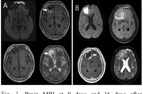
Fig. 1. Brain MRI at 9 days and 16 days after headache (A) FLAIR and T2-weighted images showed high signal intensity(Arrow) in the right frontal cortex at 9 days after headache. (B) Diffusion-weighted and T2-weighted images showed high signal intensity in the right frontal cortex, Gadolinium-enhanced images showed ring enhancement (Arrow) in right frontal cortex at 16 days after headache.
(정상 100-180 mmH20)로 뇌압이 상승 되었고, 육안상 뇌척수액 색은 깨끗하였지만, 백혈구는 320 /㎕(정상 0-5 /㎕), 적혈구는 20 /㎕(정상 0 /㎕)로 다핵구가 20 %였으 며, 단백질은 117.7 ㎎/㎗(정상 15-45 ㎎/㎗)로 전부 증 가되었고, 포도당은 65 mg/dL(혈당은 134 ㎎/㎗) 정상 이었다.
외부에서 가져온 뇌자기공명영상에서는 우측 전두엽 에 과거 뇌출혈에 의한 뇌연화(cerebromalacia)가 보였 다(Fig. 1A). 항생제 치료 및 보존적 치료 후 두통 호전 이 없어 내원1주일 후 뇌농양의 진행 확인을 위해 시행 한 뇌자기공명영상에서 우측 전두엽에 뇌농양 진행 소견 이 확인되었다(Fig. 1B).
항생제 치료에 반응이 없어, 신경외과와 협진을 통해 두개강 뇌농양 배액술(needle aspiration)을 시행하였고, 세균배양검사로 원인균은Streptococcus anginosus로 확 인되었다. 두 개강 뇌농양 배액술 후 ceftriaxone, metronidazole, vancomycin 항생제 치료를 8주간 유지 한 후 환자는 두통이 호전되었고, 치료 확인을 위해 촬영 한 뇌전산단층촬영에서 특이소견이 없었다.
퇴원 후 3개월 뒤에 경과관찰 위해 외래에서 촬영한 조영증강 뇌전산단층촬영에서 뇌연화 외에는 특이소견 없었고, 환자도 두통 등 특이 증상도 없었고, 일상생활을 잘 유지하였다(Fig 2). 그러나 코막힘, 기침 등의 비특이 적인 증상은 있었다.
Fig. 2. Brain CT with contrast at 3 months after discharge form hospital. Brain CT with contrast showed encephalomalacia lesion in right frontal lobe.
Fig. 3. Follow-up Brain CT with contrast and Brain MRI at re-admisson. (A) Follow-up Brain CT demonstrated 2 ring enhancement (Arrow) in right frontal lobe. (B) (C), Diffusion-weighted and T2-weighted images showed high signal intensity in right frontal lobe. (D) T1-weighted images with gadolium showed ring enhancement in right frontal lobe.
퇴원5개월 후 환자는 다시 두통과 의식을 잃고 온몸 을 떠는 경기 증상을 주소로 응급실 통해 내원하였고, 시 행한 뇌전산화단층 및 뇌자기공명영상에서 뇌농양 재발 을 확인하였다(Fig. 3).
현재 뇌농양 배액술 2회와 항생제 치료를 하였지만 환자는 의식상태는 강한 외부자극에도 전혀 반응을 하지 않는 혼수상태로 악화되었다.
4. 고 찰
뇌농양의 발생기전은, 원인균이 혈류에 의해 뇌에 도 달하거나, 뇌-혈관 장벽 (blood-brain barrier)를 통과하 여 뇌척수액에서 증식하거나, 뇌에 직접적인 외부 손상 에 의해 발생할 수 있다[14, 15].
뇌농양의 진단에는 뇌자기공명영상과 뇌전산단층 촬 영이 가장 중요한 진단방법이다.
뇌농양의 치료는 항생제 치료와 바늘흡입이나 외과절 제술 등의 수술적 치료가 있다. 항생제 치료는 수술적 접 근이 어려운 위치거나 크기가 3cm 미만이거나 피막이 없는 뇌염초기 단계, 작은 다발성 뇌농양일 경우 등 일 때 시행할 수 있다.[16, 17]
뇌농양은 일반적으로 단발성 즉 대부분 재발을 하지 않는다[1]. 뇌농양 재발은 특정 조건, 즉, 농양 부위에 이 물질이 남아 있거나 만성 부비동염, 동정맥루, 좌우션트 등이 있을 때 생길 수 있다. [8-13]
뇌농양 완치된 환자에서8%에서 재발한다는 보고도 있고[1], 특정 조건 없어도 그 자체가 재발할 수 있다는 연구도 있다[18]. 특정 조건이 있고, 뇌농양의 원인균을 찾지 못할 때는 25%까지 재발한다는 연구도 있다[19].
일반적인 뇌농양의 재발은 항생제 치료를 충분하게 하지 않거나, 두개강 뇌농양 배액술을 충분히 하지 않았 을 때 재발할 수 있다[20]. 위에서 언급한 특정 조건에서 는 뇌농양 재발이 더 자주 있기 때문에 원인균 감별이나 치료에 더 관심을 가져야 한다.
이러한 특정 조건에서 만성 부비동염은 흔한 질환이 다. 코 주위의 얼굴 뼈 속에 있는 빈공간인 부비동에 환 기 및 배설되지 않아 이차적으로 부비동에 염증이 발생 하고, 3개월 이상 지속될 경우 만성 부비동염이라고 한 다. 만성부비동염은 뇌실질에 인접한 구조물로 감염으로 부터 직접 파급 및 확산되어 뇌농양이 대개 단일병터로 나타난다. 만성부비동염의 증상이 두통, 코막힘, 콧물 등 비특이적이고, 뇌농양의 증상과 비슷하여 간과하기 쉽다.
뇌농양의 재발을 낮추기 위해 두 개강 뇌농양 배액술 보다 수술적 절제가 효과적이라는 연구도 있다[21, 22].
다른 연구에서는 두개강 뇌농양 배액술 시 double lumen cather 사용이 뇌농양의 재발율을 낮추었다라고 보고하고 있다[23].
뇌농양의 재발은 몇 개월에서 수년까지 할 수 있다 [20]. 한 증례에서는 처음 치료 후 7년 뒤 뇌농양이 재발 한 경우도 있었다[24]. 그래서 뇌농양 초기 치료가 완료
되어도 꾸준한 경과관찰이 필요하고, 위에서 언급한 특 정 조건 환자들은 더욱더 경각심을 가지고 경과관찰이 필요하다.
뇌농양의 예후는 환자의 의식이 명료할 때 치료를 시 작할 때는 사망률이 5-10%이지만, 혼수상태에서 치료 시에는50% 이상이다. 재발 시에는 이보다 예후가 나쁘 다[25].
본 증례의 경우에도 만성부비동염을 가진 환자에서 이러한 원인을 해결하지 않았을 때 뇌농양의 재발을 확 인할 수 있었다.
5. 결 론
뇌농양은 자체로 위험한 질환이며 빠른 진단과 치료 가 필요하다. 뇌농양 완치 후에도 고찰에서 언급한 특정 조건들을 제거하지 않는다면 재발할 수도 있다. 재발한 뇌농양 예후는 좋지 않아서 뇌농양 치료 시 앞에서 언급 한 특정 조건들을 확인할 필요가 있다.
저자들은 만성부비동염을 가진59세 남자에서 뇌농양 재발을 경험하였기에 관련된 문헌고찰과 함께 증례를 보 고한다.
References
[1] Beller AJ, Sahar A, Praiss I, “Brain abscess. Review of 89 cases over a period of 30 year,” J Neurol Neurosurg Psychiatry, vol. 36, pp. 757-768, 1973.
DOI: https://doi.org/10.1136/jnnp.36.5.757
[2] Carpenter J, Stapleton S, Holliman R, “Retrospective analysis of 49 cases of brain abscess and review of the literature,” Eur J Clin Microbiol Infect Dis, vol. 26, pp.
1-11, 2007.
DOI: https://doi.org/10.1007/s10096-006-0236-6 [3] Park SH, Lee SW, Kang DH, Hwang JH, “Sung JK,
Hwang SK : The role of f-fluorodeoxyglucose positron emission tomography in the treatment of brain abscess,”
J Korean Neurosurg Soc, vol. 49, pp. 278-283, 2011.
DOI: https://doi.org/10.3340/jkns.2011.49.5.278 [4] Rosenblum ML, Hoff JT, Norman D, “Weinstein PR,
Pitts L : Decreased mortality from brain abscesses since advent of computerized tomography,” J Neurosurg, vol.
49, pp. 658-668, 1978.
DOI: https://doi.org/10.3171/jns.1978.49.5.0658 [5] Yang SY, “Brain abscess : a review of 400 cases,” J
Neurosurg, vol. 55, pp. 794-799, 1981.
DOI: https://doi.org/10.3171/jns.1981.55.5.0794
[6] Yang SY, Zhao CS, “Review of 140 Patients with Brain Abscess,” Surg Neurol, vol. 39, pp. 290-296, 1993.
DOI: https://doi.org/10.1016/0090-3019(93)90008-O [7] Adams RD, Victor M, “Nonviral infections of the
nervous system, In: Adams RD, Victor M, eds.
Principles of neurology,” New York, McGraw-Hill Inc pp. 629-630, 1993.
[8] Mathisen GE, Johnson JP, “ Brain abscess,” Clin Infect Dis, vol. 25, pp. 763-779, 1997.
DOI: https://doi.org/10.1086/515541
[9] LaBarbera M, Berkowitz MJ, Shah A, Slater J,
“Percutaneous PFO closure for the prevention of recurrent brain abscess,” Catheter Cardiovasc Interv, vol. 68, pp. 957-960, 2006.
DOI: https://doi.org/10.1002/ccd.20869
[10] Kikuchi K, Kowada M, Shioya H, Shioya T, “Recurrent brain abscess associated with hereditary hemorrhagic telangiectasia (Rendu-Osler-Weber disease)--case report,”
Neurol Med Chir (Tokyo) Vol. 32, pp. 891-895, 1992.
[11] Kikuchi K, Kowada M, Shioya H, Shioya T, “Recurrent brain abscess associated with hereditary hemorrhagic telangiectasia (Rendu-Osler-Weber disease)--case report,”
Neurol Med Chir (Tokyo), vol. 32, pp. 891-895, 1992.
DOI: https://doi.org/10.2176/nmc.32.891
[12] Vandertop WP, de Vries WB, van Swieten J, Ramos LM, “Recurrent brain abscess due to an unexpected foreign body,” Surg Neurol, vol. 37, pp. 39-41, 1992.
DOI: https://doi.org/10.1016/0090-3019(92)90063-S [13] Caroli M, Arienta C, Rampini PM, Balbi S, “Recurrence
of brain abscess associated with asymptomatic arteriovenous malformation of the lung,” Neurochirurgia (Stuttg), vol. 35, pp. 167-170, 1992.
DOI: https://doi.org/10.1055/s-2008-1052272
[14] Quagliarello V, Scheld WM, “Bacterial meningitis : Pathogenesis, pathophysiology, and progress,” NEJM, vol. 327, pp. 864-872, 1992.
DOI: https://doi.org/10.1056/NEJM199209173271208 [15] Park TH, Kim MK, “ Bacterial Menigitis and Brain
Abscess,” J Neurocrit Care Suppl. 1, pp S63-S68, 2008.
[16] Fulgham JR, Wijdicks EF, Wright AJ, “Cure of a solitary brainstem abscess with antibiotic therapy: case report,” Neurology, vol. 46, pp. 1451-1454, 1996.
DOI: https://doi.org/10.1212/WNL.46.5.1451
[17] Carpenter JL, “Brain stem abscesses: cure with medical therapy, case report, and review,” Clin Infect Dis, vol.
18, pp. 219-226, 1994.
DOI: https://doi.org/10.1093/clinids/18.2.219
[18] Yousaf I, McInstry CS, Choudhari RA, “Recurrent intracranial abscess after a long latency,” Br J Neurosurg, vol. 18, pp. 396-397, 2004.
DOI: https://doi.org/10.1080/02688690400005271 [19] Ciurea AV, Stoica F, Vasilescu G, Nuteanu L,
“Neurosurgical management of brain abscesses in children,” Childs Nerv Syst, vol. 15, pp. 309 – 317, 1999.
DOI: https://doi.org/10.1007/s003810050400
[20] Patel K, Clifford DB, “Bacterial brain abscess,”
Neurohospitalist, vol. 4, pp. 196-204, 2014.
DOI: https://doi.org/10.1177/1941874414540684
[21] Cavusoglu H, Kaya RA, Turkmenoglu ON, Colak I,
Aydin Y, “Brain abscess: analysis of results in a series of 51 patients with a combined surgical and medical approach during an 11-year period,” Neurosurg Focus, vol. 24, E9, 2008.
[22] Gadgil N, Patel AJ, Gopinath SP, “Open craniotomy for brain abscess: a forgotten experience,” Surg Neurol Int, vol. 4, pp. 34, 2013.
DOI: https://doi.org/10.4103/2152-7806.109522 [23] Park JH, Yoo DS, Kim DS, Huh PW, Cho KS, Kang
JK, “Continuos irrigation of brain abscess using a double lumen catheter,” J Korean Neurosurg Soc, vol. 29, pp.
1328-1322, 2000.
[24] Sharma R, Mohandas K, Cooke RP, “Intracranial abscesses: changes in epidemiology and management over five decades in Merseyside,” Infection, vol. 37, pp.
39-43, 2009.
DOI: https://doi.org/10.1007/s15010-008-7359-x [25] Ko SJ, Park KJ, Park DH, Kang SH, Park JY, Chung
YG, “Risk factors associated with poor outcomes in patients with brain abscesses,” J Korean Neurosurg Soc, vol. 56, pp. 34-41, 2014.
DOI: https://doi.org/10.3340/jkns.2014.56.1.34
이 상 학(SangHak Yi) [정회원]
•2006년 2월 : 원광대학교 의과대학
의학과 (의학사)
•2015년 2월 : 원광대학교 의과대학
의학과 (의학석사)
•2015년 2월 : 대한신경과학회 전문
의 취득 (신경과 전문의)
•2015년 3월 ~ 2017년 2월 : 분당 서울대병원 신경과 임상강사
•2017년 2월 ~ 현재 : 원광대학교 의과대학 임상 조교수
<관심분야>
의/생명 공학, 임상의학
황 용(Yong Hwang) [종신회원]
•2009년 2월 : 원광대학교 의과대학 원 의학과(의학사)
•2010년 2월 : 대한응급의학회 전문 의 취득(응급의학전문의)
•2010년 3월 ~ 2013년 8월 : 원광 대학교 의과대학 임상 조교수
•2013년 9월 ~ 현재 : 원광대학교 의과대학 교수
<관심분야>
의/생명 공학, 임상의학
이 학 승(Hak Seung Lee) [정회원]