• 검색 결과가 없습니다.

A study on the inflammatory response induced by LPS of the Arthrospira platensis ethanol extract

N/A
N/A
Protected

Academic year: 2021

Share "A study on the inflammatory response induced by LPS of the Arthrospira platensis ethanol extract"

Copied!
9
0
0

로드 중.... (전체 텍스트 보기)

전체 글

(1)

http://dx.doi.org/10.12925/jkocs.2019.36.3.966

A study on the inflammatory response induced by LPS of the Arthrospira platensis ethanol extract

Shi Jie Zhang

*

․Jae-Chan Yang․Bo-Ae Kim

Mokwon University, College of Sciences & Technology, Division of Biomedicinal & Cosmetics, Doanbuk-ro 88, Seo-gu, Daejeon 302-729 Korea

(Received September 6, 2019; Revised September 28, 2019; Accepted September 28, 2019)

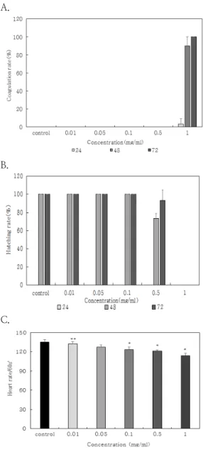
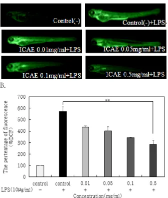
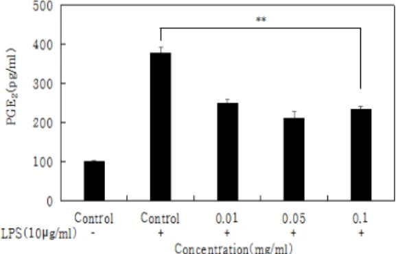
Abstract : Arthrospira platensis has been reported to contain a variety of substances such as phycocyanin, β-carotene, vitamin E and other carotenoids. In this study, zebrafish were treated with indoor cultivation spirulina ethanol extracts(ICAE) to determine toxicity(coagulation rate, hatching rate, heart rate). We used the DCFH-DA staining method to detect the effect of reactive oxygen species(ROS) generation on lipopolysaccharide(LPS)-induced zebrafish embryos ROS various concentrations(0.01, 0.05, 0.1, 0.5mg/ml) of ICAE. Cell toxicity was measured by WST-1 assay on RAW 264.7 macrophage cell line. Also, measured the inhibitory effect of nitric oxide(NO) and prostaglandin E₂(PGE₂) production in RAW264.7 macrophages induced by LPS at various concentrations of ICAE. The results of embryo coagulation rate, hatching rate, heart rate of zebrafish at various(0.01, 0.05, 0.1mg/ml) of ICAE was no toxicity. The ICAE treated group had an inhibitory effect on NO and PGE₂ production compared and decreased with concentration. The results of this study ethanol extract of Arthrospira platensis has an anti-inflammatory effect and suggest that is worthy of use cosmetics for skin protection.

Keywords : Arthrospira platensis, zebrafish, anti-inflammation, toxicity, DCFH-DA, reactive oxygen species, nitric oxide, prostaglandin E₂

1. INTRODUCTION

Recently, as people have an interest in skincare has increased, research on skincare materials focusing on existing anti- inflammatory mechanisms has diversified. Skin is an organ that has barrier function to prevent infiltration of external infectious substances including various pathogens that



Corresponding author

(E-mail: [email protected])

keep moisture in the epidermis, dermis and subcutaneous tissue[1]. The skin also has a chemical barrier function and is maintained by antibacterial peptides in the stratum corneum[2]. Antimicrobial peptides secreted from the skin perform a primary defense function, increased by infection or inflammatory reactions[3,4]. The exogenous factor that affects the skin most is the infiltration of infectious agents.

Inflammation is a complex biological

response of harmful stimuli, such as pathogens

and irritants. When inflammatory reactions

(2)

occur, cause pain, heat, itching, swelling, and redness, etc symptoms[5]. Aslo, inflammation is a protective response to skin damage caused by external invasion or tissue infection.

However, if this reaction occurs excessively, it damages normal tissues and causes inflammatory diseases[6]. When inflammatory reactions occur, the stimulation of lipopolysaccharide(LPS) produces inflammatory mediator such as nitric oxide(NO) and prostaglandin E₂(PGE₂) inflammatory factors are produced by the expression of inducible nitric oxide synthase(iNOS) and cyclooxygenase-2(COX-2)[7,8].

Arthrospira platensis is one of the oldest algae on Earth and is used as a functional food for humans such as chlorophyll in fresh water[9,10]. Arthrospira platensis consists of 60-70% protein, 6-9% lipid, 15-20%

carbohydrate, various vitamin, mineral and fiber content[11]. The physiological activity of Arthrospira platensis has been reported to improve anti-inflammatory effect[12], anti-allergic substances that inhibit histamine and TNF-α secretion[13], immunomodulatory effects[14]. It has also been reported to have a positive effect on skin elasticity, pigmentation and fine wrinkle improvement when used as a newly developed material in cosmetics[15].

The purpose of this study was to investigate the anti-inflammatory effects of ethanol extracts of Arthrospira platensis and to evaluate the possibility of using an alternative animal zebrafish model for natural functional cosmetic raw materials.

2. MATERIALS AND METHODS

2.1. Materials

Lipopolysaccharide(LPS), WST-1(Water soluble tetrazolium salt-1) assay kit, 2‘,7’-dichlorofluorescein diacetate(DCFH-DA), Griess reagent were purchased from Sigma-Aldrich(St. Louis, MO, USA). Sea salt water was purchased from Genomic-Design

(Bioengineering Company, Deajeon, Korea).

Dulbecco’s modified Eagle's medium (DMEM), Phosphate-buffered saline(PBS), 10% fetal bovine serum (FBS), 1% penicillin-streptomycin, and trypsin-ethylenediaminetetraacetic acid were purchased from Gibco(Grand Island, NY, USA). PGE₂ assay kit was purchased from Amersham pharmacia (Biotech Co., USA).

2.2. Extraction method

Arthrospira platensis was cultured in a 20L transparent water tank using an LED lamp.

Washed with purified water, dried at 40℃ for 24 hours, pulverized using a pulverizer having an opening diameter of 100μm, and lyophilized. 95% ethanol (Estrad, Korea) was added and the mixture was extracted by stirring at room temperature for 24 hours. The extract was filtered through a vacuum filter and concentrated under reduced pressure (EYELA N-1110, EYELA, Korea).

2.3. Zebrafish maintenance

Adult zebrafish were obtained 8 fish were kept in a 3L recirculating tank system using local tap water(pH 7.2~7.6). Zebrafish were maintained at 28.5℃ with a 14h light : 10h dark cycle and fed 3 times a day with live Artemia. The embryos were collected from natural spawning within 30min, induced in the morning by turning on the light. Embryos were maintained at a density of 50~70 embryos per in a Petri dish containing media as previously reported.

2.4. Toxicity measurement of ICAE in zebrafish embryo and larvae

6hpf(hour-post-fertilization) zebrafish

embryos were seeded at 10 embryos/well in

24-well plate and treated with various

concentrations(0.01, 0.05, 0.1, 0.5, 1mg/ml) of

ICAE for 72h. Coagulation rate and hatching

rate were measured by zebrafish embryos

toxicity. 3dpf(day-post-fertilization) zebrafish

larvae were seeded at 5 larvae/well in 24-well

plate and treated with various concentrations

(3)

(0.01, 0.05, 0.1, 0.5, 1mg/ml) of ICAE for 90min. After incubation, the heartbeat of zebrafish larvae after treated ICAE. Heart rate was measured by zebrafish larvae toxicity.

2.5. Measurement of LPS-induced ROS production in zebrafish

6h post-fertilization(6hpf) embryos were transferred into 24-well plate and treated with various concentration(0.01, 0.05, 0.1, 0.5mg/ml) of ICAE. Then the embryos were stimulated with LPS(10µg/ml) and incubated for 3dpf. After incubation, 3dpf zebrafish larvae were treated with DCFH-DA(20µg/ml) and for 1h at the incubator. 1h later, the larvae were rinsed in embryo medium(sea salt water) before visualization. Stained larvae were observed with a fluorescence microscope (AM4115T-GFBW, Dino-Lite, Taipei) and determined by Image J(Image J 1.51j8, Wayne Rasband NIH, USA) program[16,17].

2.6. Cell culture

Murine RAW 264.7 macrophages were obtained from the Korean Cell Line Bank(Seoul, Korea). RAW 264.7 macrophages were cultured at DMEM supplemented with 10% FBS, 1% penicillin-streptomycin in 5%

CO

2

37℃ incubator(MCL-20A, BioBiz, Korea).

2.7. Cell viability assay

Cell toxicity was measured by WST-1 assay kit(DOJEN Bio, Korea) on RAW 264.7 macrophages cell line. Cells were seeded in 96-well plates at a density of 1×10

4

cells/ml.

After 24h, the cells were treated with various concentration(0.01, 0.05, 0.1, 0.5, 1mg/ml) of ICAE for 24h at 37℃. After incubation, the WST-1 solution was added to each well and incubated for 50min at 37℃. The optical density was then read at 450nm using a microplate reader to determine the cell viability.

2.8. Measurement of NO production in RAW 264.7 macrophages

To determine the inhibitory properties of ICAE on LPS-induced NO production in RAW 264.7 cells. Cells were seeded in 96-well plates at a density of 1×10⁴cells/ml for 24h.

After 24h at 37℃, the cells were stimulated with LPS(10µg/ml) for 1h and then pretreated with the indicated concentrations(0.01, 0.05, 0.1, 0.5, 1mg/ml) of ICAE for 18h at 37℃

5% CO₂ incubator. After incubation, the levels of NO in the culture supernatants were loaded in a new 96-well plate and determined by Griess reagent(modified) assay[17]. Griess reagent assay was added to each well and incubated for 30min at 37℃. The optical density was then read at 540nm using a microplate reader to the measurement of NO production in RAW 264.7 macrophages[17].

2.9. Measurement of PGE

2

production in RAW 264.7 macrophages

To determine the inhibitory properties of ICAE on LPS-induced PGE₂ production in RAW 264.7 cells by PGE₂ assay kit. Cells were seeded in 96-well plates at a density of 1×10⁴cells/ml for 24h. After 24h at 37℃, the cells were stimulated with LPS(10µg/ml) for 1h and then pretreated with the indicated concentrations(0.01, 0.05, 0.1, 0.5, 1mg/ml) of ICAE for 18h at 37℃, 5% CO₂ incubator.

After incubation, the levels of PGE₂ in the culture supernatants were seeded in a new 96-well plate and determined by primary antibody solution PGE₂ conjugate was added to each well and incubated for 12h at 4℃.

After washing 4 times with Washing buffer,

substrate solution was added and reacted for

20 minutes. After the reaction, the stop

solution was added and the absorbance was

measured at 450nm using a microplate reader

to the measurement of PGE₂ production in

RAW 264.7 macrophages[18].

(4)

2.10. Statistical analysis

All data are presented as the mean ± standard deviation(SD) of at least three replicates. Significant differences among the groups were determined by using the unpaired Student's t-test. A value of p < 0.05 was considered statistically significant.

3. RESULTS AND DISCUSSION

3.1. Toxicity of ICAE in zebrafish

Zebrafish embryo treated with ICAE to determine the toxicity of the coagulation rate and hatching rate. The investigated results of embryo coagulation rate at various concentration(0.01, 0.05, 0.1, 0.5mg/ml) of ICAE was 0%. After incubation of the embryos for 24h and 48h, the toxicity of ICAE was showed at a concentration of 1mg/ml(Fig. 1A). Zebrafish embryo hatching rate of 0.01, 0.05, 0.1mg/ml of ICAE was 100%. After incubation for 48h, the hatching rate of the embryos at 1mg/ml concentration of ICAE was 0%(Fig. 1B). Zebrafish larvae were treated with ICAE to determine the heart rate toxicity. The heart rate of zebrafish larvae was 132.67, 127.33, 123.8times/1min at 0.01, 0.05, 0.1mg/ml of ICAE. There was little difference and no toxicity compared with the control group(135.07times/1min). Partial toxicity at concentration 1mg/ml of ICAE was observed in zebrafish larvae(Fig. 1C). In Geffroy and Simon's studies, zebrafish egg production was lower, but the survival rate higher and no toxicity in the group fed Spirulina platensis [19]. This study also showed that no toxicity at a concentration lower than 1mg/ml of ICAE was observed in zebrafish.

Therefore, it is considered that ICAE does not significantly affect the coagulation rate, hatching rate, and heart rate at concentrations lower than 0.5mg/ml. This is a possibility that skin safety is considered to be excellent for cosmetic development.

A.

B.

C.

Fig. 1. Coagulation rate(A), Hatching rate(B)

and Heart rate(C) were measured by

zebrafish toxicity. embryos were

seeded at 24-well plate and in order

to measure with various concentrations

of ICAE toxicity. The coagulation

rate, hatching rate and heart rate of

individual zebrafish were quantified,

and values represent the means ±

SD of three independent experiments

(5)

A

.

B.

Fig. 2. Intra zebrafish embryo anti-inflammatory activity of ICAE under various cultivation. Intra zebrafish embryo ROS levels generated by LPS induction was detected using a fluorometer microscope after staining with DCFH-DA(A and B). The fluorescence intensities of ROS levels in individual zebrafish were quantified, and values represent the means ± SD of three independent experiments performed in duplicate. ** : p < 0.01.

performed in duplicate. * : p < 0.05,

** : p < 0.01.

3.2. ROS production in LPS-induced zebrafish

To investigate the protective effects of ICAE against LPS-induced ROS generation, DCFH-DA staining in zebrafish was visualized(Fig. 2). DCFH-DA crosses the cell membrane and forms DCFH in the cells. In the presence of ROS, DCF to generate fluorescence[20]. ICAE increased fluorescence intensity in a dose-dependent manner compared to negative control but decreased fluorescence intensity in a dose-dependent manner compared to LPS-induced positive control(Fig. 2A). The level of ROS was 571.37% in LPS-induced zebrafish compared

to normal control zebrafish. The amount of ROS in zebrafish treated with ICAE after LPS induction was 284.77% at a concentration of 0.5mg/ml(Fig. 2B). In Hironobu Watanuki's studies has shown the anti-inflammatory effects of Spirulina platensis in Cyprinus carpio [21]. This study showed that Spirulina platensis in the zebrafish inflammatory reaction ROS production. Therefore, it was confirmed that as the concentration of ICAE increases, the amount of ROS produced decreases and an anti-inflammatory effect is obtained.

3.3 Cell viability

RAW 264.7 cells were treated with 0.01,

0.05, 0.1, 0.5, and 1mg/ml of each sample,

and cell viability was confirmed using WST-1

(6)

assay after 24 hours. In this study, the cell viability of ICAE were over 85% up at a concentration of 0.01mg/ml and 0.05mg/ml in RAW 264.7 cells. The cell viability of ICAE in RAW 264.7 cells was 68.01% at the concentration of 0.1mg/ml(Fig. 3). As a result, the cell viability was decreased as the concentration of ICAE increased. Hye-Jin Jang's studies confirmed the results of the RAW 264.7 cell viability rate of 85% or higher up to a concentration of 50 ㎍/mL of spirulina distilled water extract[14]. This study also showed that RAW 264.7 cell viability was maintained above 85% at the same concentration of the Arthrospira platensis ethanol extract. So it was concluded that ICAE did not significantly affect cell viability at concentrations lower than 0.05mg/ml.

Fig. 3. Cell viability was measured by a water-soluble tetrazolium salt-1 assay.

RAW 264.7 macrophages cell were seeded at 96-well plates and treated with different concentration for 24h.

The cell viability of RAW 264.7 macrophages cells were quantified and values represent the means ± SD of three independent experiments performed in duplicate. ** : p < 0.01.

3.4. NO production in RAW 264.7 macrophages

To confirm the protective effects of ICAE against LPS-induced NO production in the RAW 264.7 macrophages Griess reagent assay.

The result of ICAE at various concentration(0.01, 0.05, 0.1mg/ml) decreased NO production compared to the control group only treated with LPS. The level of NO production was 0.147 in LPS-induced RAW 264.7 cells compared to control RAW 264.7 cells. The amount of NO production in RAW 264.7 cells treated with ICAE after LPS induction was 0.078 at a concentration of 0.05mg/ml(Fig. 4). LPS-induced NO production is increased because of low cell viability of ICAE at 0.1mg/ml in RAW 264.7 cells(Fig. 4). It's toxic at high concentrations of ICAE ethanol extract, NO production increase was confirmed. In Hyo Jin Kim's studies confirmed the results of the RAW 264.7 cell suppressing NO production a concentration of 100 ㎍/mL of Spirulina Fermentation extract[22]. This study also showed the RAW 264.7 cells suppressing NO production at the same concentration the Arthrospira platensis ethanol extract. At low concentrations of ICAE ethanol extract, the effect of suppressing NO production was confirmed and showed anti-inflammatory activity of ICAE.

Fig. 4. Protective effect of ICAE against LPS-

induced NO production in the RAW

264.7 macrophages by Griess reagent

assay. The NO production in the

RAW 264.7 cell was quantified, and

values represent the means ± SD of

three independent experiments

performed in duplicate. ** : p < 0.01.

(7)

3.5. PGE₂reduction effect of ICAE

ICAE confirmed the inhibitory effect of LPS-induced PGE₂ production in RAW 264.7 macrophages by PGE₂ assay kit. The result of ICAE at various concentration(0.01, 0.05, 0.1mg/ml) decreased PGE₂ production in a manner compared to the positive control group only treated with LPS. The level of PGE₂ production was 377.61pg/ml in LPS-induced RAW 264.7 cells compared to control RAW 264.7 cells. The amount of PGE₂ production in RAW 264.7 cells treated with ICAE after LPS induction was 211.94pg/ml at a concentration of 0.05mg/ml(Fig. 5). In Mi Jeong Kim's studies, the results expression of iNOS and COX-2 at RAW 264.7 cell inflammatory reaction[23]. This study showed that in the RAW 264.7 cell inflammatory reaction PGE₂ production. As a result, it was possible to inhibit the production of NO and PGE₂ by LPS and show anti-inflammatory activity of ICAE. Confirmed the high possibility developed as a new cosmetic material.

Fig. 5. ICAE confirmed the inhibitory effect of LPS-induced PGE₂ production in RAW 264.7 macrophages by PGE₂ assay kit. The PGE₂ production in the RAW 264.7 cell was quantified, and values represent the means ± SD of three independent experiments performed in duplicate. ** : p < 0.01

4. CONCLUSION

In this study, the toxicity assessment of microalgae Arthrospira platensis ethanol extract and the efficacy of preventing anti- inflammatory with cell damage caused by LPS was studied to confirm the results.

This study was conducted to determine the toxicity of zebrafish models, RAW 264.7 cells using Arthrospira platensis ethanol extracts.

Arthrospira platensis ethanol extracts are not toxic at concentrations of 0.01, 0.05, 0.1, 0.5mg/ml in the zebrafish models by coagulation rate, hatching rate, and heart rate experiment. The ethanol extract of Arthrospira platensis is less toxic at concentrations of 0.01 and 0.05mg/ml in RAW 264.7 cells.

In addition, the anti-inflammatory activity of Arthrospira platensis ethanol extract was investigated through inflammatory responses in LPS-induced zebrafish models and RAW 264.7 cells. In LPS-induced zebrafish, ROS production of Arthrospira platensis ethanol extract at various concentrations(0.01, 0.05, 0.1, 0.5mg/ml) was reduced than the control group. The amount of ROS production by LPS was inhibition dependent on the extract concentration. In RAW 264.7 cells, the Arthrospira platensis ethanol extract was less toxic at a concentration of 0.01, 0.05, 0.1mg/ml, and the amount of NO and PGE₂ induced by LPS were inhibition dependent on the extract concentration.

Therefore, this study shows that ethanol extract of Arthrospira platensis is considered to be excellent for cosmetic development.

Anti-inflammatory effect is worthy to use as cosmetic raw material for skin protection.

References

1. H. Y. Yu, I. J. Yang, V. R. Lincha, I. S.

Park, D. U. Lee, H. M. Shin, "The Effects

(8)

of the Fruits Foeniculum vulgare on Skin Barrier Function and hyaluronic Acid Production in HaCaT Keratinocytes", Journal of Life Science, Vol.25, No.8 pp.

880-888, (2015).

2. H. J. Kim, J. U. Shin, K. H. Lee, "Atopic dermatitis and skin barrier dysfunction", Allergy Asthma Respir Dis, Vol.1, No.1 pp. 20–28, (2013).

3. P. Y. Ong, T. Ohtake, C. Brandt, I.

Strickland, M. Boguniewicz, T. Ganz, R.

Gallo, D. Y. Leung, "Endogenous antimicrobial peptides and skin infections in atopic dermatitis", N. Engl. J. Med, Vol.347, No.15 pp. 1151-1160, (2002).

4. K. Yamasaki, R. L. Gallo, "Antimicrobial peptides in human skin disease", Eur. J.

Dermatol, Vol.18, No.1 pp. 11–21, (2008).

5. J. H. Hwang, K. J. Kim, S. J. Ryu, B. Y.

Lee, "Caffeine prevents LPS-induced inflammatory responses in RAW264.7 cells and zebrafish", Chemico-Biological Interactions, Vol.25, No.248 pp.1–7, (2016).

6. J. Y. Lee, D. H. Yoo, D. H. Joo, S. R.

Kim, H. S. Jo, S. H. Joo, J. W. Chae,

"Anti-inflammatory Effects of Amelanchier asiatica Fruits Ethanol Extract", J. Soc.

Cosmet. Sci, Vol.43, No.1 pp. 19-26, (2017).

7. I. Posadas, MC. Terencio, I. Guillén, ML.

Ferrándiz, J. Coloma, M. Payá, MJ.

Alcaraz, "Co-regulation between cyclo-oxygenase-2 and inducible nitric oxide synthase expression in the time-course of murine inflammation", Naunyn Schmiedebergs Arch Pharmacol, Vol.361, No.1 pp. 98-106, (2000).

8. W. U. Lee, "Anti-inflammaory effects of the MeOH extract of Petiolus Nelumbinis", Department of Oriental Medicine Graduate school of Daegu Hanny University, Daegu, Korea, pp. 1-14, (2009).

9. H. N. Yang, E. H. Lee, H. M. Kim,

"Spirulina platensis inhibits anaphylactic

reaction", Life Sci, Vol.61, No.13 pp.

1237-1244, (1997).

10. J. H. Choi, M. H. Kim, M. S. Cho, H. S.

Lee, W. Y. Kim, "The nutritional status and dietary pattern by BMI in Korean elderly", Korean Nutrition Sci, Vol.35, No.4 pp. 201-207, (2002).

11. J. H. Park, "Strength of Spirulina", Wonkwang University Graduate School Master's Thesis, pp. 1-30, (2009).

12. JE. Pinero Estrada, P. Bermejo Descos, AM. Villardel Fresno, "Antioxidant activity of different fractions of spirulina platensis protean extract", Farmaco, Vol.56 No.5-7 pp. 497-500, (2001).

13. H. J. Jang, E. Y. Choi, "Effects of Spirulina Platensis Extract, the Act of which are Anti-inflammation, Anti-aging, As well as Suppressing Melanin Synthesis", Kor. J. Aesthet. Cosmetol, Vol.13, No.2 pp. 243-249, (2015).

14. H. J. Jang, "Study on the physiological functions and improve of Spirulina platensisin soothing facial redness and sensitive skin", Majorin Cosmetic Biology Department of Microbial Engineering Graduate School of Konkuk University, pp. 19-44, (2015).

15. S. J. Jung, H. J. Lee, S. H. Li, "A Study on the Effect of Spirulina-containing Cosmetics Using Micro-Needle", Journal of the Korea Academia-Industrial cooperation Society, Vol.18, No.6 pp. 269-276, (2017).

16. M. C. Kang, S. Y. Kim, Y. T. Kim, E. A.

Kim, S. H. Lee, S. C. Ko, "In vitro and in vivo antioxidant activities of polysaccharide purified from aloe vera(Aloe barbadensis) gel", Carbohydr Polym, Vol.99, pp. 365-371. (2014).

17. J. S. Kim, Y. K. Choi1, J. H. Lee, S. Y.

Kim, H. S. Kim, Y. J. Jeon, S. J. Heo,

"Anti-inflammatory Activity on

LPS-stimulated in vitro RAW 264.7 Cells

and in vivo Zebrafish of Heterosigma

(9)

akshiwo", J. Chitin Chitosan, Vol.22 No.3 pp. 185-193, (2017).

18. S. Y. Lee, "A Study on Cytoprotective Effect and Application of Cosmetic Using Zostera marina Extract", Division of Biomedicinal & Cosmetics, Mokwon University, Daejeon, Korea. pp. 15-16, (2016).

19. B. Geffroy, O. Simon, "Effects of a Spirulina platensis-based diet on zebrafish female reproductive performance and larval survival rate", Cybium, Vol.37 No.1-2 pp.

31-38, (2013).

20. T. Y. Kim, J. P. Bak, "Antioxidant and Protective Effects of Gagam-Danguieumia on Ultraviolet B-induced Damage in Human Keratinocytes", J. Physiol & Pathol Korean Med, Vol.29, No.6 pp. 475-484, (2015).

21. H. Watanuki, K. Ota, A. C. M. A. R Tassakka, T. Kato, M. Sakai,

"Immunostimulant effects of dietary Spirulina platensis on carp, Cyprinus carpio", Aquaculture, Vol.258, No.1-4 pp.

157–163, (2006).

22. H. J. Kim, "Anti-inflammatory and Antioxidant Activities of GABA Enriched Spirulina by Fermentation", Department of Food and Nutrition Graduate School Chungnam National University, pp. 35-36, (2012).

23. M. J. Kim, "Extracts from Spirulina

maxima Protects Pancreatic Beta Cells

against Cytokine Mediated Cytotoxicity",

Department of Biomedical Laboratory

Science The Graduate School of Health

Science, Eulji University, pp. 19-60,

(2011).

수치

Fig.  1.  Coagulation  rate(A),  Hatching  rate(B)  and  Heart  rate(C)  were  measured  by  zebrafish  toxicity
Fig.  2.  Intra  zebrafish  embryo  anti-inflammatory  activity  of  ICAE  under  various  cultivation
Fig. 3. Cell  viability  was  measured  by  a  water-soluble  tetrazolium  salt-1  assay
Fig.  5.  ICAE  confirmed  the  inhibitory  effect  of  LPS-induced  PGE₂  production  in  RAW  264.7  macrophages  by  PGE₂  assay  kit

참조

관련 문서

The purpose of this study was to investigate the appropriateness of the learning sequence of Science(area of matters) and Chemistry contents and

The purpose of this study is to evaluate the effects of heat treatment on the load-deflection properties and transitional temperature range(TTR) of

Purpose: This study was designed to evaluate the clinical characteristics and the treatment of colonoscopic perforation and the availability of

The purpose of this study was to investigate the effects of a disability awareness program on improving the attitudes of regular elementary school

Objectives : The purpose of this study was to investigate the effects of general characteristics, health characteristics, mental health characteristics, suicidal

The purpose of this study was to investigate the impact of social capital and positive psychology capital on the will of start-up according to experience

The purpose and necessity of this study was to investigate the effects of lactic acid fatigue and body oxidation through myofascial relaxation exercise using small tools

The purpose of this study was to investigate the effects of sound-finding strategies based on phonological awareness on word recognition, oral reading