Synthesis, Characterization and Functionalization of the Coated Iron Oxide Nanostructures
Oybek Tursunkulov, Bunyod Allabergenov, Amir Abidov, Soon-Wook Jeong, and Sungjin Kim*
Department of Advanced Materials and Engineering, Kumoh National Institute of Technology, T222, Daehak-ro 61, Gumi 730-701, Korea
(Received February 13, 2013; Accepted June 18, 2013)
···
Abstract The iron oxides nanoparticles and iron oxide with other compounds are of importance in fields including biomedicine, clinical and bio-sensing applications, corrosion resistance, and magnetic properties of materials, catalyst, and geochemical processes etc. In this work we describe the preparation and investigation of the properties of coated magnetic nanoparticles consisting of the iron oxide core and organic modification of the residue. These fine iron oxide nanoparticles were prepared in air environment by the co-precipitation method using of Fe2+: Fe3+ where chemical pre- cipitation was achieved by adding ammonia aqueous solution with vigorous stirring. During the synthesis of nanoparti- cles with a narrow size distribution, the techniques of separation and powdering of nanoparticles into rather monodisperse fractions are observed. This is done using controlled precipitation of particles from surfactant stabilized solutions in the form organic components. It is desirable to maintain the particle size within pH range, temperature, solution ratio wherein the particle growth is held at a minimum. The iron oxide nanoparticles can be well dispersed in an aqueous solution were prepared by the mentioned co-precipitation method. Besides the iron oxide nanowires were prepared by using similar method. These iron oxide nanoparticles and nanowires have controlled average size and the obtained products were investigated by X-ray diffraction, FESEM and other methods.
Keywords: Iron oxide, Nanoparticles, Nanowires, Co-precipitation method, Surfactant, Agglomeration
···
1. Introduction
It is well known that nanoparticles on the base of metal structure is not only a strategic research material, but also one of the most used nanomaterials for various applica- tions in many scientific and industrial fields. Among many of known nanomaterials, the special position belong to those, in which isolated magnetic nanoparticles consisted of several magnetic molecular clusters are divided by dielectric nonmagnetic medium. Currently, unique physi- cal properties of nanoparticles are under intensive research [1-3]. In particular, the physical characteristics of nano- particles are known to be substantially dependent on their dimensions. However, most of the currently known meth- ods of synthesis afford nanoparticles with rather broad size distributions (where dispersion > 10%). The thor-
ough control of reaction parameters of time, tempera- ture, stirring velocity, and concentrations of reactants and stabilizing ligand does not always allow one to narrow down this distribution to the required range. Therefore, together with the development of methods for synthesis of nanoparticles with a narrow size distribution, the tech- niques of separation of nanoparticles into rather monodis- perse fractions are perfected. This is done using controlled precipitation of particles from surfactant stabilized solu- tions. The process is repeated until nanoparticle fractions with specified sizes and dispersion degrees are obtained.
Thus iron oxide nanomaterials has shown an increas- ing number of applications in different fields of informa- tion, mechanics, and biomedicine due to their multifunctional properties such as small size effect, superparamagnetism, inherently biocompatibility, etc. [4-7]. Iron oxide nano-
*Corresponding Author : Sungjin Kim, TEL: +82-54-478-7759, FAX: +82-54-478-7769, E-mail: [email protected]
<PM리뷰>
particles that have excellent magnetic saturation due to:
• the strong ferromagnetic behavior,
• less sensitivity to oxidation and
• low toxicity compared to many other materials (e.g., iron, nickel and cobalt). Additionally, magnetite nano- particles are
• prepared easily
• chemically stable
• biodegradable.
That is why iron oxide nanoparticles have attracted researchers in various fields such as physics, medicine, and biology due to their multifunctional properties such as small size, superparamagnetism and low toxicity, etc.
Magnetic nanoparticles are used for many applications such as drug delivery, cell tracking, hyperthermia. How- ever, iron oxide nanoparticles tend to aggregate due to strong magnetic dipole-dipole attractions between parti- cles. So, modification of the surface of them with vari- ous biocompatible and biodegradable polymer have been widely investigated [8-10]. These particles consist of iron oxide magnetic cores coated with a protective layer. Sta- bility of the coated particles depends on the strength of the bond between core and coating. Earlier polymer based materials [11], polyethylene glycol [12], oleic acid [13] has been coated on iron oxide nanoparticle surfaces to disperse the nanoparticles, drug delivery hyperther- mia, for rapid diagnosis and effective treatment of some diseases and render the nanoparticles capable of crossing the cell membrane. According this, it is interesting to investigated iron oxide nanoparticles coated with organic layers. The fabrication of coated and none coated mag- netic iron based nanostructures has also attracted much interest because of their application in high density media, sensors, and other devices [14, 15]. Although nan- orods or nanowires have smaller surface area than coated nanoparticles, they offer a great advantage in device fab- rication. Uniform sized nanowires with unidirectional alignment are particularly useful. Iron oxide nanostruc- tured materials have drawn a great attention for nano- structure array and nanowire fabrication due to their low costs, size controllability, and ease of fabrication [16].
Recently, the nanowire of iron oxide has been prepared by direct oxidation of iron [17, 18], and the properties of it have been studied in some aspects [19-20]. This paper describes the synthesis of iron oxide nanoparticles with
layer of organic addition to the solution and synthesis complex nanostructured materials. Modifying the co-pre- cipitation method different form iron oxide nanowire arrays with different diameters and shapes were fabri- cated. Iron oxide nanoparticles and nanowires have con- trolled average size and the obtained products were investigated by X-ray diffraction (XRD) and scanning electron microscopy (SEM). The co-precipitated solution was aqueous solution with uniform dispersed iron nano- structures. The size, form and distribution of these struc- tures were investigated.
2. Experimental
The fine magnetic nanoparticles Fe3O4 were prepared using co-precipitation method. Procedures leading to iron oxide nanoparticles were prepared by co-precipitating Fe+2 and Fe+3 ions by ammonia solution. Aqueous solu- tions of 0.1 M FeCl3 6H2O and 0.05 M FeCl2 4H2O were mixed by stirring on a magnetic stir plate. Then chemi- cal precipitation was achieved at 30ºC by adding ammo- nia aqueous solution (NH4OH) with vigorous stirring [21] and described by following reaction:
FeCl2 + 2FeCl3 + 8NH4OH → Fe3O4(colloid) + 8NH4Cl + 4H2O
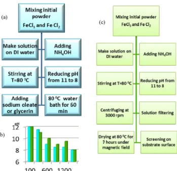
Then experimental steps divided in two ways: at first by adding glycerin to the solution; second - continue stirring throughout the slow addition of fine powder of sodium oleate. The precipitants were heated at 80ºC for 60 minutes in water bath. Finally, the Fe3O4 nanoparticles was collected by magnetic field separation and washed several times with deionized water and dried in continuous drier at 80°C for 2-5 hours. A pH of 8 was maintained during the synthesis process to reduce ferrous and ferric chlorides. We observed pH of the reaction solution. Therefore, the concentration of glyserin in the reaction system was chosen as 2 wt%, the reaction temperature of the system was chosen as 80ºC, and the stirring rate of the reaction system was varied 400-600 rpm. The experimental flowchart for preparation of Fe3O4 nanoparticles was shown in Fig. 1(a). Then concentration of sodium oleate in the reaction system was chosen as 0,5 wt%, the reaction temperature of the reaction system was chosen as 80ºC, and the stirring rate of the reaction
system was chosen 400-600 rpm. The particle distribution of Fe3O4 nanoparticles is uniformly when the pH is lower than 9. In all cases, at higher pH’s, slight increases in solution basicity corresponded to large increases in particle size. At lower pH’s, however, changes in solution basicity yielded only slight to moderate increases in particle size. This phenomenon can be explained by the forming of iron solution where particle size control and dependence on solution pH [22-23]. Besides the differ- ent iron oxide- nanowires were synthesized and charac- terized by modifying co-precipitation methods. In this case the FeCl2 · 4H2O and FeCl3· 6H2O were dissolved in deionized water with vigorous stirring with adding ammonia aqueous solution, similar as the previous exper- iment. Also pH of 8 was maintained during the synthesis process by washing solution in DI water several times.
Then obtained solution filtered and centrifuged with 3000 rmp speed and dried at 80oC for 7 hours under magnetic field (big magnetic bar set under the beaker). From Fig.
1(b), is shown reduce of the pH with the increasing treatment of solution with adding different amount deionized water for both experiments. The flowchart of nanowires preparation was shown in Fig. 1(c).
According this image the pH is lower than 8.5 when solution washed seven times with deionized water. The
other part of initial solution after centrifuging was spe- cially cleaned for making nanowire structure and it is take longer time. In particular, during the precipitation reaction, the pH value was maintained at 8.
3. Results & Discussions
3.1. Coated nanoparticles
Glycerin added solution. Optical microscope and SEM experiments were carried out to investigate the micro- structure obtained solutions. As additive compound to solution we used glycerin a colorless, viscous liquid that is widely used in pharmaceutical formulations. It is com- pletely soluble in water and alcohol but is only slightly soluble in many common solvents, such as ether, ethyl acetate, and dioxane. Earlier glycerin used as solution component for synthesis different nanostructure materi- als [24]. Glycerin is a reactive molecule that undergoes all the usual reactions of alcohols. It boils at 290°C at atmospheric pressure and melts at 17.9°C. It was shown that adding glycerin to solution is led to formation of bubbled structure with large surface heterogeneity Fig.
2(a). This is caused that continuously stirring process of glycerin in aqueous solution lead to formation surface foam Fig. 2(a) and (b). Fig. 2(c) and (d) shows the SEM image of the iron solution with adding of glycerin.
According to the SEM images, the glycerin become hard- ened and prevented to formation and agglomeration of the iron nanoparticles. Probably, this is caused that dur- Fig. 1. Experimental flowchart for preparation of iron oxide
nanoparticles (a); block diagram of effect of deionized water treatment on reducing solution pH (b); flowchart for preparation of iron oxide nanowires.
Fig. 2. Surface images of iron solution SEM with adding of glycerin: (a) and (b) optical microscope image; (c) and (d) SEM images.
ing drying and drying at 80°C glycerin uncontrolled accumulate in solution and do not promote to formation of nanoparticles. That is why we are not observed any nanoparticles in these images. However in specific area we observed partially formation of crystallization of iron compound Fig. 2(d).
Nanoparticles coated with sodium oleate. Sodium ole- ate is white powder with a tallow aroma with melting point 232~235°C, soluble in alcohol and water, with par- tial decomposition. The resulting solutions contain mod- erate concentrations of hydroxide ions and have pH's greater than 7. Thus Fig. 3(a-d) shows the optical image and SEM image of the structure of Fe3O4 nanoparticles coated with sodium oleate. According to the SEM images, the uniform distribution of particles is very strong in the group of these Fe3O4 nanoparticles. In this case sodium oleate conjugate to the nanoparticles sur- faces through chemical bonding. We suppose that sodium oleate is stabilized to the nanoparticles surface to pre- vent the nanoparticles from agglomeration and make them more uniformity Fig. 3(b,c). It is shown that Fe3O4 nanoparticles are partially agglomerated in shape of balls which reveal the particle distribution and uniformity in the bulk of solution consisted from stabilized particles.
Besides morphology of the samples with additional sodium oleate component show the particles with aver-
age size 40-60 nm. The selected area observed particle accumulation during process of solution formation. An EDX analysis of the same nanoparticles is shown in the spectrum of Fig. 3(d). The result confirms that the nanoparticles consists iron and oxygen components.
X-ray diffraction was used to characterize the structure and composition of the iron nanostructures and the modified nanoparticles. In particular the crystal structure of the iron nanostructures was analyzed by X-ray diffrac- tometer (Rigaku (Japan), SWXD). The voltage and cur- rent were set to be 40 kV and 30 mA, respectively. The diffraction angle 2θ and scanning speed were 20-100°
and 0.020 per 0.8 seconds, respectively. The XRD patterns of the as-prepared nanoparticles were shown in Fig. 4. The XRD peaks of the Fe3O4 are compared with those of standard one in JCPDS file (PDF No. 80-2377).
A series of characteristic peaks at 2θ = 33.17°, 35.63°, 43.5°, 57.45°, and 62.43°, which corresponds to (104), (110), (202), (122) and (214) Bragg reflection, respectively. Image in figure agree with Fe3O4 standard XRD patterns, identify that the Fe3O4 nanoparticles system hexagonal structure are indexed to lattice rhomb-centered Fig. 4.
Nanowires synthesized by co-precipitation method. The typical SEM images and EDX spectrum of Fe3O4 nanow- ires with different diameters released from precipitation solution are shown in Fig. 5(a-c). It is observed that there are two main shapes of Fe3O4 nanowires: needle-tip and large-tip. Most of nanowires are compact and uniform with large aspect of ratios. During the precipitation reac- tion, the pH value was maintained at 8. The crystalline
Fig. 3. The optical microscope and FESEM of Fe3O4 nanopar- ticles: (a) the bare initial solution particles, (b) and (c) separately Fe3O4 nanoparticles coated with oleate sodium on carbon tape, (d) EDX spectrum nanoparticles coated with oleate sodium (on the top – formation Fe3O4 nanoparticles covered by sodium oleate).
Fig. 4. XRD patterns of Fe3O4 prepared via co-precipitation methods.
structures after chemical process were collected and washed by DI water, centrifuged and dried. It is also shown that the nanowires cover the surface of the sub- strate with a high surface density up to 106-108 cm−1. Fig. 5(a) and (b) is images of Fe3O4 nanowires with higher magnification which reveal the smoothness and uniformity of the nanowires. The short nanowires are due thermal treatment process of solution formation Fig. 5(b).
An EDX analysis of the same nanowire is shown in the image of Fig. 5.
The result confirms that the nanowire consists of Fe, O and Cl. The chlorine peaks in EDX spectrum is caused by low thermal treatment of nanowires after precipita- tion in solution. The mechanism of nanowire growth is a very complicated process. So far, it has not had a uni- form theory to explain it. From the above three cases, large-tip and, needle-tip nanowires, it seems that the growing of the Fe3O4 nanowires through the diffusion of Fe from the bottom to tip under magnetic field. If the tip is sealed, Fe will gather in the tip and form a large-tip nanowire. Although we don’t know the condition, some- times, the large-tip will continuously grow and form a needle-tip nanowire.
4. Conclusion
In this work we investigated the effect of growing iron
oxide nanoparticles with adding organic components. In particular, nano-sized iron oxide particles was synthe- sized by the co-precipitation method from FeCl2· 4H2O and FeCl3· 6H2O dissolved in deionized water with vigor- ous stirring. Then at first initial solution was added glyc- erin; and second solution continued stirring throughout the slow addition of fine powder of sodium oleate which was chosen as the surfactant. Because sodium oleate is attached to the nanoparticles surface to prevent the nano- particles from agglomeration. It is shown that the ferrof- luid with additional glycerin component and it is contained heterogeneous agglomerations of iron solution with bubbled structure. While to solution is added sodium oleate component it has uniform distribution of ball shaped iron oxide nanoparticles in the liquid solution.
Morphology of the samples with addition of oleate shows the uniform distribution of particles with average size of nanoparticles 40-50 nm. This may be due to the fact that sodium oleate prevents magnetic particles reuniting so as to obtain particles with nanosize. It reveals that the sizes of Fe3O4 nanoparticles can be well controlled as the dis- persion action of sodium oleate, which can well control the growth of Fe3O4 nucleus. Besides, it was shown that control of solution pH also important for synthesis fine nanoparticles. By adjusting the pH, large particles and agglomerations of particles are prevented from forming of large crystalline structure which caused precipitation within the solution. For this reason, it is desirable to maintain the particle size within a pH range wherein the particle growth is held at a minimum. The Fe3O4 nano- particles can be well dispersed in an aqueous solution were prepared by the mentioned co-precipitation method.
Besides we also fabricated different form Fe3O4 nanow- ire arrays by co-precipitation method by adjusting synthe- sizes condition and growth under magnetic field. Synthesized iron nanowires array well distributed and visible and rel- atively low SEM magnification with which reveal the smoothness and uniformity of the nanowires. We pro- pose novel approach to directly synthesize nanoparticles and grow nanowire arrays at low temperature. This is allowed to produce multi dispersed iron nanoparticles and nanowires on one technologic step without using additional technical procedures. The synthesis in one step by co-precipitation process also allows to growth differ- ent type of nanowires from nanoparticle solution include Fig. 5. FE-SEM image and EDX spectrum for Fe3O4 nanowires:
(a) highly ordered nanowire arrays; (b) short nanowire arrays (covered with iron nanoparticles); (c) needle-tip type of nanowire and EDX spectrum of needle tip nanowire arrays.
iron precipitates from spherical particle under magnetic field. These nanowires with length more than 3 µm and diameter around 10-50 nm with shape of large-tip and needle-tip nanowires are successfully synthesized.
Acknowledgement
This work was supported by a Research Fund of Kumoh National Institute of Technology.
References
[1] Magnetic Nanoparticles Edited by Sergey P. Gubin.
WILEY-VCH Verlang GmbH & KGaA, Weinheim, ISBN:
978-3-527-40790-3 (2009) 466.
[2] Stepanka Klimkova, Miroslav Cernik, Lenka Lacinova, Jan Filip and Dalibor Jancik: Chemosphere, 82 (2011) 1178.
[3] Alejandro Simón de Dios and Marta Elena Díaz-García:
Anal. Chim. Acta., 666 (2010) 1.
[4] A. Dyal, K. Loos, M. Noto, S. W. Chang, C. Spagnoli, K.
V. P. M. Shafi et al.: J. Am. Chem. Soc, 125 (2003) 1684.
[5] S. P. Gubin, Yu A. Koksharov, G. B. Khomutov and G. Yu Yurkov: Russian Chemical Reviews, 74(6) (2005) 489.
[6] Y. J. Song, P. Y. Jin and T. Zhang, Mater. Lett., 64 (2010) 1789.
[7] Miha Drofenik, Gregor Ferk, Matjaž Kristl and Darko Makovec: Mater. Lett., 65 (2011) 439.
[8] Daniel Amara, Israel Felner, Israel Nowik and Shlomo Margel: Colloids and Surfaces A: Physicochem. Eng.
Aspects, 339 (2009) 106.
[9] P. C. Morais, R. L. Santos, A. C. M. Pimenta, R. B. Aze- vedo and E. C. D. Lima: Thin Solid Films, 515 (2006) 266.
[10] J. S. Suh, J. Y. Lee, Y. S. Choi, S. J. Lee, C. P. Chung and Y. J. Park: Biochemical and Biophysical Research Com-
munications, 379 (2009) 669.
[11] Yi, An, Tielong, Li, Zhaohui, Jin, Meiying, Dong, Hongcai, Xia, and Xue, Wang: Bioresour. Technol., 101 (2010) 9825.
[12] M. J. Bonder, Y. Zhanga, K. L. Kiickb and V. Papaefthy- miouc, G. C. Hadjipanayisa J. Magn. Magn. Mater., 311 (2007) 658.
[13] Di, Li, Deli Jiang, Min Chen, Jimin Xie, Yinyin Wu, Shengchun Dang and Jianxin Zhang: Mater. Lett., 64 (2010) 2462.
[14] Liying Zhang, YafeiZhang: Fabrication and magnetic proper- ties of Fe3O4 nanowire arrays in different diameters. J.
Magn. Magn. Mater., 321 (2009) L15.
[15] N. Wang, Y. Cai, R. Q. Zhang: Growth of nanowires.
Mater. Sci. Eng. R, 60 (2008) 1.
[16] Qin Han, Zhenghui Liu and Yingying Xu et al: Growth and Properties of Single-Crystalline g-Fe2O3 Nanowires J.
Phys. Chem. C, 111 (2007), 5034.
[17] Q. Han, H. Zhang et al.: Defects and growing mechanisms of a-Fe2O3 nanowires. Chem. Phys. Lett., 431 (2006) 100.
[18] H.-W. Wang et al.: J. Magn. Magn. Mater., 310 (2007) 2425.
[19] Ning Du, Yanfang Xu and Hui Zhang: Nanoscale Res Lett., 5 (2010) 1295.
[20] Yong Ding, Jenny Ruth Morber and Robert L. Snyder:
Adv. Funct. Mater., 17, (2007), 1172.
[21] R. Massart and S. Neveu: J. Magn. Magn. Mater., 149 (1995) 6.
[22] Le Thi Mai Hoa, Tran Thi Dung, Tran Mau Danh, Nguyen Huu Duc and Dang Mau Chien: J. Physics: Conference Series (Workshop on Advanced Materials Science and Nanotechnology (AMSN08), 187 (2009), 012.
[23] Cullen T. Vogelson and Andrew R. Barron: J. Non-Crys- talline Solids, 290 (2001) 216.
[24] Tran Trung, Won-Jei Cho and C.-S. Ha: Mater. Lett., 57 (2003) 2746.