1. 서 론
나노입자는 연료전지, 유기촉매, 센서, 광전자, 전자, 바이오, 의료 등에 여러 분야에 응용되고 있다. 이러한 나노입자들은 모양이나 크기에 따라 독특한 특성들 때 문에, 나노입자의 모양을 조절하기 위한 연구가 진행되 고 있다 [1-7]. 특히 팔라듐 나노입자는 유기반응의 촉 매, 연료전지, SERS (표면 라만 증강), 수소 저장 등에
✉ Young Wook Lee; [email protected], Tae Ho Shin; [email protected]
Copyright ©2019 KIEEME. All rights reserved.
This is an Open-Access article distributed under the terms of the Creative Commons Attribution Non-Commercial License (http://creativecommons.org/licenses/by-nc/3.0) which permits unrestricted non-commercial use, distribution, and reproduction in any medium, provided the original work is properly cited.
여러 분야에 응용되고 있다 [8-11]. 이렇게 이용되는 나노입자는 모양이나 크기에 따라 표면에 다른 에너지 를 있어 반응성이 다르게 나타난다. 그리고 이 입자는 면심입방구조(face-centered cubic) 형태로 구성되어 있고, 모양에 따라 다른 지수를 갖게 되는데, 저지수 결정면(low index facet) 갖는데 팔면체인 경우나 구인 경우에는 (111) 결정면을 갖고 정육면체인 경우 (100) 으로 이루어져 있으며, (110)인 경우는 마름모 십이면 체나 정십이면체 모양을 가지고 있다. 이렇게 이루어진 결정면에 따라 에너지 세기는 111<100<110이며, 이러 한 이유는 에너지의 세기가 클수록 배위 결합수가 작 아져 에너지의 세기가 증가하는 것이다 [12]. 이러한 나 노입자의 모양 조절은 쉬운 일이 아니다. 나노입자의 성 장을 잘 이해하고 나노입자의 성장 반응속도를 조절할
모양이 조절된 팔라듐 나노입자의 합성과 4-나이트로벤젠 사이올의 광환원 반응
이영욱 , 신태호
한국세라믹기술원 에너지환경본부
Synthesis of Shape Controlled Pd Nanoparticles and Surface-Induced Photoreduction of 4-Nitrobenzenethiol on Pd
Young Wook Lee and Tae Ho Shin
Energy & Environment Division, Korea Institute of Ceramic Engineering and Technology, Jinju 52851, Korea
(Received August 2, 2019; Revised August 7, 2019; Accepted August 9, 2019)
Abstract: The facile synthesis of shape-controlled Pd nanoparticles (PdNPs) with ascorbic acid as a reducing agent and cetyltrimethylammonium bromide (CTAB) as a capping agent is presented in this study. The synthesized PdNPs were characterized by UV-vis spectroscopy, transmission electron microscopy (TEM), scanning electron microscopy (SEM), X-ray diffraction (XRD), and Raman Spectroscopy. The prepared PdNPs show efficient surface-enhanced Raman scattering (SERS) properties. SERS studies on the adsorption characteristics of 1,4-phenylene diisocyanide (1,4-PDI) on colloidal PdNPs have revealed that the relative peak intensity of the (NC)free and (NC)bound modes distinctly depends on the 1,4-PDI concentration as well as the shape of the PdNPs. Furthermore, we found that the PdNPs are also efficient photoelectron emitters such that the SERS spectrum of 4-nitrobenzenethiol (4-NBT) on PdNPs is readily converted to that of 4-aminobenzenethiol (4-ABT) under 632.8 nm radiation.
Keywords: Photoreduction, SERS, Shape control, Pd nanoparticles
Regular Paper 458
J. Korean Inst. Electr. Electron. Mater. Eng.
Vol. 32, No. 6, pp. 458-461 November 2019 DOI: https://doi.org/10.4313/JKEM.2019.32.6.458 ISSN 1226-7945(Print), 2288-3258(Online)
전기전자재료학회논문지, 제32권 제6호 pp. 458-461, 2019년 11월: 이영욱 등 459
줄 알아야 가능하다. 이 연구에서는 나노입자의 모양을 쉽게 합성하고, 모양에 따른 결정면에 따라 SERS의 반 응이 어떻게 적용하는가에 대해 알아보았다.
2. 실험 방법 2.1 실험 시약
K2PdCl4, Ascorbic acid (비타민 C), 3차 증류수 계면 활성제: CTAB (Centyltrimethylammonium bromide) 표면 라만 증강 물질: 4-NBT (4-nitrobenzenethiol), 1,4- PDI (1,4-phenylene diisocyanide) 4-ABT (4-aminobenzenethiol)
시약 회상: 알드리치 2.2 합성 방법
정육면체 팔라듐 나노입자(Pd NCs) 삼각 플라스크 에 3 차 증류수 47 mL 를 주사하여 강력하게 자반시킨 상태에서 5 mM K2PdCl4의 용액을 1 mL 주사한다.
다음 계면 활성제인 30 mM CTAB 1 mL 주사하고 환 원제인 100 mM 아스코르빅 산을 1 mL 주사한 후에 5 분 동안 반응을 시킨다.
멀티 암드 팔라듐 나노입자(Pd MANPs) 삼각 플라 스크에 3 차 증류수 47 mL 를 주사하여 강력하게 스트 링시킨 상태에서 5 mM K2PdCl4의 용액을 1 mL 주 사한다. 다음 계면 활성제인 30 mM CTAB 1 mL 와 환원제인 100 mM 아스코르빅 산 1 mL 를 합친 용액 을 한 번에 주사한 후에 5 분 동안 반응을 시킨다.
가지돌기 팔라듐 나노입자(Pd NDs) 삼각 플라스크 에 3 차 증류수 47 mL 를 주사하여 강력하게 자반시킨 상태에서 5 mM K2PdCl4의 용액을 1 mL 주사한 다 음 환원제인 100 mM 아스코르빅 산을 1 mL 주사하 고 계면 활성제인 30 mM CTAB 를 1 mL 주사하여 5 분 동안 반응을 시킨다. 계면활성제는 원심분리를 통 해 제거한다.
2.3 사용 기기
사용 기기는 Uv-vis absorption spectrometer (SINCO S-3100)을 사용하였다. TEM (투과주사현미경)은 JEOL JEM-2010 을 사용하였으며, EM (주사현미경)은 Phillips Model XL30 S FEG 사용하였다. XRD (X-ray diffraction) 는 AXS D8 DISCOVER CuKα (0.1542 nm)를 사용하
였다. 라만 기기는 Jobin Yvon/HORIBA Lab RAM 을 사용했으며, 레이저의 파장은 He/Ne 633 nm 를 사용 하였다.
3. 결과 및 고찰
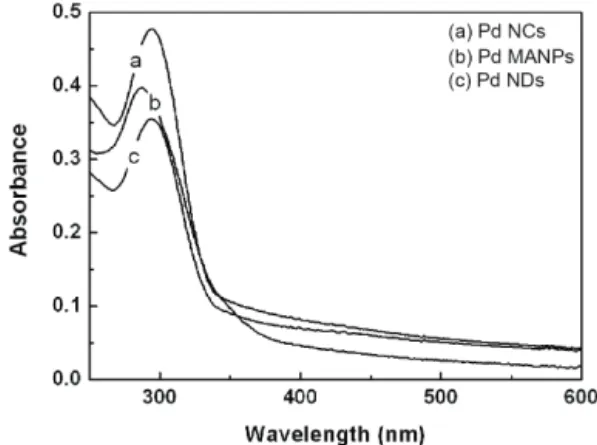
나노입자는 합성한 뒤 나노입자의 형성이나 특성을 가장 알기 쉬운 방법은 Uv-vis을 측정하는 방법이다.
Uv-vis 데이터를 보면, Pd NPs가 모양에 따라 흡광도 가 다른 것을 알 수 있으며, 이것은 입자마다 다른 에 너지를 가지고 있음을 증명한다. 나노입자가 다른 에너 지를 가지고 있기에, 촉매 반응에서 다른 효과가 나타 낼 수 있다는 것을 짐작할 수 있다 (그림 1).
그림 2는 나노입자가 모서리 길이가 21±2.5 nm인 Pd NCs 나노입자이며 [그림 2(a)], Pd MANPs는 18
±4.5 nm이며 [그림 2(b)], Pd NDs는 54±7.6 nm [그 림 2(c)]이다. 이 나노입자가 형성되는 조건은 계면 활 성제와 환원제 간의 속도에 의해서 결정된다. CTAB라 는 계면 활성제를 먼저 주사하면, 팔라듐 이온 같이 콤 플렉스를 이루기 때문에, 팔라듐의 환원속도에 관여하게 되며, 그것은 결정면 형성에 영향을 준다. 또한, 환원제 를 먼저 주사하거나 계면 활성제와 동시에 주사할 경우, 환원속도가 달라져 모양 형성에 영향을 주게 된다.
이 방식을 바탕으로 실험한 결과 실험 순서를 조절 하여 쉽게 팔라듐 입자의 모양을 조절하였다 (그림 2).
양이 다른 입자는 결정면 구성이 다르게 형성되어 있다. 그래서 XRD data를 보면 111/100 비율로 보면 Pd NCs 경우 100 결정면이 훨씬 많이 존재하는 것을 관찰할 수 있으며, PdNCs, PdMANPs, PdNDs의 100/
111의 비율을 보면 0.79>0.44>0.40으로 나타나며, 이
Fig. 1. Uv-vis spectroscopy of Pd NCs, Pd MANPs and Pd NDs.
J. Korean Inst. Electr. Electron. Mater. Eng., Vol. 32, No. 6, pp. 458-461, November 2019: Y.-W. Lee et al.
460
것으로 PdNCs가 100 결정면을 많이 갖는 것을 알 수 있었다 (그림 3).
결정면에 대한 표면 에너지의 세기를 비교하기 위해 서 쉽게 에너지 결합 비교에 이용할 수 있는 물질인 1,4-PDI 물질로 SERS 측정을 하였다 [13]. 1,4-PDI라 는 물질로 측정하는데 CN의 피크의 위치에 따라 표면 에너지가 다르다는 것을 알 수 있었다. 그림 4를 보면 Pd NCs의 라만의 CN 피크의 위치가 15 cm-1 정도 이동하여 위치한 것을 알 수 있다. 이것은 “CN-Pd의 결합 에너지의 세기가 다르다”라는 것을 증명하였다 [그림 4(b)].
라만 증강의 표면에서 일어나는 광반응을 측정하기 위해 나이트로 물질을 가진 4-NBT 물질을 사용하였으
며, 그 물질이 분해되는 것을 보기 위해 4-ABT 물질 을 사용하여 라만 세기와 속도를 비교하는 실험을 진 행하였다 [14]. 그림 5를 보면 4-NBT로 SERS 실험을 한 결과를 보면, Pd NCs가 다른 팔라듐 나노입자보다 니트로화합물에서 아민 화합물로 시간에 따른 아민 피 크를 비교하였을 때, 훨씬 빠른 속도로 진행되고 있는 것을 알았다 (그림 5). 반응속도로 보아 Pd 나노입자의 Fig. 3. (a) XRD patterns and (b) (111) and (100) diffraction peak positions of the PdNCs, PdPNCs and PdMNPs on silicon wafer.
Fig. 4. SERS spectra of 1,4-PDI on (a) Pd NCs, (b) Pd MANPs, and (c) Pd NSDs. All spectra were normalized with 8a mode peak at 1,600 cm-1 for convenience.
Fig. 5. (a) SERS spectra of (A) Normal Raman spectrum of 4-NBT in the neat solid state and 4-NBT adsorbed on (B) Pd NCs, (C) Pd MANPs, (D) Pd NDs and (E) PdMNPs on 4-ABT. (b) SERS intensity of the reactant and product band located 1,335 and 1,445 cm-1. All spectra were obtained usig a He/Ne laser at 632.8 nm as the excitation source and normalized with NO2 peak at 1,335 cm-1 for convenience.
(a)
(b)
(c)
Fig. 2. TEM images of (a) PdNCs, (b) PdPNCs, and (c) PdMNPs.
전기전자재료학회논문지, 제32권 제6호 pp. 458-461, 2019년 11월: 이영욱 등 461
표면에 의해 화합물의 그림 5(b)를 보면 환원이 다르 게 일어난다는 것을 알 수 있었으며, 이러한 표면 에 너지는 나노입자의 모양에 의해 결정되기 때문에, 중요 한 요소로 작용할 수 있다는 것을 알 수 있었다.
4. 결 론
Pd 나노입자의 모양 조절은 시약의 주사 순서를 조 절함으로 인해 반응속도가 다르게 작용하여, 손쉽게 나 노입자의 모양을 조절할 수 있었다. 또한, 모양이 조절 된 Pd 나노입자를 SERS에 적용하여, 이 나노입자들이 반응성을 분석한 결과, 나노입자의 모양 조절은 중요하 다는 것을 알 수 있었다. 나노입자의 모양 조절은 결 정면에 의해 표면 에너지가 다를 수 있으며, 에너지의 세기에 의해 반응성이 다르게 나타날 수 있다는 것을 알 수 있었다. 이 연구를 통해 나노입자의 모양 조절 과 반응성에 연구가 진행되었으면 한다.
ORCID
Young Wook Lee https://orcid.org/0000-0002-5663-5713 Tae Ho Shin https://orcid.org/0000-0002-9001-7863
감사의 글
This work was supported by the International R
& D Program (grant no.P0004433) funded By the Ministry or Trade, Industry & Energy (MI, Korea) and the Korea Institute of Energy Technology Evaluation and Planning (KETEP) and the Ministry or Trade, Industry & Energy (MOTIE) of the Republic of Korea (No.20182010600400).
REFERENCES
[1] Y.W.C. Cao, R. Jin, and C. A. Mirkin, Science, 297, 1536 (2002). [DOI: https://doi.org/10.1126/science.297.5586.1536]
[2] W. P. Halperin, Rev. Mod. Phys., 58, 533 (1986). [DOI: https://
doi.org/10.1103/RevModPhys.58.533]
[3] L. N. Lewis, Chem. Rev., 93, 2693 (1993). [DOI: https://
doi.org/10.1021/cr00024a006]
[4] T. S. Ahmadi, Z. L. Wang, T. C. Green, A. Henglein, and M.
A. El-Sayed, Science, 272, 1924 (1996). [DOI: https://doi.org/
10.1126/science.272.5270.1924]
[5] S. A. Maier, M. L. Brongersma, P. G. Kik, S. Meltzer, A.A.G. Requicha, and H. A. Atwater, Adv. Mater., 13, 1501 (2001). [DOI: https://doi.org/10.1002/1521-4095(200110)13:19
<1501::AID-ADMA1501>3.0.CO;2-Z]
[6] S. Chen and Y. Yang, J. Am. Chem. Soc., 124, 5280 (2002).
[DOI: https://doi.org/10.1021/ja025897+]
[7] X. Zhang, M. A. Young, O. Lyandres, and R. P. Van Duyne, J. Am. Chem. Soc., 127, 4484 (2005). [DOI: https://doi.org/
10.1021/ja043623b]
[8] M. Kim, Y. Kim, J. W. Hong, S. Ahn, W. Y. Kim, and S.
W. Han, Chem. Commun., 50, 9454 (2014). [DOI: https://
doi.org/10.1039/C4CC02494J]
[9] S. Choi, H. Jeong, K. H. Choi, J. Y. Song, and J. Kim, ACS Appl. Mater. Interfaces, 6, 3002 (2014). [DOI: https://
doi.org/10.1021/am405601g]
[10] X. Zhang, M. A. Young, O. Lyandres, and R. P. Van Duyne, J. Am. Chem. Soc., 127, 4484 (2005). [DOI: https://doi.org/
10.1021/ja043623b]
[11] P. Xi, F. Chen, G. Xie, C. Ma, H. Liu, C. Shao, J. Wang, Z. Xu, X. Xu, and Z. Zeng, Nanoscale, 4, 5597 (2012).
[DOI: https://doi.org/10.1039/C2NR31010D]
[12] T. K. Sau and A. L. Rogach, Adv. Mater., 22, 1781 (2010).
[DOI: https://doi.org/10.1002/adma.200901271]
[13] N. H. Kim and K. Kim, J. Phys. Chem. B, 110, 1837 (2006).
[DOI: https://doi.org/10.1021/jp055541v]
[14] B. O. Skadtchenkoand and R. Aroca, Spectrochim. Acta, Part A, 57, 1009 (2001). [DOI: https://doi.org/10.1016/S1386-1425 (00)00415-7]