Mori Fructus Induces Cell Death through ROS-dependent Mitochondrial Apoptotic Pathway in Human Glioma Cells
Sang Won Jang, Ji Cheon Jeong*
Department of Internal Medicine, College of Korean Medicine, Dongguk University
Mulberry has been reported to contain wide range of polyphenols and have chemopreventive activity. However, little has been known regarding the effect of mulberry fruits on cell viability in human glioma cells. The present study was undertaken to examine the effect of mulberry fruit (Mori Fructus) on cell viability and to determine its underlying mechanism in human glioma cells. Cell viability and cell death were estimated by MTT assay and trypanblue exclusion assay, respectively. Reactive oxygen species (ROS) generation was measured using the fluorescence probe DCFH-DA. The mitochondrial transmembrane potential was measured with DiOC6(3). Bax expression and cytochrome c release were measured by Western blot analysis. Caspase activity was estimated using colorimetric kit. Mori Fructus resulted in apoptotic cell death in a dose- and time-dependent manner. Mori Fructus increased ROS generation and the Mori Fructus-induced cell death was also prevented by antioxidants, suggesting that ROS generation plays a critical role in Mori Fructus-induced cell death. Western blot analysis showed that Mori Fructus treatment caused an increase in Bax expression, which was inhibited by the antioxidant N-acetylcysteine (NAC). Mori Fructus induced depolarization of mitochondrial membrane potential and its effect was inhibited by the antioxidants NAC and catalase.
Mori Fructus induced cytochrome c release, which was inhibited by NAC. Caspase activity was stimulated by Mori Fructus and caspase inhibitors prevented the Mori Fructus-induced cell death. These findings suggest that Mori Fructus results in human glioma cell death through ROS-dependent mitochondrial pathway in human glioma cells.
Key words : Mori Fructus, cell viability, mitochondrial membrane potential, caspase, human glioma cells
* To whom correspondence should be addressed at : Ji Cheon Jeong, Department of Internal Medicine, Hospital of Korean Medicine, Dongguk University, Sukjang-Dong 1090-1, Gyeong-Ju City, Gyeong-Buk, Korea
․E-mail : [email protected], ․Tel : 054-770-1265
․Received : 2008/07/09 ․Revised : 2008/08/04 ․Accepted : 2008/08/29
Introduction
Tumors of glial origin, gliomas, are the most frequent primary tumors that arise in the brain. The most malignant form of glioma, glioblastoma multiforme, is one of the most aggressive human cancers, with a median survival of less than 1 year1). Despite aggressive treatment including surgery, radiation, and chemotherapy, this statistic has not changed significantly over the past years2,3).
Natural products derived from plants have recently received much attention as potential chemopreventive and chemotherapeutic agents. Among them great attention has been given to natural products with polyphenols. These substances appear very promising for cancer prevention and treatment in preclinical models and clinical trials4-6). Considering that many chemotherapeutic agents have serious
side effects and development of multidrug resistance further limits chemotherapy in cancer, oriental medicinal drugs may be a very promising group of compounds exerting the chemopreventive and chemotherapeutic effects.
Mulberry is widely distributed in Asia and has been used in traditional medicine in Korea. Mulberry fruit (Mori Fructus, MF) have attracted much attention due to their potential human health benefits. Berries contain a diverse range of phytochemicals with biological properties such as antioxidant, anticancer, anti-neurodegenerative, and anti-inflammatory activities on account of its apparent anti-inflammatory, antibiotic, antioxidant, and hepatoprotective effects, and anti-diabetic effect7-10). Phenolic compounds isolated from berries and crude extracts inhibited the growth of human oral, colon, and prostate cancer cells11-13). Recently, it has been reported that anthocyanins, major active constituents of mulberry, inhibit migration of human glioblastoma cells14). However, little has been known regarding the effect of mulberry fruit on viability of human glioma cells.
The present study was undertaken to investigate whether MF has anti-proliferative in human glioma cells and to determine
its molecular mechanism in A 172 human glioma cells.
Materials and Methods
1. Reagents
N-acetylcysteine (NAC), catalase, 3-[4,5-dimethylthiazol-2-yl]-2,5-diphenyltetrazolium bromide (MTT), and propidium iodide were purchased from Sigma-Aldrich Chemical (St. Louis, MO, USA). Tween 20, VAD-FMK, and DEVD-CHO were purchased from Calbiochem (San Diego, CA, USA). 3,3’-dihexyloxacarbocyamide [DiOC6(3)]
and 2’,7’-dichlorofluorescein diacetate (DCFH-DA) were obtained from Molecular Probes (Eugene, OR, USA).
Antibodies were obtained from Cell Signaling Technology Inc.
(Beverly, MA, USA). All other chemicals were of the highest commercial grade available.
2. Extraction of MF
The crushed Mori Fructus (300 g) was extracted 3 times, each time with 3 volumes of methyl alcohol at 60℃ for 24 h.
The extract was filtered and evaporated under a reduced pressure using a rotary evaporator to yield 85.20 g(yield 28.4%).
3. Cell culture
A172 cells were obtained from the American Type Culture Collection (Rockville, MD, USA) and maintained by serial passages in 75-cm2 culture flasks (Costar, Cambridge, MA, USA). The cells were grown in Dulbecco’s modified Eagle’s medium (DMEM, Gibco BRL, Invitrogen, Carsbad, CA, USA) containing 10% heat inactivated fetal bovine serum (HyClone, Logan, UT, USA) at 37°C in humidified 95% air/5%
CO2 incubator. When the cultures reached confluence, subculture was prepared using a 0.02% EDTA-0.05% trypsin solution. The cells were grown on well tissue culture plates and used 1-2 days after plating when a confluent monolayer culture was achieved. Unless otherwise stated, cells were treated with MF in serum-free medium.
4. Measurement of cell viability and cell death
Cell viability was evaluated using a MTT assay15). After washing the cells, culture medium containing 0.5 mg/ml of MTT was added to each well. The cells were incubated for 2 hr at 37℃, the supernatant was removed and the formed formazan crystals in viable cells were solubilized with 0.11 ml of dimethyl sulfoxide. A 0.1 ml aliquot of each sample was then translated to 96-well plates and the absorbance of each well was measured at 550 nm with ELISA Reader (FLUOstar OPTIMA, BMG LABTECH, Offenburg, Germany). Data were
expressed as a percentage of control measured in the absence of MF. Unless otherwise stated, the cells were exposed to 50 μ M MF for 48 hr. Test reagents were added to the medium 30 min before MF exposure.
Cell death was estimated by counting the cell number and trypan blue exclusion assay, respectively. The cells were harvested using 0.025% trypsin and incubated with 4% trypan blue solution. The number of viable and nonviable cells was counted using a hemocytometer under light microscopy. Cells failing to exclude the dye were considered nonviable.
5. Measurement of reactive oxygen species (ROS)
The intracellular generation of ROS was measured using DCFH-DA. The nonfluorescent ester penetrates into the cells and is hydrolyzed to DCFH by the cellular esterases. The probe (DCFH) is rapidly oxidized to the highly fluorescent compound 2’,7’-dichlorofluorescein (DCF) in the presence of cellular peroxidase and ROS such as hydrogen peroxide or fatty acid peroxides. Cells cultured in 24-well plate were preincubated in the culture medium with 30 mM DCFH-DA for 1 hr at 37℃. After the preincubation, the cells were exposed to 50 μM MF for various durations. Changes in DCF fluorescence was assayed using FACSort Becton Dickinson Flow Cytometer (Becton-Dickinson Bioscience, San Jose, CA, USA) and data were analyzed with CELLQuest Software.
6. Measurement of mitochondrial membrane potential The mitochondrial transmembrane potential was measured with DiOC6(3), a fluorochrome that is incorporated into cells depending upon the mitochondrial membrane potential16). Loss in DiOC6(3) staining indicates disruption of the mitochondrial inner transmembrane potential. Cells were stained with DiOC6(3) at a final concentration of 50 nM for 20 min at 37℃ in the dark. Cells were washed and resuspended in Hank’s balanced salts solution containing Ca2+ and Mg2+. The fluorescence intensity was analyzed with a FACScan flow cytometer using the fluorescence signal 1 channel.
7. Western blot analysis
Cells were harvest at various times after MF treatment and disrupted in lysis buffer (1% Triton X-100, 1 mM EGTA, 1 mM EDTA, 10 mM Tris-HCl, pH 7.4). Cell debris was removed by centrifugation at 10,000 g for 10 min at 4℃. The resulting supernatants were resolved on a 12% SDS-PAGE under denatured reducing conditions and transferred to nitrocellulose membranes. The membranes were blocked with 5% non-fat dried milk at room temperature for 30 min and incubated with primary antibodies. The membranes were
washed and incubated with horseradish peroxidase-conjugated secondary antibody. The signal was visualized using an enhanced chemiluminescence(Amersham, Buckinghamshire, UK).
8. Measurement of cytochrome c release
Cells were harvested and washed twice with PBS. The Cells were incubated with extraction buffer (10 mM Hepes, 250 mM sucrose, 10 mM KCl, 1.5 mM MgCl2, 1 mM EDTA, 1 mM EGTA, 0.05% digitonin, and 1 mM phenylmethylsulfonyl fluoride) at 4℃ for 10 min, then centrifuged at 100,000 g for 10 min at 4℃. The supernatant represented the cytosolic protein.
The fraction was loaded onto a 12% SDS-polyacrylamide gels and transferred to nitrocellulose membranes. After blocking in 5% non-fat dried milk at room temperature for 30 min, membranes were probed with rabbit polyclonal anti-cytochrome, followed by horseradish peroxidase-conjugated secondary antibodies. Bands were visualized using the ECL detection system (Amersham, Buckinghamshire, UK).
9. Measurement of caspase-3 activity
Caspase-3 activity was measured by the caspase-3 colorimetric assay kit (R&D Systems, Minneapolis, MN, USA) according to the manufacturer’s instructions. Cells were collected by centrifugation at 250 g, and the supernatant was gently removed and discarded while the cell pellet was lysed by the addition of the cell lysis buffer at 4℃ for 10 min. Then the cell lysate was incubated with caspase-3 colorimetric substrate, DEVD-pNA at 37℃ for 1hr. The cleavage of the peptide was quantified spectrophotometrically at a wavelength of 405 nm.
10. Statistical analysis
The data are expressed as means±SEM and the difference between two groups was evaluated using Student's t-test.
Multiple group comparison was done using one-way analysis of variance followed by the Tukey post hoc test. A probability level of 0.05 was used to establish significance.
Results
1. Effect of MF on cell viability and cell death
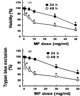
Cell viability was measured in cells exposed to 1-50 mg/ml for 24 and 48 hr. MF resulted in loss of cell viability in dose- and time-dependent manner(Fig. 1A). The cell viability was approximately 75 and 48% of control after addition of 30 and 50 mg/ml MF for 24 hr, respectively. When cells were exposed to MF for 48 hr, the cell viability was reduced to 53, 42, and 26% of control after addition of 10, 30,
and 50 mg/ml. To determine if reduction in cell viability was attributed to cell death, trypan blue exclusion assay was performed. As shown in Fig. 1B, changes in cell death by MF were similar to those estimated by MTT assay, suggesting that the reduction in cell viability by MF was mainly due to induction of cell death.
To ascertain whether MF-induced cell death was due to apoptosis, cells were exposed to 30 mg/ml MF for 24 hr.
Apoptosis was estimated by flow cytometric analysis and annexin-V staining assay. The DNA content analysis by flow cytometry showed that the proportion of the cells in the sub-G1 phase (apoptotic cells) was increased 3.73% of control to 34.38% after MF treatment(Fig. 2A). Similar results also were obtained from cytometric analysis of annexin-V positive cells(Fig. 2B). These results indicate that MF induces loss of cell viability largely through apoptosis.
Fig. 1. Effect of Mori Fructus (MF) on cell viability. Cells were exposed to various concentrations of MF for 24 and 48 hr (A). Cell viability was estimated by MTT reduction assay. Data are mean±SEM of four independent experiments performed in duplicate. *p<0.05 compared with control without MF. (B) Effect of MF on cell death. Cells were exposed to various concentrations of MF for 24 and 48 hr. Cell death was estimated by trypan blue exclusion assay. Data are mean± SEM of four independent experiments performed in duplicate. *p<0.05 compared with control without MF.
2. Role of ROS in MF-induced cell death
To determine whether MF induces ROS generation in A172 cells, the cells were exposed to MF and changes in DCF fluorescence were measured by flow cytometry. In cells were exposed to 30 mg/ml for 60 min, ROS generation was increased (Fig. 3A). The increase in ROS generation was present as early as 10 min after MF treatment and was time-dependent up to 180 min(Fig. 3B)
To determine whether ROS generation is involved in the MF-induced loss of cell viability, the effect of antioxidants NAC and catalase on the cell viability was examined.
MF-induced loss of cell viability was prevented by these antioxidants(Fig. 3C), indicating that MF-induced cell death is associated with ROS generation in A172 cells.
Fig. 2. Effect of Mori Fructus (MF) on apoptosis. Cells were exposed to 30 mg/ml MF for 24 hr. Apoptosis was estimated by flow cytometric analysis (A) and Annexin-V binding assay (B). In flow cytometric analysis, numbers indicate the percentage of cells with the sub-G1 peak (apoptotic cells). In Annexin-v binding assay, numbers indicate the percentage of cells in each quadrant. Earrly apoptotic and late apoptotic cells were shown in right olwer and righ upper quadrants, respectively.
3. Role of Bax and mitochondria in MF-induced cell death Since numerous death signals converge on mitochondria through the activation of pro-apoptotic members of the Bcl-2 family such as Bax17), mitochondria play an important role in apoptotic and necrotic cell death18,19). MF may induce cell death through a Bax-dependent pathway. To test this possibility, the effect of MF on Bax expression was examined.
MF treatment caused an increase in Bax expression in a time-dependent manner(Fig. 4A). Such an effect was inhibited by the antioxidant NAC(Fig. 4B), suggesting that ROS generation is responsible for Bax expression.
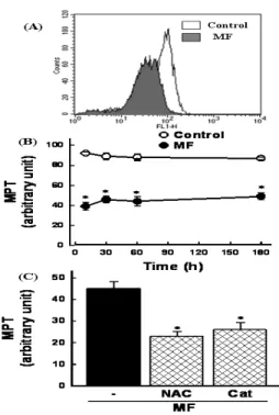
The increase in Bax expression may cause mitochondrial permeability transition (MPT) to induce cell death. Cells were exposed to 30 mg/ml MF and mitochondrial membrane potential was measured using the fluorescence dye. After 60 min of MF treatment, disruption of mitochondrial membrane potential was observed as evidenced by an increase in the proportion of cells with lower fluorescence intensity(Fig. 5A).
A reduction in mitochondrial membrane potential was present
as early as 10 min and remained unchanged up to 180 min(Fig. 5B). To investigate if MPT was attributed to ROS generation, cells were exposed to MF in the presence of antioxidants. MF-induced MPT was prevented by NAC and catalase(Fig. 5C), suggesting that ROS generation is responsible for the MF-induced MPT in A172 human glioma cells.
Fig. 3. Role of reactive oxygen species generation in Mori Fructus (MF)-induced cell death. Cells were exposed to 30 mg/ml MF for 60 min (A) and various times (B) and the DCF fluorescence intensity was measured by a flow cytometer. Data in (B) are mean±SEM of four independent experiments performed in duplicate. *p<0.05 compared with control without MF. (C) Effects of antioxidants on MF-induced cell death. Cells were exposed to 30 mg/ml MF for 48 hr in the presence or absence of 2 mM N-acetylcysteine (NAC) and 500 units/ml catalase (Cat). Cell viability was estimated by MTT reduction assay. Data are mean±SEM of four independent experiments performed in duplicate. *p<0.05 compared with MF alone.
Fig. 4. Effects of Mori Fructus (MF) on Bax expression. (A) Representative results of Bax expression. Cells were exposed to 30 mg/ml MF for various times. a-Actin was employed as a loading control. (B) Effect of antioxidant on MF-induced Bax expression. Cells were exposed to 30 mg/ml MF for 24 hr in the presence or absence of each 2 mM N-acetylcysteine (NAC).
Fig. 5. Effect of Mori Fructus (MF) on mitochondrial membrane potential transition (MPT). Cells were exposed to 30 mg/ml MF for 60 min (A) and various times (B). Mitochondrial membrane potential was estimated by the uptake of a membrane potential-sensitive fluorescence dye DiCO6(3). The fluorescence intensity was analyzed with a flow cytometer. Data in (B) are mean±SEM of three independent experiments performed in duplicate. *p<0.05 compared with control without MF. (C) Effect of antioxidants on MF-induced MPT.
Cells were exposed to 30 mg/ml MF for 60 min in the presence or absence of 2 mM N-acetylcysteine (NAC) and 500 units/ml catalase (Cat). Data are mean±SEM of four independent experiments performed in duplicate. *p<0.05 compared with MF alone.
4. Effect of MF on cytochrome c release
To determine whether MF induces cytochrome c release from mitochondria, Cells were exposed to 30 mg/ml MF for various times and cytochrome c release was measured. As shown in Fig. 6, MF treatment induced cytochrome c release, which was inhibited by the antioxidant NAC (Fig. 6).
Fig. 6. Effect of Mori Fructus (MF) on cytochrome c release. Cells were exposed to 30 mg/ml MF for various times (A) and for 24 hr in the presence or absence of 2 mM N-acetylcysteine (NAC).
5. Role of caspase in MF-induced cell death
Caspases play a key role during the execution phase in
apoptosis and the caspase-3 is one of the executioners of apoptosis20). To examine if caspase activation is involved in MF-induced cell death, activity of caspase-3 was measured using assay kit in cells exposed to 30 mg/ml MF for various times. MF increased the caspase activity in a time-dependent manner(Fig. 7A) and its effect was inhibited by the caspase-3 inhibitor DEVD-CHO and the general caspase inhibitor VAD-FMK. To evaluate if caspase is involved in the MF-induced cell death, the effect of caspase inhibitors on the cell viability was examined. Cells were exposed to MF in the presence of DEVD-CHO and VAD-FMK and. The MF-induced cell death was prevented by these inhibitors(Fig. 7C). These data indicate that MF induces cell death through a caspase-dependent mechanism.
Fig. 7. Role of caspase activation on Mori Fructus (MF)-induced cell death. Cells were exposed to 30 mg/ml MF for various times (A) and for 24 hr in the presence or absence of each 20 μM of DEVD-CHO (CHO) and z-DEVD-FMK (FMK). Caspase-3 activation was estimated by assay kit. Data are mean±SEM of four independent experiments performed in duplicate. *p<0.05 compared with zero time (A) and control without MF (B); #p<0.05 compared with MF alone. (C) Effects of caspase inhibitors on MF-induced cell death. Cells were exposed to 30 mg/ml MF for 48 hr in the presence or absence of each 20 μM of DEVD-CHO (CHO) and z-DEVD-FMK (FMK). Cell viability was estimated by MTT assay. Data are mean±SEM of four independent experiments performed in duplicate. *p<0.05 compared with MF alone.
Discussion
The present study demonstrated that MF caused loss of cell viability in a dose-and time-dependent manner(Fig. 1A).
MF treatment also resulted in cell death with fashion similar to loss of cell viability(Fig. 1B). Flow cytometric analysis and annexin-V binding assay showed that MF-induced cell death
was largely attributed to apoptosis(Fig. 2). These data indicated that MF induces loss cell viability through apoptotic cell death in human glioma cells.
Flavonoids, the major component of oriental medicinal herbs, behave as an antioxidant21) or a pro-oxidant generating ROS22,23). ROS generation by flavonoids is responsible for cell death in some cancer cells23,24). In the present study, MF increased ROS generation and its effect was inhibited by the antioxidants NAC and catalase(Fig. 3A and B). MF-induced cell death was also prevented by the antioxidants, indicating that the MF-induced cell death is attributed to ROS generation.
Bax is a member of the Bcl-2 family of proteins that has been associated with apoptotic cell death in cell culture25) and in intact animals26). Bax is localized to mitochondria27) and appears to be an essential gateway to mitochondrial dysfunction required for cell death in response to diverse stimuli16,28). In the present study, MF treatment increased Bax expression, and the effect was prevented by the antioxidant NAC(Fig. 4).
A decrease in mitochondrial membrane potential plays an important role in cell death18). Enhanced Bax expression also suggests that MPT is involved in the MF-induced cell death.
Indeed, in the present study, MF caused a significant reduction in mitochondrial membrane potential(Fig. 5A and B) and its effect was prevented by the antioxidants, suggesting that the MF-induced MPT is due to ROS generation.
Two distinct apoptotic pathways have been proposed in mammalian cells : receptor-mediated and mitochondria- mediated pathways. The receptor-mediated pathway is triggered by an activation of cell death receptors (Fas and tumor necrosis factor) followed by an activation of caspase-8, which in turn cleaves and activates downstream caspase-329-31). The mitochondrial pathway is initiated by cytochrome c release that promotes the activation of caspase-9 through Apaf-1. The activated caspase-9 then activates the downstream caspase-332-34). Since MF treatment induces enhanced Bax expression and MPT, MF may induce apoptosis through mitochondrial pathway. To test the possibility, MF induces cytochrome c release and caspase activation. The results indicate that MF treatment induced cytochrome c release and caspase-3 activation (Fig. 6 and 7). The MF-induced cytochrome c release was inhibited by the antioxidant(Fig. 6B) and the MF-induced cell death was prevented by caspase inhibitors(Fig. 7C). These data indicate that MF induces apoptotic cell death through ROS-dependent mitochondrial pathway involving MPT, cytochrome c release, and caspase activation in human glioma cells.
Although the present study did not define the major
active constituent of MF which exerts apoptotic effect in human glioma cells, mulberry fruits contain high levels of anthocyanins35,36), which are polyphenolic compounds that can be used as food colorants such as red, blue, and purple. They have many physiological benefits, such as antioxidant37-39) and anticancer activity40-43). These data suggest that the antitumor activities of MF could be related to their high levels of anthocyanins. However, the additive and/or synergistic effects resulting from multiple phytochemicals found in berry fruits may be responsible for observed chemopreventive properties rather than effects being due to a single constituent alone10). In conclusion, the present study demonstrated that MF results in human glioma cell death through ROS-dependent mitochondrial pathway. Induction of cell death may be a promising therapeutic approach in cancer therapy. Our results suggest that MF may be considered a potential candidate for both glioblastoma prevention and treatment. Further investigation is needed to validate the contribution of MF to tumor therapy in vivo.
Conclusions
Mulberry has been reported to contain wide range of polyphenols and have chemopreventive activity. However, little has been known regarding the effect of mulberry fruits on cell viability in human glioma cells. The present study was undertaken to examine the effect of mulberry fruit (Moris Fructus, MF) on cell viability and to determine its underlying mechanism in human glioma cells. MF resulted in apoptotic cell death in a dose- and time-dependent manner. MF increased reactive oxygen species (ROS) generation and the MF-induced cell death was also prevented by antioxidants, suggesting that ROS generation plays a critical role in MF-induced cell death. Western blot analysis showed that MF treatment caused an increase in Bax expression, which was inhibited by the antioxidant NAC. MF induced depolarization of mitochondrial membrane potential and its effect was inhibited by the antioxidants NAC and catalase. MF induced cytochrome c release, which was inhibited by NAC. Caspase activity was stimulated by MF and caspase inhibitors prevented the MF-induced cell death. Taken together, these findings suggest that MF results in human glioma cell death through ROS-dependent mitochondrial pathway in human glioma cells.
References
1. Ohgaki, H., Kleihues, P. Population-based studies on
incidence, survival rates and genetic alterations in astrocytic and oligodendroglial gliomas. J Neuropathol Exp Neurol. 64: 479-489, 2005.
2. DeAngelis, L.M. Brain tumors. N Engl J Med. 344: 114-123, 2001.
3. Sanai, N., Alvarez-Buylla, A., Berger, M.S. Neural stem cells and the origin of gliomas. N Engl J Med. 353: 811-822, 2005.
4. Feng, R., Lu, Y., Bowman, L.L., Qian, Y., Castranova, V., Ding, M. Inhibition of activator protein-1, NF-kappa B, and MAPKs and induction of phase 2 detoxifying enzyme activity by chlorogenic acid. J Biol Chem. 280: 27888-27895, 2005.
5. Feng, R., Ni, H.M., Wang, S.Y., Tourkova, I.L., Shurin, M.R., Harada, H., Yin, X.M. Cyanidin-3-rutinoside, a natural polyphenol antioxidant, selectively kills leukemic cells by induction of oxidative stress. J Biol Chem. 282:
13468-13476, 2007.
6. Surh, Y.J. Cancer chemoprevention with dietary phytochemicals. Nat Rev Cancer. 3: 768-780, 2003.
7. Jin, Y.S., Kim, M.K., Heo, S.I., Han, W., Wang, M.H.
Identification and properties of 2,5-dihydroxy-4,3'-di (beta-D-glucopyranosyloxy)-trans-stilbene from Morus bombycis Koidzumi roots. Phytother Res. 21: 605-608, 2007.
8. Kimura, T., Nakagawa, K., Kubota, H., Kojima, Y., Goto, Y., Yamagishi, K., Oita, S., Oikawa, S., Miyazawa, T.
Food-grade mulberry powder enriched with 1-deoxynojirimycin suppresses the elevation of postprandial blood glucose in humans. J Agric Food Chem. 55: 5869-5874, 2007.
9. Park, M.Y., Lee, K.S., Sung, M.K. Effects of dietary mulberry, Korean red ginseng, and banaba on glucose homeostasis in relation to PPAR-alpha, PPAR-gamma, and LPL mRNA expressions. Life Sci. 77: 3344-3354, 2005.
10. Seeram, N.P. Berry fruits : compositional elements, biochemical activities, and the impact of their intake on human health, performance, and disease. J Agric Food Chem. 56: 627-629, 2008.
11. Wu, Q.K., Koponen, J.M., Mykkanen, H.M., Torronen, A.R.
Berry phenolic extracts modulate the expression of p21 (WAF1) and Bax but not Bcl-2 in HT-29 colon cancer cells.
J Agric Food Chem. 55: 1156-1163, 2007.
12. Yi, W., Fischer, J., Krewer, G., Akoh, C.C. Phenolic compounds from blueberries can inhibit colon cancer cell proliferation and induce apoptosis. J Agric Food Chem. 53:
7320-7329, 2005.
13. Zhang, Y., Seeram, N.P., Lee, R., Feng, L., Heber, D.
Isolation and identification of strawberry phenolics with antioxidant and human cancer cell antiproliferative properties. J Agric Food Chem. 56: 670-675, 2008.
14. Lamy, S., Lafleur, R., Bedard, V., Moghrabi, A., Barrette, S.,
Gingras, D., Beliveau, R. Anthocyanidins inhibit migration of glioblastoma cells: structure-activity relationship and involvement of the plasminolytic system. J Cell Biochem.
100: 100-111, 2007.
15. Denizot, F., Lang, R. Rapid colorimetric assay for cell growth and survival. Modifications to the tetrazolium dye procedure giving improved sensitivity and reliability. J Immunol Methods. 89: 271-277, 1986.
16. Pastorino, J.G., Chen, S.T., Tafani, M., Snyder, J.W., Farber, J.L. The overexpression of Bax produces cell death upon induction of the mitochondrial permeability transition. J Biol Chem. 273: 7770-7775, 1998.
17. Birbes, H., Bawab, S.E., Obeid, L.M., Hannun, Y.A.
Mitochondria and ceramide: intertwined roles in regulation of apoptosis. Adv Enzyme Regul. 42: 113-129, 2002.
18. Kroemer, G., Dallaporta, B., Resche-Rigon, M. The mitochondrial death/life regulator in apoptosis and necrosis. Annu Rev Physiol. 60: 619-642, 1998.
19. Pastorino, J.G., Snyder, J.W., Serroni, A., Hoek, J.B., Farber, J.L. Cyclosporin and carnitine prevent the anoxic death of cultured hepatocytes by inhibiting the mitochondrial permeability transition. J Biol Chem. 268: 13791-13798, 1993.
20. Cohen, G.M. Caspases : the executioners of apoptosis.
Biochem J. 326: 1-16, 1997.
21. Ren, W., Qiao, Z., Wang, H., Zhu, L., Zhang, L. Flavonoids:
promising anticancer agents. Med Res Rev. 23: 519-534, 2003.
22. Miura, Y.H., Tomita, I., Watanabe, T., Hirayama, T., Fukui, S. Active oxygens generation by flavonoids. Biol Pharm Bull. 21: 93-96, 1998.
23. Wang, I.K., Lin-Shiau, S.Y., Lin, J.K. Induction of apoptosis by apigenin and related flavonoids through cytochrome c release and activation of caspase-9 and caspase-3 in leukaemia HL-60 cells. Eur J Cancer. 35: 1517-1525, 1999.
24. Chen, Y.C., Shen, S.C., Chow, J.M., Ko, C.H., Tseng, S.W.
Flavone inhibition of tumor growth via apoptosis in vitro and in vivo. Int J Oncol. 25: 661-670, 2004.
25. Hassouna, I., Wickert, H., Zimmermann, M., Gillardon, F.
Increase in bax expression in substantia nigra following 1-methyl-4-phenyl-1,2,3,6-tetrahydropyridine (MPTP) treatment of mice. Neurosci Lett. 204: 85-88, 1996.
26. Yin, C., Knudson, C.M., Korsmeyer, S.J., Van Dyke, T. Bax suppresses tumorigenesis and stimulates apoptosis in vivo.
Nature. 385: 637-640, 1997.
27. Oltvai, Z.N., Milliman, C.L., Korsmeyer, S.J. Bcl-2 heterodimerizes in vivo with a conserved homolog, Bax, that accelerates programmed cell death. Cell. 74: 609-619, 1993.
28. Wei, M.C., Zong, W.X., Cheng, E.H., Lindsten, T.,
Panoutsakopoulou, V., Ross, A.J., Roth, K.A., MacGregor, G.R., Thompson, C.B., Korsmeyer, S.J. Proapoptotic BAX and BAK: a requisite gateway to mitochondrial dysfunction and death. Science. 292: 727-730, 2001.
29. Ashkenazi, A., Dixit, V.M. Death receptors: signaling and modulation. Science. 281: 1305-1308, 1998.
30. Boldin, M.P., Goncharov, T.M., Goltsev, Y.V., Wallach, D.
Involvement of MACH, a novel MORT1/FADD-interacting protease, in Fas/APO-1- and TNF receptor-induced cell death. Cell. 85: 803-815, 1996.
31. Kischkel, F.C., Hellbardt, S., Behrmann, I., Germer, M., Pawlita, M., Krammer, P.H., Peter, M.E.
Cytotoxicity-dependent APO-1 (Fas/CD95)-associated proteins form a death- inducing signaling complex (DISC) with the receptor. Embo J. 14: 5579-5588, 1995.
32. Chandra, D., Liu, J.W., Tang, D.G. Early mitochondrial activation and cytochrome c up-regulation during apoptosis. J Biol Chem. 52: 50842-50854, 2002.
33. Green, D.R., Reed, J.C. Mitochondria and apoptosis.
Science. 281: 1309-1312, 1998.
34. Zou, H., Henzel, W.J., Liu, X., Lutschg, A., Wang, X.
Apaf-1, a human protein homologous to C. elegans CED-4, participates in cytochrome c-dependent activation of caspase-3. Cell. 90: 405-413, 1997.
35. Liu, X., Xiao, G., Chen, W., Xu, Y., Wu, J. Quantification and Purification of Mulberry Anthocyanins with Macroporous Resins. J Biomed Biotechnol. 2004(5): 326-331, 2004.
36. Neto, C.C. Cranberry and blueberry: evidence for protective effects against cancer and vascular diseases. Mol Nutr
Food Res. 51: 652-664, 2007.
37. Elisia, I., Kitts, D.D. Anthocyanins inhibit peroxyl radical-induced apoptosis in Caco-2 cells. Mol Cell Biochem. 312: 139-145, 2008.
38. Rossetto, M., Vanzani, P., Lunelli, M., Scarpa, M., Mattivi, F., Rigo, A. Peroxyl radical trapping activity of anthocyanins and generation of free radical intermediates.
Free Radic Res. 41: 854-859, 2007.
39. Wang, C.Y., Wang, S.Y., Chen, C. Increasing antioxidant activity and reducing decay of blueberries by essential oils.
J Agric Food Chem. 56: 3587-3592, 2008.
40. Fimognari, C., Berti, F., Nusse, M., Cantelli-Forti, G., Hrelia, P. Induction of apoptosis in two human leukemia cell lines as well as differentiation in human promyelocytic cells by cyanidin-3-O-beta-glucopyranoside. Biochem Pharmacol. 67:
2047-2056, 2004.
41. Katsuzaki, H., Hibasami, H., Ohwaki, S., Ishikawa, K., Imai, K., Date, K., Kimura, Y., Komiya, T. Cyanidin 3-O-beta-D-glucoside isolated from skin of black Glycine max and other anthocyanins isolated from skin of red grape induce apoptosis in human lymphoid leukemia Molt 4B cells. Oncol Rep. 10: 297-300, 2003.
42. Lazze, M.C., Savio, M., Pizzala, R., Cazzalini, O., Perucca, P., Scovassi, A.I., Stivala, L.A., Bianchi, L. Anthocyanins induce cell cycle perturbations and apoptosis in different human cell lines. Carcinogenesis. 25: 1427-1433, 2004.
43. Wenzel, U., Kuntz, S., Brendel, M.D., Daniel, H. Dietary flavone is a potent apoptosis inducer in human colon carcinoma cells. Cancer Res. 60: 3823-3831, 2000.