S-allylcysteine-mediated Activation of Caspases and Inactivation of PARP to Inhibit Proliferation of HeLa
Hyun Hee Kim1, Il-Keun Kong1 and Gyesik Min2*
1Department of Animal Science, Division of Applied Life Science (BK21 plus), Gyeongsang National University, Jinju 52828, Korea
2Department of Nursing, College of Life Science, Gyeongnam National University of Science & Technology, Jinju 52725, Korea Received December 23, 2016 /Revised February 7, 2017 /Accepted February 10, 2017
Our previous study suggested that S-allylcysteine (SAC) inhibits the proliferation of the human cer- vical cancer cell line, HeLa, at least in part through the induction of apoptosis and cell cycle arrest.
To further analyze the specific molecular mechanism(s) by which SAC mediates its antiproliferative effects, this study examined the role of SAC in regulating the protein expression of initiator caspase (caspase-9), effector caspases (caspase-3 and caspase-7), and poly-ADP-ribose polymerase (PARP) in HeLa. Western blot analysis showed that when cells were treated with 50 mM SAC for 48 hr, the expression of procaspase-3, -7, and -9 and PARP was reduced by 94%, 38%, 95%, and 64%, re- spectively, as compared to the untreated control. In contrast, the expression of caspase-3, -7, and -9 and cleaved-PARP was markedly increased by SAC treatment. The SAC-mediated changes in the ex- pression of these proteins were correlated with the concomitant inhibition of cellular proliferation by SAC. The cell proliferation assay showed that HeLa treatment with more than 20 mM SAC for 6–48 hr resulted in both concentration- and time-dependent inhibition of cellular proliferation. These results indicate that the SAC-induced antiproliferative effect in HeLa may be mediated at least in part through the activation of caspase-9, followed by the activation of caspase-3 and caspase-7 as well as the inactivation of PARP, thus leading to cellular apoptosis.
Key words : Apoptosis, caspase, cell proliferation, poly-ADP-ribose polymerase (PARP), S-allylcysteine (SAC)
*Corresponding author
*Tel : +82-55-751-3651, Fax : +82-55-751-3659
*E-mail : [email protected]
This is an Open-Access article distributed under the terms of the Creative Commons Attribution Non-Commercial License (http://creativecommons.org/licenses/by-nc/3.0) which permits unrestricted non-commercial use, distribution, and reproduction in any medium, provided the original work is properly cited.
Journal of Life Science 2017 Vol. 27. No. 2. 164~171 DOI : https://doi.org/10.5352/JLS.2017.27.2.164
서 론
마늘(Allium sativum)로부터 유래된 다양한 유기황 화합물 들은 크게 diallyl sulfide, diallyl disulfide 및 diallyl trisulfide 를 포함하는 지용성 군과 S-allylcysteine (SAC) 및 S-allylmer- captocysteine을 포함하는 수용성 군으로 나뉜다[35]. 그 중 SAC는 allyl기에 황 원자가 첨가된 cysteine의 유도체로서 숙 성된 마늘에 다량 함유되어 있으며, 지난 수년간의 여러 연구 를 통하여 항산화, 항염증, 신경보호 및 항암기능 등을 포함한 다양한 생리적 활성효과를 갖는 것으로 보고되어 왔다[15, 21, 35]. 그리고, 특히 마늘의 섭취가 암 발생의 감소뿐만 아니라 암의 진행을 억제하는 효과에 기여하는 것으로 보고됨에 따라 [4, 36], 암의 예방과 치료를 위한 잠재적인 식품유래 화학요법 제로서 SAC를 포함하는 유기황화합물들이 다양한 유형의 암 세포에 대한 특이적 항암활성을 갖는 지에 대한 연구들이 진
행되어 왔다[9]. 최근까지의 연구결과들을 분석한 보고에 따르 면 SAC가 세포주기의 억제, 암세포 전이 억제 및 독성물질로 부터 유도된 암화 과정을 억제하여 암의 발생 및 진행을 감소 시킬 뿐만 아니라, 암세포의 자멸을 유도하여 암의 성장을 억 제하는 것으로 제시되었다[14].
세포자멸(apoptosis)은 손상 또는 노화되거나 변이된 세포 를 제거함으로써 조직의 정상적인 발달과 항상성 유지에 필수 적인 생리적 과정들 중의 하나이다[6, 13, 17, 26, 35]. 세포자멸 은 DNA 손상, 바이러스 감염, 독성물질 또는 호르몬과 같은 다양한 자극에 의해 촉발된 내인성 또는 외인성 경로의 활성 화를 통하여 Bax 및 Bak와 같은 세포자멸 촉진인자와 Bcl-2, Bcl-xL 및 Bcl-W와 같은 세포자멸 억제인자의 균형을 조절하 는 일련의 다단계 과정으로, 두 경로 모두 주로 caspase (cysteine aspartic acid protease)라고 불리는 단백질가수분해 효소군의 활성화를 통해 진행된다[23, 30]. Caspase는 일반적 으로 caspase-2, -8, -9 및 -10을 포함하는 initiator caspase와 caspase-3, -6 및 -7을 포함하는 effector caspase로 분류되며, 세포자멸과정 중에 자가활성화된 initiator caspase는 effector caspase 단백질 내부의 특정 Asp 잔기를 절단함으로써 비활성 전구체 형태의 procaspase를 활성 형태의 effector caspase로 전환시킨다[31]. 활성화된 effector caspase는 세포의 생존과 관련된 단백질의 활성을 억제할 뿐만 아니라, 유전체 DNA를
분해하는 핵산내부가수분해효소(endonuclease)를 활성화시 켜 세포자멸을 유도한다[11]. 최근의 연구에서, SAC 처리가 난소암[35], 간암[24] 및 안드로겐-비의존성 전립선암세포[5]
에서 세포의 증식억제와 자멸을 유도하는 것으로 보고되었다.
Xu 등[35]에 의하면, 인간 난소암세포주에서 SAC는 Bcl-2 및 poly-ADP-ribose polymerase (PARP)-1의 발현억제와 Bax 및 caspase-3의 활성을 통한 세포자멸을 유도하였으며, Ng 등 [24]의 보고에 따르면, SAC가 Bcl-2 및 Bcl-xL의 발현억제와 caspase-3 및 caspase-9의 활성을 유도하여 인간 간암세포주의 사멸을 일으켰다. 또한, in vivo의 안드로겐-비의존성 전립선암 세포에서 SAC는 Ki-67 및 PCNA와 같은 proliferation marker 의 면역세포화학적 반응성을 감소시켜 항증식효과를 나타냈 을 뿐만 아니라 caspase-3의 활성화 및 Bcl-2의 발현억제를 통 해 세포자멸을 유도함으로써[5], SAC가 다양한 조직의 암 치 료를 위한 새로운 식물유래 항암요법제로 주목 받아 오고 있 다. 이에 따라, 본 연구실에서 수행된 이전 연구 결과, 인간 자궁경부암세포주(HeLa)에서 SAC의 처리가 DNA 분절과 세 포주기 억제를 통하여 세포의 형태학적 변화뿐만 아니라 세포 의 viability 감소 및 세포자멸을 유도함으로써 세포증식이 억 제됨을 보고하였다[15]. 그러나, 자궁경부암세포에서 SAC에 의한 세포자멸의 유도가 어떠한 분자적 기전을 통하여 매개되 는지는 알려져 있지 않다. 따라서, 본 연구에서는 인간 자궁경 부암세포주에서 SAC가 세포자멸경로에서 중요한 역할을 담 당하는 initiator caspase의 하나인 caspase-9와 effector cas- pase에 속하는 caspase-3 및 caspase-7 그리고 세포의 증식, 분화 및 자멸 과정에서 DNA의 복구를 수행하는 DNA-bind- ing zinc finger protein 중 하나인 PARP의 발현조절에 미치는 영향을 조사하였다.
재료 및 방법
세포배양
HeLa 세포주는 Korean Cell Line Bank (Seoul, Korea)로부 터 구입되었으며, 5회의 계대배양을 통하여 실험에 사용되었 다. 배양을 위한 Dulbecco’s Modified Eagle’s Medium (DMEM) 및 태아송아지 혈청은 GIBCO Invitrogen (Grand Island, NY, U.S.A.)으로부터, 그리고 세포배양 plate는 Nunc A/S (Roskilde, Denmark)로부터 구입되었다. SAC는 Tokyo Chemical Industry (Tokyo, Japan)로부터 구입되어 500 mM 의 농도로 phosphate buffered saline (PBS)에 녹여 -20℃에 보관되었다. HeLa 세포는 5% 태아송아지 혈청, 100 unit/ml 의 penicillin 및 100 μg/ml의 streptomycin이 첨가된 DMEM 내에서 5% CO2 및 37℃에서 배양되었다. 배양된 세포는 PBS 에 용해된 0.05% trypsin 및 2 mM EDTA로 37℃에서 5분간 처리 후 10 cm 배양 plate로부터 분리시키고, trypan blue 염색 및 hemocytometer를 사용하여 세포를 계수한 다음, 새로운
96-well 또는 10 cm plate에 각각 분주하여 SAC 처리 전 12시 간 동안 5% CO2, 37℃에서 배양되었다.
Cell proliferation assay
각 96-well plate에 균등히 분주된 HeLa 배양세포(6,000 cells/well)에 다양한 농도의 SAC (0, 1, 10, 20 및 50 mM in PBS)를 첨가한 다음, 각각 6, 12, 24 및 48 hr 동안 5% CO2, 37℃에서 배양하였다. SAC 처리에 의한 HeLa 세포의 증식에 미치는 효과는 시약공급자의 protocol에 따라 CellTiter-Glo® Luminescent cell viability assay (Promega, Madison, WI, U.S.A.)에 의해 분석되었다. 간단히 말하면, 세포를 함유하는 plate의 배양액을 제거한 후, 100 μl의 신선한 배양액(DMEM) 과 동일한 부피의 CellTiter-Glo® reagent를 혼합하여 각 well 에 200 μl의 반응혼합액을 첨가한 다음, orbital shaker에서 2분 그리고 정지상태에서 8분 동안 반응시킨 후 luminometer를 이용하여 발광양을 측정하였다. 이러한 실험은 각각 3회 반복 되었으며, 각 실험군의 세포증식률은 대조군(PBS)에 대한 상 대적인 백분율로 표시되었다.
Gel Electrophoresis
다양한 농도의 SAC (0, 10, 20 및 50 mM in PBS)를 10 cm plate에 균등히 분주된 HeLa 배양세포(3,000,000 cells/plate) 에 첨가한 다음, 각각 6, 12, 24 및 48 hr 동안 5% CO2, 37℃에서 배양하였다. 각각의 배양시간 이후 세포를 PBS로 3회 수세한 다음 1X SDS sample buffer [62.5 mM Tris-HCl (pH 6.8), 2%
SDS, 10% glycerol, 50 mM DTT 및 0.001% bromophenol blue]를 첨가하여 95℃에서 10분간 가열한 후 용해된 단백질 을 함유한 상등액을 원심분리하였다. 세포로부터 추출된 총 단백질을 10% SDS-polyacrylamide gel 상에서 전기영동을 통 해 분자량에 따라 분리시킨 다음 Immobilon®-P PVDF trans- fer membrane (EMD Millipore, Billerica, MA, U.S.A.)으로 이 동, 고정시켰다.
Western Blot Assay
단백질이 고정된 PVDF membrane을 이용하여 다음과 같 이 Western immunoblotting을 실시하였다. 먼저, immun- oglobulin G (IgG)의 비특이적 결합을 방지하기 위하여 PVDF membrane을 blocking solution (5% skim milk 및 0.05%
Tween-20 in TBS) 내에서 25℃, 1시간 동안 처리하였다. 이후, SAC의 처리농도 및 시간에 따라 발현된 caspase-3, caspase-7, caspase-9 및 PARP 단백질의 양을 탐지하기 위하여, 각각 rab- bit anti-caspase-3 IgG, rabbit anti-caspase-7 IgG, rabbit an- ti-caspase-9 IgG, rabbit anti-PARP IgG 및 rabbit anti- cleaved-PARP IgG (Cell Signaling Technology, Danvers, MA, U.S.A.)를 primary antibody로 사용하였다. 그리고, 각 처리군 사이의 상대적인 단백질의 발현 양을 비교하기 위한
A B
C D
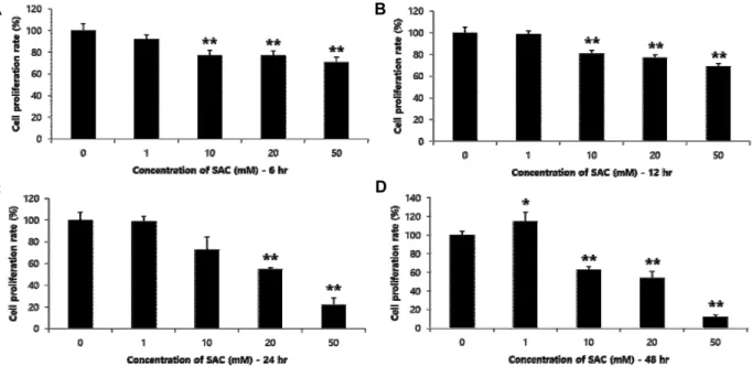
Fig. 1. Inhibitory effects of SAC on cellular proliferation of HeLa. Hela cells were treated for 6 hr (A), 12 hr (B), 24 hr (C) or 48 hr (D) with different concentrations (0, 1, 10, 20 and 50 mM) of SAC. The value of each bar represents the mean percentage of PBS control ± S.E. from three independent experiments. * indicates p<0.05 and ** indicates p<0.01 as compared to the PBS control.
표준 대조단백질의 발현탐지는 rabbit anti-alpha tubulin IgG (Cell Signaling Technology) 또는 mouse anti-beta actin IgG (Sigma-Aldrich, St. Louis, MO, U.S.A.)를 primary antibody 로 사용하였다. 각각의 primary antibody는 blocking solution 에서 모두 1:1,000의 농도로 희석하여 PVDF membrane과 함 께 4℃ over-night 및 25℃ 1시간 동안 반응시킨 다음, TBST (0.05% Tween-20 in TBS) wash buffer로 각 10분간 3회 반복 수세하였다. 각 항원에 결합된 primary antibody를 탐지하기 위하여 사용된 secondary antibody는 goat anti-rabbit IgG- HRP (Cell Signaling Technology) 또는 goat anti-mouse IgG- HRP (ThermoFisher, Rockford, IL, U.S.A.)로서 blocking sol- ution에 모두 1:3,000의 농도로 희석하여 25℃에서 1시간 동안 반응시켰으며, TBST wash buffer로 각 10분간 3회 반복 수세 하였다. 그리고, secondary antibody에 결합된 HRP의 촉매반 응을 통한 chemiluminescent 생성물을 유도하기 위하여, HRP 특이적 기질이 포함된 EzWestLumi plus (Atto, Tokyo, Japan) A, B 용액을 1:1 혼합하여 2분간 반응시켰으며, 발광 생성물의 양은 암실에서 x-ray 필름에 감광시켜 발현된 각 단백질의 양 을 정성적으로 분석하였다. 각 필름에 노출된 단백질 band의 강도는 Image studio lite program (LI-COR, Lincoln, NE, U.S.A.)을 이용하여 정량적으로 분석되었다.
통계분석
실험결과는 평균±S.E.로 표시되었으며, 처리군 사이의 통계 적 유의성은 Student’s t-test에 의해 결정되었다. p<0.05의 값 은 통계적으로 유의성이 있음을 의미한다.
결 과
SAC 처리에 의한 HeLa 세포주의 증식억제 효과
각 SAC 농도(0, 1, 10, 20 및 50 mM)에 따른 처리시간별 세포증식에 미치는 영향을 cell proliferation assay를 이용하여 비교 분석하였다. Fig. 1에 나타나 있는 바와 같이, 모든 처리시 간(6~48 hr)에서 10 mM 이상의 SAC는 HeLa 세포주의 증식 을 억제하였으며 특히, 20 mM 이상의 농도에서는 농도 의존 적인 억제효과를 나타내었다. 구체적으로, 20 및 50 mM의 SAC 처리는 6 hr의 경우 각각 23% 및 29%, 12 hr의 경우 각각 23%, 및 31%, 24 hr의 경우 각각 45% 및 78% 그리고 48 hr의 경우 각각 46% 및 88%의 현저한 증식억제 효과를 보임으로써, SAC가 HeLa 세포주에 대하여 농도 의존적일 뿐만 아니라 처 리시간 의존적인 증식억제 효과를 나타냄을 보였다.
SAC 처리에 의한 procaspase-3 및 caspase-3 단백질 발현조절
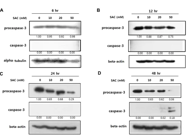
배양된 HeLa 세포를 각각 다른 농도(0, 10, 20 및 50 mM) 및 시간(6, 12, 24 및 48 hr)으로 SAC를 처리하여 procaspase-3 및 caspase-3 단백질의 발현변화를 분석한 결과, 대부분의 처 리농도(10, 20 및 50 mM) 및 시간(12~48 hr)에서 SAC가 pro- caspase-3의 발현을 감소시켰다(Fig. 2). 특히, 50 mM의 SAC 처리는 12, 24 및 48 hr에서 각각 25%, 71% 및 94%의 시간 의존적인 발현감소를 유도하였다. 이와는 대조적으로, 활성형 caspase-3 단백질의 양은 48 hr의 SAC 처리에 의해 현저한 증가를 나타내었다(Fig. 2D).
A B
C D
Fig. 2. Effects of SAC on expression of procaspase-3 and caspase-3 in HeLa. HeLa cells were treated with SAC in different concen- trations (0, 10, 20 and 50 mM) for 6 hr (A), 12 hr (B), 24 hr (C) or 48 hr (D). The panels show the results from Western blot analysis of procaspase-3 and caspase-3 expression with alpha-tubulin or beta-actin as a loading control. The numbers below each panel show the relative intensities of procaspase-3 or caspase-3 to the loading control which were calculated by dividing the background-subtracted density value of procaspase-3 or caspase-3 by the corresponding value of the loading control at the respective concentration, and comparision to the PBS control.
SAC 처리에 의한 procaspase-7 및 caspase-7 단백질 발현조절
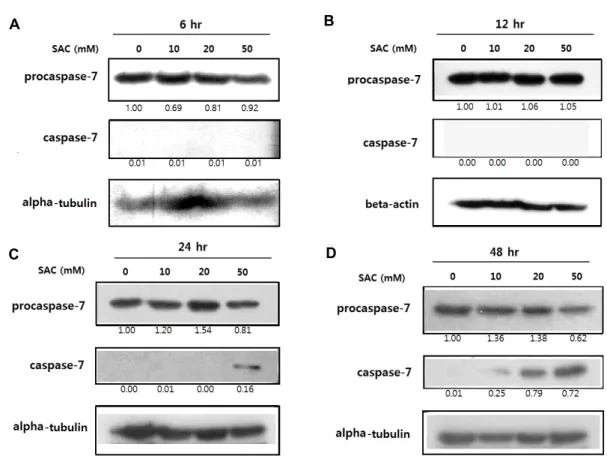
배양된 HeLa 세포를 각각 다른 농도(0, 10, 20 및 50 mM) 및 시간(6, 12, 24 및 48 hr)으로 SAC를 처리하여 procaspase-7 및 caspase-7 단백질의 발현변화를 분석한 결과, procaspase-7 의 발현은 특히 50 mM의 24 및 48 hr 처리에서 각각 19%
및 38%만큼 감소되었다(Fig 3C, Fig 3D). 반면에, 활성형 cas- pase-7 단백질의 양은 24 및 48 hr SAC 처리에 의해 현저한 증가를 보였으며, 특히 50 mM의 처리에 의해 두드러진 증가 를 나타내었다(Fig. 3C, Fig. 3D).
SAC 처리에 의한 procaspase-9 및 caspase-9 단백질 발현조절
배양된 HeLa 세포를 각각 다른 농도(0, 10, 20 및 50 mM) 및 시간(6, 12, 24 및 48 hr)으로 SAC를 처리하여 procaspase-9 및 caspase-9 단백질의 발현변화를 분석한 결과, 대부분의 처 리농도(10, 20 및 50 mM)에서 SAC가 procaspase-9의 발현을 시간 의존적으로 감소시켰다(Fig. 4). 특히, 50 mM의 SAC 처 리는 24 및 48 hr에서 모두 95%의 현저한 발현감소를 유도하 였다. 이와는 대조적으로, 활성형 caspase-9 단백질의 양은
12~48 hr의 SAC 처리에 의해 현저히 증가되었으며, 특히 50 mM의 처리에 의해 두드러진 증가를 나타내었다(Fig. 4B-Fig.
4D).
SAC 처리에 의한 PARP 및 cleaved-PARP 단백질 발 현조절
배양된 HeLa 세포를 48 hr 동안 다른 농도(0, 20 및 50 mM) 로 SAC를 처리하여 PARP 및 cleaved-PARP 단백질의 발현변 화를 분석한 결과, 농도 의존적인 PARP의 발현감소를 나타내 었다(Fig. 5). 이와는 대조적으로, 불활성형 cleaved-PARP 단백질 의 양은 50 mM SAC 처리에 의해 현저히 증가되었다(Fig. 5).
고 찰
자궁경부암은 여성 암 사망률의 7.5%에 해당하며[2], 현재 치료제로 사용되는 cisplatin 및 paclitaxel 등의 화학요법제는 독성과 내성의 부작용을 함유함에 따라[29], 정상세포에 대한 영향은 최소화하면서 항암효과를 갖는 천연화합물 유래 항암 제의 개발을 위한 많은 연구가 진행되고 있다. 숙성된 마늘로 부터 유래된 수용성 유기황화합물 중 하나인 SAC는 다양한
A B
C D
Fig. 3. Effects of SAC on expression of procaspase-7 and caspase-7 in HeLa. HeLa cells were treated with SAC in different concen- trations (0, 10, 20 and 50 mM) for 6 hr (A), 12 hr (B), 24 hr (C) or 48 hr (D). The panels show the results from Western blot analysis of procaspase-7 and caspase-7 expression with alpha-tubulin or beta-actin as a loading control. The numbers below each panel show the relative intensities of procaspase-7 or caspase-7 to the loading control which were calculated by dividing the background-subtracted density value of procaspase-7 or caspase-7 by the corresponding value of the loading control at the respective concentration, and comparision to the PBS control.
암세포 연구에서 정상세포에 대한 낮은 독성 및 높은 항암효 과를 가지는 것으로 보고되어 왔다[35]. 특히, 지난 보고에서 SAC가 세포사멸의 유도와 세포주기의 억제를 통하여 자궁경 부암세포주의 증식을 억제함을 제시하였다[15]. 본 연구에서 는 SAC에 의한 자궁경부암세포주의 세포사멸 효과가 어떠한 분자적 경로를 통하여 매개되는지를 확인하기 위하여, 세포자 멸관련 단백질인 caspase-3, caspase-7 및 caspase-9 그리고 DNA 복구 단백질인 PARP의 SAC 처리농도 및 시간에 따른 발현변화를 조사하였다.
SAC에 의한 세포자멸 관련 단백질 및 DNA 복구 단백질의 발현변화가 세포증식억제를 통한 기능적 작용과 일치하는지 를 확인하기 위하여, 먼저 농도 및 시간에 따른 SAC 처리에 의한 자궁경부암세포주의 세포증식 억제효과를 분석하였다.
SAC는 0~50 mM의 농도 및 각 시간대별로(6~48 hr) 처리된 HeLa 세포주에서, 대체로 농도 및 시간 의존적인 세포증식 억제효과를 보였다(Fig. 1). 이러한 결과는 이전에 보고된 자궁 경부암세포주[15] 뿐만 아니라, 유방암[7], 구강암[32], 난소암 [35], 간암세포주[24] 및 안드로겐-비의존성 전립선암세포[21]
등에서 유사한 농도범위의 SAC 매개 세포증식 억제효과 보고
들과 일치한다. 따라서, 자궁경부암세포주에서 SAC에 의한 세포자멸 및 DNA 복구 관련 단백질의 발현변화는 기능적 세 포증식억제 작용에 영향을 미칠 수 있는 하나의 분자적 신호 전달기전으로 간주될 수 있다.
세포자멸은 일련의 다단계 과정으로 내인성 또는 외인성 경로를 통하여 세포자멸 촉진인자와 억제인자를 조절함으로 써, 변이되거나 손상된 세포를 조직으로부터 염증반응 없이 제거한다[14]. 내인성 경로에서 세포자멸 신호는 미토콘드리 아 외막의 투과성을 증가시켜 cytochrome c의 방출을 유도한 다[8, 18, 34]. 세포기질에서 cytochrome c는 adaptor 단백질 Aparf-1 및 비활성 상태의 procaspase-9와 함께 apoptosome 을 형성하여 활성형 caspase-9를 생성하고, 활성화된 cas- pase-9는 다시 effector caspase인 caspase-3 및 caspase-7의 활 성화를 매개한다[19, 27]. 외인성 경로는 Fas와 같은 세포 표면 에 위치한 사멸수용체에 외부의 리간드가 결합함으로써 활성 화되며, 리간드와 사멸수용체의 결합은 Fas-associated pro- tein with death domain (FADD)과 caspase-8로 구성된 death- inducing signaling complex (DISC)의 형성을 유도한다[3, 16, 28]. 조립된 DISC는 caspase-3의 활성화를 유도하여 세포의
A B
C D
Fig. 4. Effects of SAC on expression of procaspase-9 and caspase-9 in HeLa. HeLa cells were treated with SAC in different concen- trations (0, 10, 20 and 50 mM) for 6 hr (A), 12 hr (B), 24 hr (C) or 48 hr (D). The panels show the results from Western blot analysis of procaspase-9 and caspase-9 expression with alpha-tubulin or beta-actin as a loading control. The numbers below each panel show the relative intensities of procaspase-9 or caspase-9 to the loading control which were calculated by dividing the background-subtracted density value of procaspase-9 or caspase-9 by the corresponding value of the loading control at the respective concentration, and comparision to the PBS control.
자멸을 초래한다[1, 20, 33].
본 연구에서는 SAC가 HeLa 세포주의 procaspase-3, pro- caspase-7 및 procaspase-9의 단백질 발현을 하향조절할 뿐만 아니라, 활성형 caspase-3, caspase-7 및 caspase-9의 단백질 양을 상향조절한 것으로 나타났다(Fig. 2 - Fig. 4). 이러한 SAC 에 의해 유도된 HeLa 세포주의 caspase 발현조절은 이전에 보고된 난소암[35] 및 간암세포주[24]에서의 procaspase-3 및 procaspase-9의 발현 감소와 더불어 활성화된 caspase-3 및 caspase-9의 증가와 일치한다. 이는 SAC가 비활성형 procas- pases (procaspase-3, procaspase-7 및 procaspase-9)를 활성형 caspases (caspase-3, caspase-7 및 caspase-9)로의 전환을 매개 하는 것으로 보인다. 따라서, 이러한 결과들은 SAC 처리에 의해 initiator caspase인 caspase-9가 비활성형 상태(procas- pase-9)로부터 활성형 상태(caspase-9)로 전환되며, 활성화된 caspase-9는 다시 effector caspase인 procaspase-3 및 procas- pase-7의 분절을 통한 활성화(caspase-3 및 caspase-7)를 매개 하는 것으로 사료된다.
또한 DNA-binding zinc finger protein 중 하나로서, 세포 의 증식, 분화 및 자멸과정에서 DNA의 복구를 수행하는 PARP 단백질의[10] 발현이 SAC 처리에 의해 감소된 반면, 불활성 상태의 cleaved-PARP의 단백질 양은 증가되었다(Fig.
5). PARP는 caspase-3과 caspase-7의 기질로 사용되며[12, 22], 난소암세포주에서 SAC 처리를 통해 활성화된 caspase-3이
Fig. 5. Effects of SAC on expression of PARP and cleaved-PARP in HeLa. HeLa cells were treated with SAC in different concentrations (0, 20 and 50 mM) for 48 hr. The panel shows the results from Western blot analysis of PARP and cleaved-PARP expression with alpha-tubulin as a loading control. The numbers below each panel show the relative intensities of PARP or cleaved-PARP to the loading control which were calculated by dividing the background-subtracted density value of PARP or cleaved-PARP by the corresponding value of the loading control at the respective concentration, and comparision to the PBS control.
PARP의 분절을 유도한다는 Xu 등[35]의 보고는 본 연구에서 나타난 SAC에 의한 procaspase-3과 procaspase-7의 감소, cas- pase-3과 caspase-7의 증가, 그리고 활성형 PARP 감소와 불활 성화된 cleaved-PARP의 증가 등의 결과와 일치한다. 따라서, 이러한 결과는 자궁경부암세포주에서 SAC 처리에 의한 세포 증식 억제효과가 최소한 부분적으로 caspase-3 그리고/또는 caspase-7의 활성에 의하여 절단되어 불활성화된 PARP에 의 해 초래된 DNA repair 기능의 소실에 기인할 수 있음을 의미 한다. 그리고, SAC에 의한 PARP의 불활성화는 이전에 보고된 DNA fragmentation assay 및 TUNEL (terminal deoxy- nucleotidyl transferase-dUTP nick and labeling) assay에서 나타난 바와 같이 SAC에 의해 유도되는 유전체 DNA의 분절 효과를 뒷받침한다[15]. 뿐만 아니라, caspase-3 및 caspase-7 의 활성화는 유전체 DNA를 분해하는 endonuclease의 활성화 를 유도하여 세포사멸을 초래하는 것으로 보고되었다[11]. 따 라서 이러한 결과들은 자궁경부암세포주에서 SAC가 effector caspase인 caspase-3과 caspase-7의 활성화를 통하여 DNA 복 구를 촉매하는 PARP의 불활성화를 초래할 뿐만 아니라, DNA의 분해를 촉진시키는 endonuclease의 활성화를 유도함 으로써 DNA 분절을 일으키고 이는 결국 세포사멸을 초래하 는데 기여하는 것으로 사료된다.
결론적으로, 본 연구에서는 SAC 처리가 자궁경부암세포주 의 세포증식을 억제하며, 이에 대한 가능한 분자적 작용기전 들 중의 하나로 세포자멸과정의 상위경로에 해당하는 ini- tiator caspase 중 하나인 caspase-9의 활성을 유도하고 이에 따른 하위경로의 effector caspase인 caspase-3과 caspase-7의 활성화를 초래할 뿐만 아니라, DNA 복구에 관여하는 PARP 를 불활성화시킴으로써 세포의 자멸을 초래함을 제시한다.
SAC의 세포증식억제효과에 대한 보다 더 구체적인 분자적 신호경로 또는 다른 조절기전은 향후 추가적인 연구를 통해 규명되어야 할 것으로 사료된다.
감사의 글
이 논문은 2015년도 경남과학기술대학교 대학회계 연구비 지원에 의하여 연구되었으며 이에 감사드립니다.
References
1. Algeciras-Schimnich, A., Pietras, E. M., Barnhart, B. C., Legembre, P., Vijayan, S., Holbeck, S. L. and Peter, M. E.
2003. Two CD95 tumor classes with different sensitivities to antitumor drugs. Proc. Natl. Acad. Sci. USA 100, 11445- 11450.
2. Ariga, K., Li, J., Fei, J., Ji, Q. and Hill, J. P. 2016. Nanoarchi- tectonics for dynamic functional materials from atom- ic-/molecular-level manipulation to macroscopic action.
Adv. Mater. 28, 1251-1286.
3. Ashkenazi, A. and Dixit, V. M. 1998. Death receptors: signal- ing and modulation. Science 281, 1305-1308.
4. Butt, M. S., Sultan, M. T., Butt, M. S. and Iqbal, J. 2009.
Garlic: nature's protection against physiological threats.
Crit. Rev. Food Sci. Nutr. 49, 538-551.
5. Chu, Q., Lee, D. T., Tsao, S. W., Wang, X. and Wong, Y.
C. 2007. S-allylcysteine, a water-soluble garlic derivative, suppresses the growth of a human androgen-independent prostate cancer xenograft, CWR22R, under in vivo conditions.
BJU Int. 99, 925-932.
6. Desagher, S. and Martinou, J. C. 2000. Mitochondria as the central control point of apoptosis. Trends Cell Biol. 10, 369- 377.
7. Gapter, L. A., Yuin, O. Z. and Ng, K. Y. 2008. S-Allylcysteine reduces breast tumor cell adhesion and invasion. Biochem.
Biophys. Res. Commun. 367, 446-451.
8. Green, D. R. and Reed, J. C. 1998. Mitochondria and apoptosis. Science 281, 1309-1312.
9. Iciek, M., Kwiecien, I., Chwatko, G., Sokolowska-Jezewicz, M., Kowalczyk-Pachel, D. and Rokita, H. 2011. The effects of garlic-derived sulfur compounds on cell proliferation, caspase 3 activity, thiol levels and anaerobic sulfur metabo- lism in human hepatoblastoma HepG2 cells. Cell Biochem.
Funct. 30, 198-204.
10. Isabelle, M., Moreel, X., Gagné, J. P., Rouleau, M., Ethier, C., Gagné, P., Hendzel, M. J. and Poirier, G. G. 2010.
Investigation of PARP-1, PARP-2, and PARG interactomes by affinity-purification mass spectrometry. Proteome Sci. 8, 22.
11. Jia, J., Furlan, A., Gonzalez-Hilarion, S., Leroy, C., Gruenert, D. C., Tulasne, D. and Lejeune, F. 2015. Caspases shutdown nonsense-mediated mRNA decay during apoptosis. Cell Death Differ. 22, 1754-1763.
12. Jimenez, F., Aiba-Masago, S., AI, Hashimi. I., Vela-Roch, N., Fernandes, G., Yeh, C. K,, Talal, N. and Dang, H. 2002.
Activated caspase 3 and cleaved poly(ADP-ribose)polymer- ase in salivary epithelium suggest a pathogenetic mecha- nism for Sjögren's syndrome. Rheumatology (Oxford) 41, 338-342.
13. Jin, Z. and El-Deiry, W. S. 2005. Overview of cell death sig- naling pathways. Cancer Biol. Ther. 4, 139-163.
14. Kim, H. H., Kong, I. K. and Min, G. 2015. Anticarcinogenic effect of S-allylcysteine (SAC). J. Life Sci. 25, 101-107.
15. Kim, H. H. and Min, G. 2015. Inhibitory effects of S-allylcys- teine on cell proliferation of human cervical cancer cell line, HeLa. J. Life Sci. 25, 101-109.
16. Kischkel, F. C., Hellbardt, S., Behrmann, I., Germer, M., Pawlita, M., Krammer, P. H. and Peter, M. E. 1995. Cytotox- icity-dependent APO-1 (Fas/CD95)-associated proteins form a death-inducing signaling complex (DISC) with the receptor. EMBO J. 14, 5579-5588.
17. Klener, P. Jr., Andera, L., Klener, P., Necas, E. and Zivny, J. 2006. Cell death signalling pathways in the pathogenesis and therapy of haematologic malignancies: overview of therapeutic approaches. Folia Biol (Praha). 52, 119-136.
18. Kroemer, G., Dallaporta, B. and Resche-Rigon, M. 1998. The
초록:S-allylcysteine 매개 caspases의 활성화 및 PARP의 불활성화를 통한 HeLa 세포주의 증식 억제효과
김현희1․공일근1․민계식2*
(1경상대학교 응용생명과학부, 2경남과학기술대학교 생명과학대학 간호학과)
본 연구에서는 인간 자궁경부암세포주에서 S-allylcysteine (SAC)이 세포자멸경로에 중요한 역할을 담당하는 initiator caspase의 하나인 caspase-9와 effector caspase에 속하는 caspase-3 및 caspase-7 그리고 DNA 복구에 관 여하는 poly ADP-ribose polymerase (PARP)의 발현조절에 미치는 영향과, SAC에 의한 이러한 세포자멸 및 DNA 복구 관련 단백질의 발현변화가 세포증식억제를 통한 기능적 작용을 유발하는지를 조사하였다. 단백질 발 현분석 결과, 특히 50 mM의 SAC로 48시간 동안 처리하였을 경우, procaspase-3, -7, -9 및 PARP의 발현은 각각 94%, 38%, 95% 및 64% 감소되었으며, 이와 반대로 caspase-3, -7, -9 및 cleaved-PARP의 발현은 현저히 증가되었 다. 또한 cell proliferation assay 결과, 20 mM 이상의 SAC 처리는 6, 12, 24 및 48시간에서 농도 및 시간 의존적인 세포증식 억제효과를 나타내었다. 이러한 결과는 SAC 처리가 자궁경부암세포의 증식을 억제하며, 이에 대한 가능 한 분자적 작용기전들 중의 하나로 세포자멸과정 중 initiator caspase의 하나인 caspase-9의 활성을 유도하고 이에 따른 effector caspase인 caspase-3과 caspase-7의 활성을 촉진시킬 뿐만 아니라 DNA 복구에 관여하는 PARP의 불활성화를 초래함으로써 세포자멸 유도에 관여하는 것으로 사료된다.
mitochondrial death/life regulator in apoptosis and necrosis. Annu. Rev. Physiol. 60, 619-642.
19. Kwon, S. B., Kim, M. J., Yang, J. M., Lee, H. P., Hong, J.
T., Jeong, H. S., Kim, E. S. and Yoon, D. Y. 2016. Cudrania tricuspidata stem extract induces apoptosis via the extrinsic pathway in SiHa cervical cancer cells. PLoS One 11, e0150235.
20. Lavrik, I., Golks, A. and Krammer, P. H. 2005. Death re- ceptor signaling. J. Cell Sci. 118, 265-267.
21. Liu, Z., Li, M., Chen, K., Yang, J., Chen, R., Wang, T., Liu, J., Yang, W. and Ye, Z. 2012. S-allylcysteine induces cell cy- cle arrest and apoptosis in androgen-independent human prostate cancer cells. Mol. Med. Rep. 5, 439-443.
22. Los, M., Mozoluk, M., Ferrari, D., Stepczynska, A., Stroh, C., Renz, A., Herceg, Z., Wang, Z. Q. and Schulze-Osthoff, K. 2002. Activation and caspase-mediated inhibition of PARP: a molecular switch between fibroblast necrosis and apoptosis in death receptor signaling. Mol. Biol. Cell 13, 978- 988.
23. MacKenzie, S. H. and Clark, A. C. 2012. Death by caspase dimerization. Adv. Exp. Med. Biol. 747, 55-73.
24. Ng, K. T., Guo, D. Y., Cheng, Q., Geng, W., Ling, C. C., Li, C. X., Liu, X. B., Ma, Y. Y., Lo, C. M., Poon, R. T., Fan, S. T. and Man, K. 2012. A garlic derivative, S-allylcysteine (SAC), suppresses proliferation and metastasis of hep- atocellular carcinoma. PLoS One 7, e31655.
25. Nobili, S., Lippi, D., Witort, E., Donnini, M., Bausi, L., Mini, E. and Capaccioli, S. 2009. Natural compounds for cancer treatment and prevention. Pharmacol. Res. 59, 365-378.
26. Plati, J., Bucur, O. and Khosravi-Far, R. 2011. Apoptotic cell signaling in cancer progression and therapy. Integr. Biol (Camb). 3, 279-296.
27. Rodriguez, J. and Lazebnik, Y. 1999. Caspase-9 and APAF-1 form an active holoenzyme. Genes Dev. 13, 3179-3194.
28. Schneider, P. and Tschopp, J. 2000. Apoptosis induced by death receptors. Pharm. Acta. Helv. 74, 281-286.
29. Seol, H. J., Ulak, R., Ki, K. D. and Lee, J. M. 2014. Cytotoxic and targeted systemic therapy in advanced and recurrent cervical cancer: experience from clinical trials. Tohoku J. Exp.
Med. 232, 269-276.
30. Shi, W. Y., Cao, C. and Liu, L. 2016. Interferon α induces the apoptosis of cervical cancer HeLa cells by activating both the intrinsic mitochondrial pathway and endoplasmic reticulum stress-induced pathway. Int. J. Mol. Sci. 17, E1832.
31. Shi, Y. 2002. Mechanisms of caspase activation and in- hibition during apoptosis. Mol. Cell 9, 459-470.
32. Tang, F. Y., Chiang, E. P., Chung, J. G., Lee, H. Z. and Hsu, C. Y. 2009. S-allylcysteine modulates the expression of E-cadherin and inhibits the malignant progression of human oral cancer. J. Nutr. Biochem. 20, 1013-1020.
33. Wilson, N. S., Dixit, V. and Ashkenazi, A. 2009. Death re- ceptor signal transducers: nodes of coordination in immune signaling networks. Nat. Immunol. 10, 348-355.
34. Xiong, S., Mu, T., Wang, G. and Jiang, X. 2014. Mitochon- dria-mediated apoptosis in mammals. Protein Cell 5, 737-749.
35. Xu, Y. S., Feng, J. G., Zhang, D., Luo, M., Su, D. and Lin, N. M. 2014. S-allylcysteine, a garlic derivative, suppresses proliferation and induces apoptosis in human ovarian can- cer cells in vitro. Acta Pharmacol. Sin. 35, 267-274.
36. Zhou, Y., Zhuang, W., Hu, W., Liu, G. J., Wu, T. X. and Wu, X. T. 2011. Consumption of large amounts of Allium vegetables reduces risk for gastric cancer in a meta-analysis.
Gastroenterology 141, 80-89.