J Korean Surg Soc 2012;83:187-195 http://dx.doi.org/10.4174/jkss.2012.83.4.187
ORIGINAL ARTICLE
Journal of the Korean Surgical Society
JKSS
pISSN 2233-7903ㆍeISSN 2093-0488
Received April 6, 2012, Revised July 16, 2012, Accepted August 16, 2012 Correspondence to: Chung Han Lee
Department of Surgery, Kosin University College of Medicine, 262 Gamcheon-ro, Seo-gu, Busan 602-702, Korea Tel: +82-51-990-6462, Fax: +82-51-246-6093, E-mail: [email protected]
cc Journal of the Korean Surgical Society is an Open Access Journal. All articles are distributed under the terms of the Creative Commons Attribution Non-Commercial License (http://creativecommons.org/licenses/by-nc/3.0/) which permits unrestricted non-commercial use, distribution, and reproduction in any medium, provided the original work is properly cited.
Outcome of triple-negative breast cancer in patients with or without markers regulating cell cycle and cell death
Dong Won Ryu, Chung Han Lee
Department of Surgery, Kosin University Gospel Hospital, Busan, Korea
Purpose: The genes p53 and B-cell lymphoma (bcl)-2 play an important role in regulating the mechanisms of apoptosis. In this paper, we retrospectively applied these factors to our series of triple negative breast cancer (TNBC) patients, in con- junction with an evaluation of the prognostic significance of these factors’ influence on TNBC survival rate. Particular focus was placed on the role of bcl-2, p53, Ki-67. Methods: The study subjects, 94 women with TNBC, were a subset of patients op- erated at Kosin University Gospel Hospital from January 2000 to December 2005. Chi-squared tests were used for statistical analysis. Results: Positive staining for cytokeratin (CK)5/6 in 23 cases (24.5%), epidermal growth factor receptor in 15 cases (16.0%), bcl-2 in 26 cases (27.7%), p53 in 55 cases (58.5%) and Ki-67 in 74 cases (78.7%) was determined. Lymph node status, tumor size and expression of CK5/6 or Ki-67 were independent prognostic factors for patients with TNBC. Conclusion:
Markers regulating cell cycle and cell death such as p53 and bcl-2 cannot be used to classify TNBCs into two subtypes with differing disease-free survival. But because our study is small in size, more abundant patient data will be needed to evaluate the factors’ predictive role in regulating cell cycle and cell death.
Key Words: Triple negative, bcl-2, p53, Ki-67
INTRODUCTION
By using DNA microarray techniques, it has been shown that breast cancers can be classified into bio- logically distinct groups based on their gene expression profiles [1]. These groups comprise luminal A (estrogen receptor [ER]-positive and human epidermal growth fac- tor receptor 2 [HER2]-negative), luminal B (ER- and HER2-positive), HER-2 (ER-negative and HER2-positive), and triple negative (ER- and HER2-negative) subtypes [2].
TNBC is a heterogeneous group and is further categorized into the basal-phenotype (BP) and non-BP groups, which are positive and negative, respectively, for myoepithelial/
basal markers such as basal cytokeratins (CKs) (i.e., CK5/6, CK14, and CK17), and epidermal growth factor receptor (EGFR) [3,4]. Although TNBCs account for only 10 to 17%
of all breast carcinomas, this subgroup is regarded as im- portant clinically because of the aggressive clinical behav- ior, poorer patient prognosis, and lack of an established therapeutic target [5]. The ratio of basal-like subtype in
Marker Species Manufacturer Dilution Incubation time (min) Cutoff value
ER Mouse mAb Dako 1:50 60, RT ≥10%
PgR Mouse mAb Dako 1:100 60, RT ≥10%
HER-2/neu Rabbit poly Dako 1:250 60, RT 3+ or FISH+
Ki-67 Mouse mAb Dako 1:50 60, RT ≥10%
p53 Mouse mAb Dako 1:50 60, RT ≥10%
CK5/6 Mouse mAb Dako 1:100 60, RT ≥10%
bcl-2 Mouse mAb Dako 1:100 60, RT ≥10%
ER, estrogen receptor; RT, room temperature; PgR, progesterone receptor; HER-2, human epidermal growth factor receptor 2; FISH, fluorescent in situ hybridization; CK, cytokeratin; bcl-2, B-cell lymphoma.
Table 1. Antibodies, dilutions, incubation times, and cutoff values TNBC was estimated to be up to 56 to 84%. Among the genes regulators of the apoptotic process, the tumor sup- pressor gene p53 and the B-cell lymphoma (bcl)-2 gene and its family members have been studied in clinical set- tings of breast cancer [1,6]. Among the several biomarkers, bcl-2 is an anti-apoptotic gene, and it is a poor prognostic factor in various malignant tumors. However, the prog- nostic significance of bcl-2 expression in breast cancer re- mains controversial [7]. We retrospectively applied these factors to our series of TNBC patients, in conjunction with an evaluation of the prognostic significance of these fac- tors influencing TNBC survival rate. Particular focus was placed on the role of CK5/6, EGFR, bcl-2, and p53.
METHODS
Tissue samples were obtained from 617 patients with invasive breast cancer who were diagnosed from 2000 to 2005 at Kosin University Gospel Hospital in Busan. A total of 617 specimens of primary invasive carcinoma were ob- tained from resected tumors. None of these cancer pa- tients received treatment prior to surgery. The patients un- derwent standard and partial mastectomies. Patients re- ceived anthracycline-containing chemotherapy if the tu- mor was node positive. Endocrine therapy was given for 5 years to patients with ER-positive tumors. Median follow up was 5.5 years (range, 0.3 to 14.8 years), during which there were 84 relapses and 32 deaths.
Immunohistochemical techniques
The expression of ER, PgR, HER-2/neu, CK5/6 and other
biological markers was determined immunohistochemi- cally in paraffin-embedded tissue specimens [1,6]. Table 1 summarizes all the antibodies, dilutions, incubation times, and cutoff values used for this analysis. All data were collected from the pathology reports. Histopatholog- ical features such as hormone receptor status and HER-2/
neu status on immunohistochemistry (Dako, Copenha- gen, Denmark) were all analyzed at the Institute of Pathology at Kosin University. Expressions of p53, ER, Ki-67 and HER-2/neu were determined immunohistoche- mically on paraffin sections using antibodies against ER (Dako), Ki- 67 (Dako), HER-2/neu (Dako), p53 (Dako) [1,8].
Tumor necrosis was defined as the presence of necrosis of any dimension in a section of invasive cancer. Histologic grading was performed using the criteria of Bloom and Richardson [9], ER status and progesterone receptor (PgR) status were taken as positive if more than 10% of tumor cells showed staining. Immunohistochemical score of 3+
or fluorescent in situ hybridization + for HER-2/neu was accepted as HER-2/neu positivity. Immunostaining for p53 and bcl-2 was performed on wax sections cut to a thickness of 5 μm, immersed in a citric acid buffer (pH 6.0) and incubated twice in a microwave oven at 750 W power for five minutes each. The sections were subsequently im- munostained using the APAAP Complex method (Dako);
the primary antibodies used for detection of p53 and bcl-2 were, respectively, the Immunotech antihuman p53 pro- tein clone DO-7 (Immunotech, Marseille, France) and the DAKO antihuman bcl-2 oncoprotein clone 124 (Dako).
Ki-67 proliferative activity was determined by the avi- din-biotin procedure. Briefly, after inhibition of endoge- nous peroxidase, slides were incubated with Ki-67 mono-
Characteristic Value Median age (yr) 54.2 (25.9-78.9) pT
T1 245 (39.7)
T2 354 (57.3)
T3 14 (2.3)
T4 4 (0.7)
pN
N0 357 (57.7)
N1 122 (19.7)
N2 70 (11.3)
N3 69 (11.2)
6th AJCC stage
I 146 (23.6)
IIA 235 (38.0)
IIB 94 (15.2)
IIIA 63 (10.2)
IIIB 12 (1.9)
IIIC 68 (11.0)
Histologic grade
G1 81 (13.1)
G2 230 (37.2)
G3 307 (49.7)
p53 status
Negative 248 (40.0)
Positive 370 (60.0)
Ki-67
Negative 87 (14.1)
Positive 531 (85.9)
Microarray
Luminal A 166 (26.9)
Luminal B 178 (28.9)
HER-2 162 (26.3)
TNBC 111 (17.9)
Values are presented as mean (range) or number (%).
AJCC, American Joint Committee on Cancer; HER-2, human epidermal growth factor receptor 2; TNBC, triple negative breast cancer.
Table 2. Patients and tumor characteristics clonal antibody (diluted 1:50 in phosphate buffered saline
[PBS]) for 1 hour at room temperature. After quickly rins- ing in PBS, the sections were incubated with biotin-con- jugated rabbit antimouse immunoglobulin G (Vector Laboratories, Burlingame, CA, USA) for 30 minutes. After an additional wash in PBS, slides were incubated with avi- din-biotin complexes conjugated with peroxidase (Vector Laboratories) for 45 minutes. The median value of Ki-67 was 10% and this was taken as the cut-off point dis- tinguishing slowly from quickly proliferating tumors. The slides were scored by the same pathologist. We considered the tumor positive for p53 and bcl-2 when 10% or more of the cells counted were stained [7,10].
Statistical analysis
Statistical tests were performed using the SPSS ver. 12.0 (SPSS Inc, Chicago, IL, USA). The survival function was calculated from the time of the pathologic diagnosis to the occurrence of death. Survival data were censored on December 31, 2011, which was the date on which the sur- vival data were correlated with the death registry for the last time or 5 years after the onset of the disease. Kaplan- Meier estimates are presented for the survival function, and differences in survival were analyzed using the log rank test. Associations between specific histopathological and clinical survival estimates and curves were estab- lished using the Kaplan-Meier method and differences in observed survival distribution among patient subgroups were tested with two-sided log-rank test. All survival rates were presented with their standard errors. We used Pearson's correlation to determine the association of pairs of explanatory variables, differences in qualitative varia- bles were evaluated by chi-squared test, where necessary.
The survival differences between patients with a basal phenotype TNBC and a non-BP TNBC were calculated with the Cox proportional hazards model. A P-value of
<0.05 was considered to indicate statistical significance.
All P-values were two-sided and a P-value of less than 0.05 was considered to indicate a statistically significant difference.
RESULTS
The main clinicopathological characteristics of the patients in our series are summarized in Table 2. Mean age was 54 years. Luminal A was reported in 166 patients (26.9%). Luminal B, HER-2/neu, and TNBC type were reported in 178 patients (28.9%), 162 patients (26.3%), 111 patients (17.9%), respectively. Among the 111 TNBC patients, seventeen had missing data as a result of a follow-up failure; thus, 94 TNBC patients are included in the final morphologic characteristics data set. Other reasons
Characteristic Non basal TNBC (n = 61) Basal TNBC (n = 33) Total P-value
pT 0.597
T1 24 (39.3) 13 (39.4) 37 (39.4)
T2 29 (47.5) 15 (45.5) 44 (46.8)
T3 8 (13.1) 5 (15.1) 13 (13.9)
pN 0.395
0 31 (50.8) 16 (48.5) 47 (50.0)
1 10 (16.4) 10 (30.3) 20 (21.3)
2 11 (18.0) 4 (12.1) 15 (16.0)
3 9 (14.8) 3 (9.1) 12 (12.8)
6th AJCC stage 0.493
I 13 (21.3) 10 (30.3) 23 (24.5)
IIA 21 (34.4) 7 (21.2) 28 (29.8)
IIB 9 (14.8) 9 (27.3) 18 (19.1)
IIIA 7 (11.5) 3 (9.1) 10 (10.6)
IIIB 2 (3.3) 0 (0) 2 (2.1)
IIIC 9 (14.7) 4 (12.1) 13 (13.8)
EGFR 0.000
Negative 61 (100) 18 (54.5) 79 (84.0)
Positive 0 (0) 15 (45.5) 15 (16.0)
CK5/6 0.000
Negative 61 (100) 10 (14.1) 71 (75.5)
Positive 0 (0) 23 (69.7) 23 (24.5)
Ki-67 0.397
Negative 14 (23.0) 6 (18.2) 20 (21.3)
Positive 47 (77.0) 27 (81.8) 74 (78.7)
Necrosis 0.015
No 30 (49.2) 8 (24.2) 38 (40.4)
Yes 31 (50.8) 25 (75.8) 56 (59.6)
Border 0.440
Pushing 5 (8.2) 4 (12.1) 9 (9.6)
Borderline 8 (13.1) 7 (21.1) 15 (16.0)
Infiltrating 48 (78.7) 22 (66.7) 70 (74.5)
Lympho 0.422
Marked continuous 3 (4.9) 1 (3.0) 4 (4.3)
Marked cuffing 2 (3.3) 1 (3.0) 3 (3.2)
Continous 19 (31.1) 16 (48.5) 35 (37.2)
No 37 (60.7) 15 (45.5) 52 (55.3)
Nucleoli 0.072
≥50% prominent 24 (39.3) 21 (63.6) 45 (47.9)
<50% prominent 25 (41.0) 9 (27.3) 34 (36.2)
No 12 (19.7) 3 (9.1) 15 (16.0)
Histologic grade 0.151
G1 5 (10.4) 1 (4.2) 6 (8.3)
G2 10 (20.8) 10 (41.7) 20 (27.8)
G3 33 (68.6) 13 (54.2) 46 (63.9)
bcl-2 0.037
Negative 40 (65.6) 28 (84.8) 68 (72.3)
Positive 21 (34.4) 5 (15.2) 26 (27.7)
p53 0.532
Negative 25 (41.0) 14 (42.4) 39 (41.5)
Positive 36 (59.0) 19 (57.6) 55 (58.5)
Table 3. Clinicopathological factors according to basal subtype in triple-negative cancer
Characteristic Non basal TNBC (n = 61) Basal TNBC (n = 33) Total P-value
Mastectomy 0.197
Total 50 (82.0) 30 (90.9) 80 (85.1)
Partial 11 (18.0) 3 (9.1) 14 (14.9)
CTx type 0.170
Non taxane 43 (70.5) 27 (81.8) 70 (74.5)
Taxane 18 (29.5) 6 (18.2) 24 (25.5)
Metastasis site 0.051
Bone 0 (0) 1 (25.0) 1 (9.1)
Lung 1 (14.3) 3 (75.0) 4 (36.4)
Liver 3 (42.9) 0 (0) 3 (27.3)
Brain 3 (42.9) 0 (0) 3 (27.3)
Values are presented as number (%).
TNBC, triple negative breast cancer; AJCC, American Joint Committee on Cancer; EGFR, epidermal growth factor receptor; CK, cytokeratin;
bcl-2, B‐cell lymphoma.
Table 3. Continued
Characteristic
bcl-2 negative
(n = 68)
bcl-2 positive
(n = 26)
Total P-value
6th AJCC stage 0.386
I 19 (27.9) 4 (15.4) 23 (24.5) IIA 19 (27.9) 9 (34.6) 28 (29.8) IIB 12 (17.6) 6 (23.1) 18 (19.1) IIIA 9 (13.2) 1 (3.8) 10 (10.6) IIIB 2 (2.9) 0 (0) 2 (2.1) IIIC 7 (10.3) 6 (23.0) 13 (13.8)
EGFR 0.149
Negative 55 (80.9) 24 (92.3) 79 (84.0) Positive 13 (19.1) 2 (7.7) 15 (16.0)
CK5/6 0.328
Negative 50 (73.5) 21 (80.8) 71 (75.5) Positive 18 (26.5) 5 (19.2) 23 (24.5)
Ki-67 0.135
Negative 12 (17.6) 8 (30.8) 20 (21.3) Positive 56 (82.4) 18 (69.2) 74 (78.7)
p53 0.368
Negative 27 (39.7) 12 (46.2) 39 (41.5) Positive 41 (60.3) 14 (53.8) 55 (58.5) Values are presented as number (%).
bcl‐2, B‐cell lymphoma; AJCC, American Joint Committee on Cancer; EGFR, epidermal growth factor receptor; CK, cytokeratin.
Table 4. Clinicopathological factors according to bcl-2 in triple- negative cancer
Characteristic p53 negative
(n = 39)
p53 positive
(n = 55)
Total P-value
6th AJCC stage 0.642
I 8 (20.5) 15 (27.3) 23 (24.5) IIA 13 (33.3) 15 (27.3) 28 (29.8) IIB 7 (17.9) 11 (20.0) 18 (19.1) IIIA 3 (7.7) 7 (12.7) 10 (10.6) IIIB 2 (5.1) 0 (0) 2 (2.1) IIIC 6 (15.4) 7 (12.7) 13 (13.8)
EGFR 0.031
Negative 29 (74.4) 50 (90.9) 79 (84.0) Positive 10 (25.6) 5 (9.1) 15 (16.0)
CK5/6 0.160
Negative 32 (82.1) 39 (70.9) 71 (75.5) Positive 7 (17.9) 16 (29.1) 23 (24.5)
Ki-67 0.051
Negative 12 (30.8) 8 (14.5) 20 (21.3) Positive 27 (69.2) 47 (85.5) 74 (78.7) G3 16 (59.3) 30 (66.7) 46 (63.9)
bcl-2 0.368
Negative 27 (69.2) 41 (74.5) 68 (72.3) Positive 12 (30.8) 14 (25.5) 26 (27.7) Values are presented as number (%).
AJCC, American Joint Committee on Cancer; EGFR, epidermal growth factor receptor; CK, cytokeratin; bcl-2, B‐cell lymphoma.
Table 5.Clinicopathological factors according to p53 in triple- negative cancer
for ineligibility included incompetent specimen. Positive staining for CK5/6 was found in 23 cases (24.5%), 15 cases (16.0%) were determined as positive staining for EGFR.
According to the positivity of CK5/6 and EGFR, 33 of the 94 patients (35.1%) had been diagnosed as basal pheno-
type TNBC and 61 patients (64.9%) had non-BP TNBC (Table 3). As compared with the non-basal TNBCs, basal TNBCs were not associated with a larger tumor size (P = 0.597), more advanced American Joint Committee on
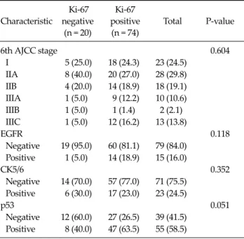
Characteristic
Ki-67 negative
(n = 20)
Ki-67 positive (n = 74)
Total P-value
6th AJCC stage 0.604
I 5 (25.0) 18 (24.3) 23 (24.5) IIA 8 (40.0) 20 (27.0) 28 (29.8) IIB 4 (20.0) 14 (18.9) 18 (19.1) IIIA 1 (5.0) 9 (12.2) 10 (10.6) IIIB 1 (5.0) 1 (1.4) 2 (2.1) IIIC 1 (5.0) 12 (16.2) 13 (13.8)
EGFR 0.118
Negative 19 (95.0) 60 (81.1) 79 (84.0) Positive 1 (5.0) 14 (18.9) 15 (16.0)
CK5/6 0.352
Negative 14 (70.0) 57 (77.0) 71 (75.5) Positive 6 (30.0) 17 (23.0) 23 (24.5)
p53 0.051
Negative 12 (60.0) 27 (26.5) 39 (41.5) Positive 8 (40.0) 47 (63.5) 55 (58.5) Values are presented as number (%).
AJCC, American Joint Committee on Cancer; EGFR, epidermal growth factor receptor; CK, cytokeratin.
Table 6. Clinicopathological factors according to Ki-67 in triple- negative cancer
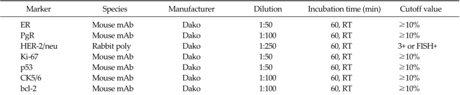
Fig. 1. Kaplan-Meier estimates of overall survival according to basal type. TNBC, triple negative breast cancer.
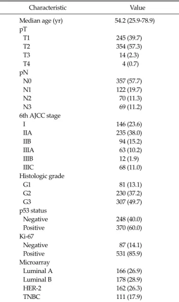
Fig. 2. Kaplan-Meier estimates of overall survival according to cytokeratin (CK)5/6.
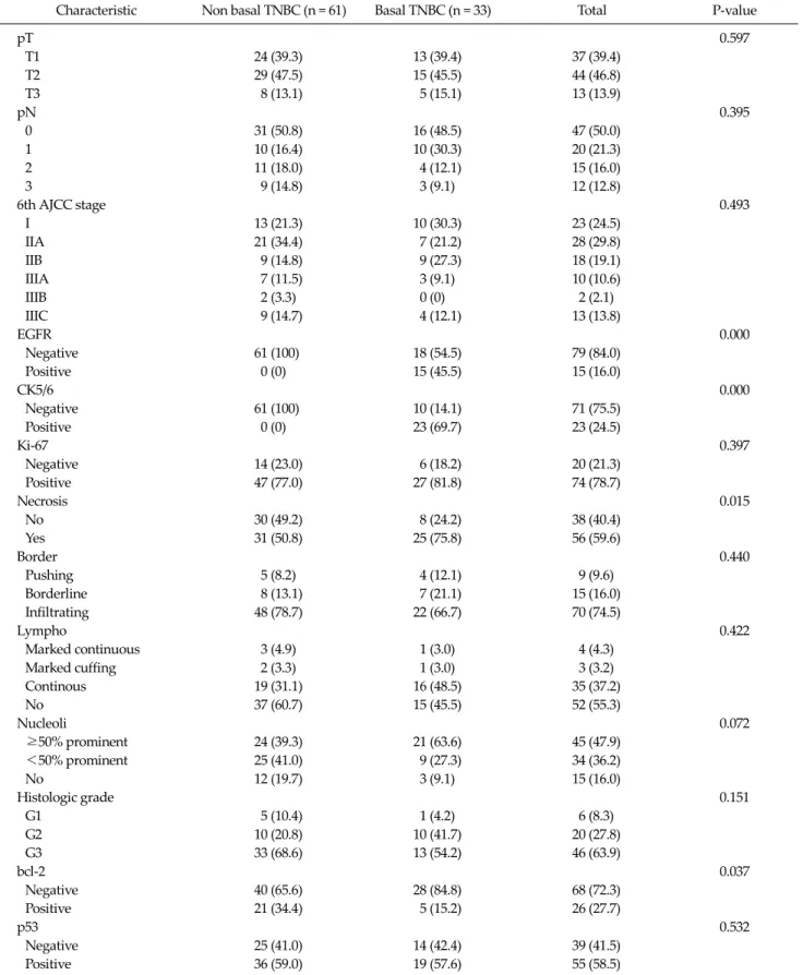
Fig. 3. Kaplan-Meier estimates of overall survival according to B-cell lymphoma (bcl)-2.
Cancer (AJCC) stage (P = 0.493) and shorter overall surviv- al (OS, P = 0.438) and disease-free survival (DFS, P = 0.260).
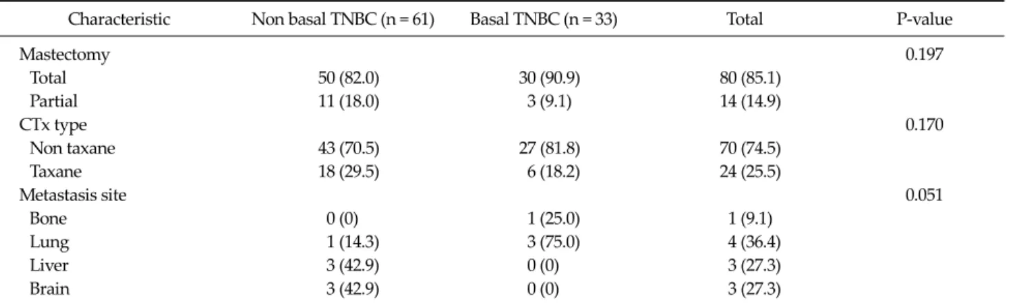
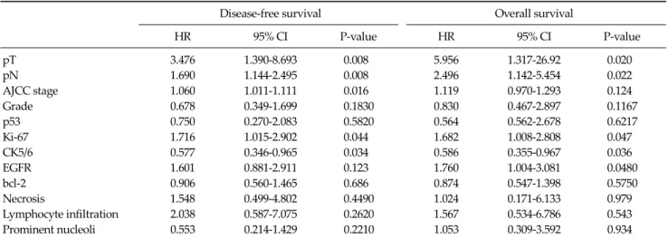
And bcl-2 in 26 cases (27.7%) and p53 in 55 cases (58.5%) was determined as positive staining. Tumor necrosis was found in 56 patients (59.6%) in TNBC. And infiltrating bor- der was found in 70 patients (74.5%). Also continuous lymphocytic distribution and prominent nucleoli (>50%) was found in 35 patients (37.2%), and 45 patients (47.9%), respectively. No statistical relationship was found be- tween bcl-2 positivity and other variables, such as tumor size, nodal status, AJCC stage, tumor grading, p53 and Ki-67 (Tables 4-6). At a median follow-up of 5.5 years, 84 patients (13.6%) had a recurrence, and 32 patients (5.2%) expired. Among TNBC, we observed 7 relapses in 61 pa- tients (11.5%) with non-BP TNBC and 4 relapses in 33 pa- tients (12.1%) with BP TNBC. The univariate analysis for prognostic factors associated with DFS revealed that the Ki-67 was statistically significant (P = 0.044). CK5/6 was al- so statistically significant (P = 0.034). Tumor group as T1, T2, T3, or T4, lymph node stage group as N0, N1, N2, or N3 was also significant (P = 0.008, P = 0.008 respectively) (Table 6). Also, the univariate analysis for prognostic factors as-
sociated with OS revealed that Ki-67, CK5/6 was statisti- cally significant (P = 0.047, P = 0.036 respectively) (Figs.
1-4). Lymph node stage group as N0, N1, N2, or N3 was al- so statistically significant (P = 0.022) (Table 7).
Disease-free survival Overall survival
HR 95% CI P-value HR 95% CI P-value
pT 3.476 1.390-8.693 0.008 5.956 1.317-26.92 0.020
pN 1.690 1.144-2.495 0.008 2.496 1.142-5.454 0.022
AJCC stage 1.060 1.011-1.111 0.016 1.119 0.970-1.293 0.124
Grade 0.678 0.349-1.699 0.1830 0.830 0.467-2.897 0.1167
p53 0.750 0.270-2.083 0.5820 0.564 0.562-2.678 0.6217
Ki-67 1.716 1.015-2.902 0.044 1.682 1.008-2.808 0.047
CK5/6 0.577 0.346-0.965 0.034 0.586 0.355-0.967 0.036
EGFR 1.601 0.881-2.911 0.123 1.760 1.004-3.081 0.0480
bcl-2 0.906 0.560-1.465 0.686 0.874 0.547-1.398 0.5750
Necrosis 1.548 0.499-4.802 0.4490 1.024 0.171-6.133 0.979
Lymphocyte infiltration 2.038 0.587-7.075 0.2620 1.567 0.534-6.786 0.543
Prominent nucleoli 0.553 0.214-1.429 0.2210 1.053 0.309-3.592 0.934
HR, hazard ratio; CI, confidence interval; AJCC, American Joint Committee on Cancer; CK, cytokeratin; EGFR, epidermal growth factor receptor; bcl‐2, B‐cell lymphoma.
Table 7. Five years overall and disease-free survival probabilities in triple negative breast cancer as calculated by Cox proportional hazards regression model (P-value for log rank test)
Fig. 4. Kaplan-Meier estimates of overall survival according to Ki-67.
DISCUSSION
Apoptosis, or programmed cell death, plays a major role not only in the formation and progression of cancer, but in the development of resistances to anticancer drugs as well [11]. The genes p53 and bcl-2 play an important role in regulating the mechanisms of apoptosis [12]. p53 is a tu- mor suppressor gene involved in the regulation of pro- liferative activity of human cells. Indeed, p53 serves a mul- tifunctional role as a transcriptional regulator, genomic stabilizer, inhibitor of cell cycle progression, facilitator of apoptosis and also perhaps as inhibitor of angiogenesis [13]. By all these mechanisms, wild type p53 slows pro-
liferation, while mutations of these gene in cancer cells may result in higher proliferation rates, higher genetic in- stability, and resistance to apoptosis [14]. Many studies have analyzed the prognostic impact of p53 alterations in breast cancer, and most of the studies have reported an as- sociation with worse outcome, either in node negative and node positive disease. However, others studies failed to demonstrate a significant association of p53 with poorer prognosis [8,11,15]. In our series, p53 over-expression was found in 60.0% of breast cancers, a value in line with the re- ported values between 11 and 60% of p53 mutation in breast cancer by immunohistochemistry, but did not cor- relate to either bcl-2 expression or to disease-free and over- all survival. It has been recently accepted that gene se- quencing is the most appropriate method to study p53 sta- tus; indeed different types of mutated p53 oncogene exist that do not determine protein accumulation and are there- fore not detected by immunohistochemistry [15,16]. The discordance between our results and other studies may be partly due to the immunohistochemical technique used to detect p53 mutations and reflects the inconsistent data re- ported in literature for p53 mutation in breast cancer. The bcl-2 gene was initially identified in human bcls and its ac- tivity as apoptosis inhibitor, either drug and p53 induced, in cancer cells has been well demonstrated [7,10]. As a con- sequence, the expression of bcl-2 in breast cancer cells
should inhibit apoptosis and therefore relate to a worse outcome [1]. On the contrary, the expression of bcl-2 in breast cancer has been found to be associated with favor- able prognostic factors such as small tumor size, ER pos- itivity, low nuclear grade and to predict a better outcome [7,10,17]. These unexpected results may be explained by the fact that the ability of bcl-2 to inhibit apoptosis de- pends on the intracellular balance among a number of its family members, with other pro-apoptotic members such as Bax counteracting its action [7,18]. Given its anti- apoptotic function, bcl-2 should be expected to correlate with worse prognosis. On the contrary, high levels of bcl-2 expression have been reported as associated to favorable tumor characteristics, such as low tumor size, low tumor grade, low proliferative activity and mainly with ex- pression of ER and PgR [19]. These associations account for the favorable prognostic impacts reported for bcl-2 ex- pression on DFS and OS in breast cancer, either node neg- ative and node positive, even if an independent prognostic role has been confirmed only in some series [20,21]. Our data do not support the association of bcl-2 expression with other favorable prognostic factors and mainly with ERs expression, even if the percentages of bcl-2 and ERs expression in the study population are in line with the oth- er series [22]. On the other hand, data regarding outcome in our series are consistent with other studies; that bcl-2 ex- pression doesn’t correlate with better outcome, either for DFS and OS. Reasons for this unfavorable prognostic val- ue of bcl-2 expression are not clear; possible explanations may include the pro- apoptotic activity of other members of the bcl-2 family, an inhibitory activity of bcl-2 on cell proliferation or the estrogen-inducibility of bcl-2 ex- pression [10]. In addition, data regarding outcome in our series are not consistent with other studies; that CK5/6 positivity correlates with better outcome, either for DFS and OS. Reasons for this favorable prognostic value of CK5/6 expression are not clear; possible explanations may include the small sample size, short-term follow-up peri- od and no differences in survival rate between TNBC groups according to basal type.
In conclusion, paradoxically, CK5/6 positive TNBCs are higher DFS and OS than CK5/6 negative TNBCs. So, our study has some limitations in that CK5/6 can be used to
classify TNBCs into at least two subtypes with differing survival rates. Also, the nuclear proliferation antigen, Ki-67, can be used to differentiate disease-free survival.
But markers regulating cell cycle and cell death such as p53 and bcl-2 cannot be used to classify TNBCs into two subtypes with differing survival rates. However, because our study is small in size, there is no survival difference be- tween TNBC groups according to basal type. So, more abundant patients’ data will be needed to evaluate the pro-apoptotic activity of other members of the bcl-2 fam- ily’s predictive role.
CONFLICTS OF INTEREST
No potential conflict of interest relevant to this article was reported.
ACKNOWLEDGEMENTS
This article was granted by Kosin Medical College 2011.
REFERENCES
1. Nakagawa M, Bando Y, Nagao T, Morimoto M, Takai C, Ohnishi T, et al. Expression of p53, Ki-67, E-cadherin, N-cadherin and TOP2A in triple-negative breast cancer.
Anticancer Res 2011;31:2389-93.
2. Elnashar AT, Ali el-SM, Gaber A. The prognostic value of triple negative in stage II/III breast cancer. J Oncol Pharm Pract 2012;18:68-75.
3. Yerushalmi R, Tyldesley S, Kennecke H, Speers C, Woods R, Knight B, et al. Tumor markers in metastatic breast can- cer subtypes: frequency of elevation and correlation with outcome. Ann Oncol 2012;23:338-45.
4. Montagna E, Bagnardi V, Rotmensz N, Viale G, Renne G, Cancello G, et al. Breast cancer subtypes and outcome after local and regional relapse. Ann Oncol 2012;23:324-31.
5. Skarlos P, Christodoulou C, Kalogeras KT, Eleftheraki AG, Bobos M, Batistatou A, et al. Triple-negative phenotype is of adverse prognostic value in patients treated with dose- dense sequential adjuvant chemotherapy: a translational research analysis in the context of a Hellenic Cooperative Oncology Group (HeCOG) randomized phase III trial.
Cancer Chemother Pharmacol 2012;69:533-46.
6. Nishimura R, Osako T, Okumura Y, Tashima R, Toyozumi Y, Arima N. Changes in the ER, PgR, HER2, p53 and Ki-67
biological markers between primary and recurrent breast cancer: discordance rates and prognosis. World J Surg Oncol 2011;9:131.
7. Kallel-Bayoudh I, Hassen HB, Khabir A, Boujelbene N, Daoud J, Frikha M, et al. Bcl-2 expression and triple neg- ative profile in breast carcinoma. Med Oncol 2011;28 Suppl 1:S55-61.
8. Chae BJ, Bae JS, Lee A, Park WC, Seo YJ, Song BJ, et al. p53 as a specific prognostic factor in triple-negative breast cancer. Jpn J Clin Oncol 2009;39:217-24.
9. Collins LC, Marotti JD, Gelber S, Cole K, Ruddy K, Kereakoglow S, et al. Pathologic features and molecular phenotype by patient age in a large cohort of young wom- en with breast cancer. Breast Cancer Res Treat 2012;131:
1061-6.
10. Tawfik K, Kimler BF, Davis MK, Fan F, Tawfik O.
Prognostic significance of Bcl-2 in invasive mammary car- cinomas: a comparative clinicopathologic study between
"triple-negative" and non-"triple-negative" tumors. Hum Pathol 2012;43:23-30.
11. Lee DS, Kim SH, Suh YJ, Kim S, Kim HK, Shim BY. Clinical implication of p53 overexpression in breast cancer patients younger than 50 years with a triple-negative subtype who undergo a modified radical mastectomy. Jpn J Clin Oncol 2011;41:854-66.
12. Jiang Z, Jones R, Liu JC, Deng T, Robinson T, Chung PE, et al. RB1 and p53 at the crossroad of EMT and triple-neg- ative breast cancer. Cell Cycle 2011;10:1563-70.
13. Lee DS, Kim SH, Kim S, Suh YJ, Kim HK, Shim BY.
Prognostic significance of breast cancer subtype and p53 overexpression in patients with locally advanced or high-risk breast cancer treated using upfront modified rad- ical mastectomy with or without post-mastectomy radia- tion therapy. Int J Clin Oncol 2011 Sep 7 [Epub]. http://
dx.doi.org/10.1007/s10147-011-0309-0.
14. Biganzoli E, Coradini D, Ambrogi F, Garibaldi JM, Lisboa P, Soria D, et al. p53 status identifies two subgroups of tri- ple-negative breast cancers with distinct biological features. Jpn J Clin Oncol 2011;41:172-9.
15. Guarneri V, Barbieri E, Piacentini F, Giovannelli S, Ficarra G, Frassoldati A, et al. Predictive and prognostic role of p53 according to tumor phenotype in breast cancer pa- tients treated with preoperative chemotherapy: a single-in- stitution analysis. Int J Biol Markers 2010;25:104-11.
16. Jung SY, Jeong J, Shin SH, Kwon Y, Kim EA, Ko KL, et al.
Accumulation of p53 determined by immunohistochem- istry as a prognostic marker in node negative breast cancer;
analysis according to St Gallen consensus and intrinsic subtypes. J Surg Oncol 2011;103:207-11.
17. Moran MS, Yang Q, Haffty BG. The Yale University experi- ence of early-stage invasive lobular carcinoma (ILC) and invasive ductal carcinoma (IDC) treated with breast con- servation treatment (BCT): analysis of clinical-pathologic features, long-term outcomes, and molecular expression of COX-2, Bcl-2, and p53 as a function of histology. Breast J 2009;15:571-8.
18. Tawfik O, Davis K, Kimler BF, Davis MK, Hull S, Fan F, et al. Clinicopathological characteristics of triple-negative in- vasive mammary carcinomas in African-American versus Caucasian women. Ann Clin Lab Sci 2010;40:315-23.
19. Broustas CG, Ross JS, Yang Q, Sheehan CE, Riggins R, Noone AM, et al. The proapoptotic molecule BLID inter- acts with Bcl-XL and its downregulation in breast cancer correlates with poor disease-free and overall survival. Clin Cancer Res 2010;16:2939-48.
20. Rhee J, Han SW, Oh DY, Kim JH, Im SA, Han W, et al. The clinicopathologic characteristics and prognostic sig- nificance of triple-negativity in node-negative breast cancer. BMC Cancer 2008;8:307.
21. Nozoe T, Mori E, Kono M, Iguchi T, Maeda T, Matsukuma A, et al. Serum appearance of anti-p53 antibody in triple negative breast cancer. Breast Cancer 2012;19:11-5.
22. Keam B, Im SA, Kim HJ, Oh DY, Kim JH, Lee SH, et al.
Prognostic impact of clinicopathologic parameters in stage II/III breast cancer treated with neoadjuvant docetaxel and doxorubicin chemotherapy: paradoxical features of the tri- ple negative breast cancer. BMC Cancer 2007;7:203.