Introduction
Scapular winging (SW), defined as a prominence of the entire medial border of the scapula, is mainly caused by insufficient activity of the serratus anterior (SA) muscle (Henry and Westervelt, 2005; Martin and Fish, 2008). The SA muscle is one of the most important muscles used for scapular stabilization be- cause the SA inserts on the scapular medial border and inferior angle and originates from the 1st to 8th ribs (Neumann, 2013). Excess upper trapezius (UT) activation or reduced activation of the lower trapezius
(LT) and SA may contribute to abnormal scapular motion, scapular dysfunction, and pain and attempt to compensate for the weak SA (Sahrmann, 2002). Thus, therapeutic exercise for recovering the scapular sta- bilizer muscle function is an important part of the re- habilitation program (Bang and Deyle, 2000).
Open and closed kinetic chain exercises to increase SA muscle activity have been examined by many previous researchers (Cools et al, 2007; Decker et al, 1999; Hardwick et al, 2006; Martins et al, 2008). The push-up plus (PUP) exercise as a key exercise in shoulder rehabilitation exercise is based on the high Corresponding author: Heon-seock Cynn [email protected]
Effect of the Abdominal Drawing-in Maneuver on the Scapular Stabilizer Muscle Activities and Scapular Winging During Push-up Plus
Exercise in Subjects With Scapular Winging
Da-eun Kim
1, BHSc, PT, A-reum Shin
1, BPT, PT, Ji-hyun Lee
1, MSc, PT, Heon-seock Cynn
2,3, PhD, PT
1Dept. of Physical Therapy, The Graduate School, Yonsei University
2Dept. of Physical Therapy, College of Health Science, Yonsei University
3Dept. of Ergonomic Therapy, The Graduate School of Health and Environment, Yonsei University
Abstract 1)
Background: Scapular winging is a prominence of the entire scapular medial border, mainly caused by insufficient activity of the serratus anterior (SA) and imbalance of scapulothoracic muscles. Push-up plus (PUP) exercise has been commonly used to increase SA muscle activity. The facilitation of abdominal muscle may affect scapular muscle activity by myofascial connections. Thus, the sequential activation of the turnk muscles is suggested to facilitate the transition of proper force from upper limb and restore force couple of scapular muscles. The abdominal drawing-in maneuver (ADIM) has been effective in improving activation of the deep trunk muscles during movement.
Objects: The aim of this study was to determine the effect of ADIM on the activity of the upper trapezius (UT), lower trapezius (LT), and SA during PUP exercises in subjects with scapular winging.
Methods: Fourteen men with scapular winging (determined as a of distance between the scapular medial border and thoracic wall over 3 ㎝) volunteered for our study. The subjects performed the PUP exercise with and without ADIM. Surface electromyography was used to collect the electromyography data of the UT, LT, and SA. A scapulometer was used to measure the amount of scapular winging.
Results: SA activity was significantly greater and scapular winging significantly lower during the PUP exercise with ADIM than during those without ADIM.
Conclusion: PUP exercise with ADIM can be used as an beneficial method to improve SA activation and to reduce the amount of scapular winging in subjects with scapular winging.
Key Words: Electromyography; Serratus anterior; Shoulder rehabilitation exercise.
activity of various shoulder muscles, including the SA (Moseley et al, 1992). Furthermore, the PUP exercise was found to elicit SA activity was greater than 20%
maximal isometric voluntary contraction as compared to a number of rehabilitation exercises (Decker et al, 1999). Therefore, the PUP exercise has been widely used to correct scapular kinematics and enhance the SA activity in patients with SW (Hardwick et al, 2006; Park et al, 2013).
Previous study reported that activation of trunk muscle may affect the scapular muscle activity by myofascial connections (Myers, 2009). Therefore, the facilitation of the abdominal muscles is recommended to restore force couples of scapular muscle (UT, LT and SA) (Kibler and Sciascia, 2010). Trunk stability is asked to transfer force and energy to the upper extremities during movement or exercise (Hirashima et al, 2002; Jang et al, 2015; Kibler et al, 2006). The scapulothoracic muscles and joints are an anatomo- functional connection of the trunk and the upper limbs (Kibler and Sciascia, 2010). Many studies re- garding the transversus abdominal muscle have fo- cused on motor control and order of muscle activa- tion during the spine stabilization (Hodges and Richardson, 1999). A previous study reported that the UT, LT, and middle trapezius muscle activities were affected by adding pelvic and thoracic supports (Jang et al, 2015). The abdominal drawing-in maneuver (ADIM), a core stabilization exercise, has been more effective than other core stabilization exercises in improving lumbopelvic stability and optimal co- activation of the deep trunk muscles during move- ments (Richardson et al, 2002). Previous investigators reported that ADIM is effective in stabilizing the lumbopelvic region during limb movements as com- pared to abdominal bracing (Suehiro et al, 2014). For proper performance of ADIM, a real-time ultrasound imaging is an effective visual feedback device and a useful clinical teaching device for physical therapists (Hodges and Richardson, 1996).
No previous studies have examined the effect of ADIM on the activities of the scapular stabilizer
muscles during PUP exercises. Thus, this study aimed to compare the muscle activities of the UT, LT, and SA, UT/LT and UT/SA ratios, and amounts of SW during the PUP exercise with and without ADIM. We hypothesized that ADIM during the PUP exercise would increase the activities of the LT and SA and reduce the UT activity and UT/LT, UT/SA ratios, and amount of SW compared with the preferred PUP exercise in subjects with SW.
Methods
Subjects
G-power Software (ver. 3.1.6; Franz Faul, University of Kiel, Germany) was used for power analyses. A necessary sample size of 9 subjects was gained from a pilot study of 4 subjects to achieve an effect size of .83, with a power of .8 and a significance level of .05.
Thus, 14 men with SW were recruited into the study (age=22.0±3.1 years, height=173.4±5.1 ㎝, weight=71.5±6.2 ㎏, body mass index=23.8±2.4 ㎏/㎡, amount of SW=3.2±.1 ㎝).
The SW side was all used in all tests. If the sub- jects had bilateral SW, we used the worse side when a bilateral comparison indicated a difference of over
<.1 ㎝ in the amount of SW.
The inclusion criteria were as follows: (1) SW was determined if the distance of between medial border of scapula and thoracic wall was over 3 ㎝ (Park et al, 2007) and (2) normal range of motion of shoulder internal rotation and horizontal adduction to rule out dominance of the deltoid (Warner et al, 1997). The exclusion criteria were as follows: (1) past to present shoulder pain or dysfunction, (2) history of shoulder or abdominal or back injury or surgery, (3) signs and symptoms of cervical pain, (4) adhesive capsu- litis, thoracic outlet syndrome, or a current complaint of numbness or tingling in the upper limbs (Park et al, 2013), and (5) presence of a winged scapula due to a long thoracic nerve denervation (Choi et al, 2016).
The participants were provided with a detailed ex-
planation of this study process and safety instructions
and were asked to sign a written consent prior to voluntary participation. The protocol was approved by the Yonsei University Wonju Institutional Review Board (approval number: 1041849-201701-BM-003-04).
EMG recording and data processing A Noraxon TeleMyo-DTS (Noraxon, Inc., Scottsdale, AZ, USA) was used to collect surface EMG data from the scapular stabilizer muscles (UT, LT, and SA) on the more prominent side of SW.
EMG data were analyzed using the Noraxon MyoRexearch 1.08 XP software. The EMG signals were amplified, band pass-filtered (20 and 450 ㎐), and notch-filtered (60 ㎐) before being recorded digi- tally at 1000 ㎐ and processed into root-mean-square data with a window of 50 ㎳. Skin preparation in- cluded shaving of the hair and scrubbing of the skin with a cotton wool dipped in alcohol to decrease im- pedance before data collection. The disposable Ag/AgCl surface electrodes were attached to each muscle at standardized placements (Criswell, 2010).
The electrodes for the UT were placed halfway be- tween the C7 spinous process and the scapular acromion. The electrodes for the LT were placed ap- proximately 5 ㎝ down from the scapular spine, next to the medial edge of the scapula at a 55° oblique angle. The electrodes for the SA were placed just be- low the axillary area, at the level of the inferior tip of the scapula, and medial to the latissimus dorsi (Criswell, 2010). Two active electrodes were attached nearly 2 ㎝ apart in the direction of the muscle fibers.
The maximal voluntary isometric contractions (MVICs) were initiated to normalize the EMG signal amplitude in the UT, LT, and SA according to standardized procedures recommended by previous research studies (Ekstrom et al, 2005; Kendall et al, 1993). To gain the MVIC for the UT, each subject was asked to perform a 90° shoulder abduction with manual resistance to the head after the neck was first side-flexed to the same side, rotated to the op- posite side, and extended in the sitting position with no back support (Ekstrom et al, 2005). To obtain the
MVIC for the LT, the subject was tested in the prone position. The subject’s arm was placed diago- nally overhead, in line with the lower fibers of the trapezius muscle during external rotation while re- sistance was applied distal to the elbow (Ekstrom et al, 2005). The MVIC for the SA was performed with the subject sitting and the shoulder rotated internally and abducted at 125° in the scapular plane. Manual resistance was applied proximal to the subject’s el- bow by the researcher (Kendall et al, 1993). EMG data for each muscle were obtained for 5 sec, and a 1 min break time was given between trials to mini- mize muscle fatigue (Vera-Garcia et al, 2010). All average amplitude values were calculated for the middle three sec of the two trials during exercise and expressed as %MVIC.
Scapular winging measurement
A scapulometer was used to determine SW after visual screening (Figure 1). The subject was in a quadruped position with 90˚ flexion of hip, knee, and shoulder joint. The investigator used a vertical ruler combined with a horizontal ruler to measure the dis- tance between the medial edge of scapula and thoracic wall (Park et al, 2007). The horizontal ruler was placed across both the right and left scapula, and the lower part of vertical ruler was fixed to the spinous process of the thoracic vertebra in the center of the scapula. SW was defined as a of distance ≥3 ㎝ (Park et al, 2007). We proved the test-retest reliability of the scapulometer for measuring the amount of SW.
Figure 1. Measurement of scapular winging.
The intra-class correlation coefficient (ICC) was .80 (95% confidence interval; CI: .48∼.93). The standard error of the measurement (SEM) was .15 ㎝, and the minimal detectable change (MDC) was .42 ㎝.
Procedures
The subjects familiarized themselves with the PUP exercise and ADIM for 20 min to perform the ex- ercises properly before data collection. After familiar- ization, a 10 min rest time was given. The EMG data on the preferred PUP exercise was collected before the data collection on the PUP exercise with ADIM.
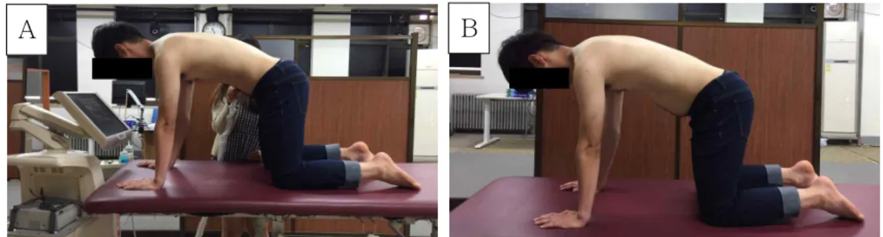
The subjects were cued to start the exercise when they heard the command “start” and maintained the quadruped position and full protraction of scapula dur- ing the exercise. The PUP exercise with and without ADIM were performed for the 5 sec of the three trials (Figure 2). SW was measured immediately after 3 tri- als of the PUP exercise with and without ADIM.
Preferred push-up plus exercise
The subject assumed a quadruped position with both the hands and knees shoulder-width apart. The hip and knee joint flexions were at 90° in the quad- ruped position with 90° of shoulder flexion, while the head and pelvis were positioned in a neutral state. The subject maintained the ankle joint in a plantarflexion position and the elbow joint in a full extension position.
To maintain the cervical spine in a neutral posture, the cervical spine and thoracic spine were aligned in a straight line. We instructed the subjects to “give an extra push” to protract the scapula maximally.
Push-up plus exercise with abdominal drawing-in maneuver
After the collection of EMG data on the preferred PUP exercise in the quadruped position, the subjects were familiarized with the typical clinical in- structions and visual feedback of ADIM from ultra- sound imaging to achieve a proper ADIM perform- ance ability. When the subjects could not maintain the ADIM during the exercise period, the test was immediately stopped. With the subject in the quad- ruped position, ultrasonography (SonaAce X8, Medison Co. Ltd., Seoul, Korea) was used for visual feedback and monitoring of the ADIM during the PUP exercise. An ultrasound transducer was located on the anterolateral abdominal wall approximately 2.5 ㎝ under the inferior tip of the ribs and approx- imately 15 ㎝ lateral to the umbilicus during ADIM (Misuri et al, 1997). A real-time ultrasound imaging is indicated on the ultrasound machine screen placed in a position that was easily visible for the subject from the quadruped position. The images were ob- tained at rest, with the subjects exhaling during the contraction, while maintaining the ADIM (Hides et al, 2007). Before starting the ADIM, the subjects were instructed to cough so they can see the move- ment of their abdominal muscles on the monitor.
The investigators instructed the subjects to pull the navel in and up without allowing any movement at the pelvis, rib, or spine and to maintain the abdomi- nal contraction while continually breathing normally after the navel has been drawn close to the spine (O’sullivan, 2000). Ultrasound images of each muscle
A B
Figure 2. Push-up plus exercise (A: without abdominal drawing-in maneuver, B: with abdominal
drawing-in maneuver).
thickness were monitored to confirm that the thick- ness of the muscle layers (transverse abdominis, in- ternal oblique abdominis, and external oblique ab- dominis) was increased by the principal investigator and, Furthermore, an image of the muscle layer was shown to the subjects to provide a visual feedback during the PUP exercise with ADIM. The principal investigator explained the transverse abdominis, in- ternal oblique abdominis, and external oblique ab- dominis layers and instructed on the maximal pref- erential activation of the transverse abdominis. A previous study reported that the correct ADIM trials in the ADIM with typical clinical feedback and re- al-time ultrasound imaging augmented feedback were greater compared to those with a typical clin- ical feedback including verbal and tactile cues only (Henry and Westervelt, 2005). We discarded the data to perform the correct ADIM when the thickness of the transverse abdominis and internal oblique ab- dominis was not increased, that of the external obli- que abdominis muscle was increased, or a pelvic movement in the posterior direction was shown, or the subjects took a deep inhalation followed by breath holding as determined by palpation (Henry and Westervelt, 2005).
Statistical Analysis
The SPSS software ver. 23.0 (SPSS Inc., Chicago, IL, USA) was used to assess all statistical analyses.
The one-sample Kolmogorov-Smirnov test was used to assess the normality of distribution. Test-retest reli- ability of SW measurement in 2 conditions (with ADIM and without ADIM) of the PUP exercise was assessed using ICC, 95% CI, SEM, and MDC. SEM was calcu- lated to assess absolute consistency (SEM=SD×√
1-ICC), MDC (95% CI) (MDC
95=SEM×1.96√2) (Ries et al, 2009). The effect size index (ESI) was calcu- lated to decide meaningful changes between the PUP exercise with ADIM and without ADIM. Because all dependent variables were confirm the normality of distribution, a paired t-test was used to assess the statistical significance of the UT, LT, and SA activ- ities, UT/LT and UT/SA ratios, and amount of SW between the preferred PUP exercise and the PUP exercise with ADIM with a significance level of .05.
Results
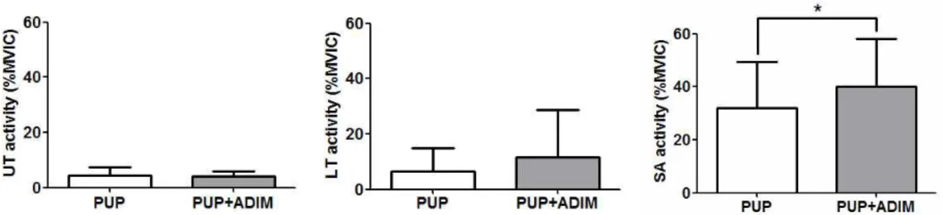
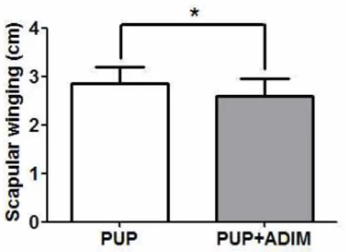
Muscle activity and muscle activity ratios Table 1 and Figure 3 shows the %MVIC of the UT, LT, and SA as well as the UT/LT and UT/SA ratios during the exercises. There were no significant differences in the UT and LT activities between the preferred PUP exercise and the PUP exercise with ADIM (p>.05). The SA activity was significantly greater during the PUP exercise with ADIM than during the preferred PUP exercise (p=.02). The
Variables Preferred PUP
aPUP with ADIM
bp
UT
c(%MVIC
d) 4.40±2.87
e4.02±1.91 .536
LT
f(%MVIC) 6.38±8.43 11.67±16.97 .098
SA
g(%MVIC) 31.88±17.64 40.15±18.06 .018*
UT/LT .18±.16 .12±.08 .132
UT/SA 1.26±1.36 .90±.79 .194
SW
h(㎝) 2.86±.33 2.6±.35 <.001*
a
push-up plus exercise,
babdominal drawing-in maneuver,
cupper trapezius,
dmaximal volutary contraction,
e