J. Exp. Biomed. Sci. 14 (2008) 211–218
Effects of Chungkookjang on Blood Glucose, Antioxidant Enzyme Activities and Histological Changes in Kidney of STZ-induced Diabetic Rats
Hye-Jeong Kim1,2 and Young-Chul Kim1,†
1Department of Public Health, Keimyung University, Daegu 704-701, Korea.
2Division of Human Blood Safety Surveillance, Korea Centers for Disease Control & Prevention, Seoul 122-701, Korea
The purpose of this study was to investigate the effects of dietary Chungkookjang (Korean fermented soybean) powder on blood glucose level, lipid profiles, antioxidant enzymes activities and histological changes in kidney of streptozotocin (STZ)-induced diabetic rats. Male Sprague-Dawley rats of three groups including nondiabetic group fed normal diet (NC), diabetic group fed normal diet (DC) and diabetic group fed Chungkookjang diet (DCH; 100 g/kg diet) were reared for 8 weeks. The serum glucose, triglyceride and total lipid levels in the DCH group were significantly lower (P<0.05) than the DC group. The renal xanthine oxidase, catalase and glutathione S-transferase activities in the DC group were significantly higher than the NC group. The xanthine oxidase, superoxide dismutase and glutathione S-transferase activities in the DCH group were significantly lower than the DC group (P<0.05). Tubular epithelial change, such as Armanni-Ebstein cells, was significantly reduced in the DCH group compared to the DC group. In conclusion, these results indicated that Chungkookjang supplement seems to be beneficial to correct the hyperglycemia and hyperlipidemia as well as to protect kidney against diabetic changes.
Key Words: Chungkookjang, Streptozotocin, Diabetic rat, Antioxidant enzymes, Armanni-Ebstein cells
INTRODUCTION
Diabetes mellitus is the complex metabolic disease caused by an absolute or relative lack of insulin and resulting in deleterious effects on both the macrovascular and micro- vascular systems (Zimmet et al., 2001). Diabetes mellitus is a chronic disease that cannot be completely cured and may develop various complications if not properly treated.
The most devastating complication of diabetes is nephro- pathy, which causes 14% of all deaths in diabetes patients (Edwads et al., 1995) and accounts for 40% of end-stage renal failure (Lasaridis and Sarafidis, 2003).
Recently, attention has been drawn to the theory that oxidative stress is involved in the development of compli-
cations associated with chronic diabetes (Maritim et al., 2003). Oxidative stress has been considered as a common pathogenetic factor in diabetic nephropathy and other complications (Tomohiro et al., 2007). Excessive excretion of glycogen through the glomeruli is reabsorbed into the cytoplasm of tubules. These phenomena are especially pro- minent in the straight part of proximal tubules. In histologic preparations, these glycogen inclusions are washed out and result in a clearing effect within the tubular epithelial cells.
This phenotype is referred to as Almanni-Ebstein cells and is a clear morphological characteristic of a diabetic kidney (Watanabe and Hotta, 1997).
There is an increasing demand to use the natural products with antidiabetic activity presently (Kim et al., 2008). Soy- bean is effective in lowering plasma cholesterol, triglyceride concentration (Anderson et al., 1995; Lee, 2006). Several studies in animals and humans have shown that the consump- tion of soybean have beneficial effects in variety of disorders including hypercholesterolemia as well as protection against cardiovascular disease, renal disease, bone resorption, certain
*Received: November 22, 2008
Accepted after revision: December 5, 2008
†Corresponding author: Young Chul Kim, Department of Public Health, Keimyung University, Dalseo-Gu, Daegu 704-701, Korea.
Tel: +82-53-580-5931, Fax: +82-53-588-5233 e-mail: [email protected]
forms of cancer and menopausal symptoms (Velasquez and Bhathena, 2001). Increasing evidence suggests that soybean may also have beneficial effect against obesity and diabetes (Bhathena and Velasquez, 2002). Soybean-based fermented foods such as the whole cooked fermented soybean products (Chungkookjang in Korea, Natto in Japan) are very popular in Korea and Japan. Chungkookjang has been made tradi- tionally with whole cooked soybean by fermenting with bacilli without salt addition.
Although the importance of antihyperglycemic effect of soybean has been recognized, effect of Chungkookjang on histopathological changes in kidney has not been reported.
Hence, the present study was planned to evaluate the effect of Korean soybean fermented foods (Chungkookjang) on blood glucose, lipid profiles, antioxidant enzyme activities and histological renal changes of streptozotocin-induced diabetic rats.
MATERIALS AND METHODS 1. Animals and diets
Thirty male Sprague-Dawley rats with body weight ranging from 200 to 220 g were obtained from animal house, Daehan Biolink (Chungbook, Korea) and were housed in plastic cages in a room with regulated temperature (22±
1℃), relative humidity (50±5%), alternating 12-hr light/dark cycle, and were allowed to have access to their respective diets ad libitum. Body weight and blood glucose levels were monitored between 09:00~10:00 hr weekly. Blood was taken from the tail vein for blood glucose determination. The whole cooked soybean fermented product (Chungkookjang) was purchased from Manpojang Food Co. (Gyeongbuk, Korea). The composition of experimental diets (Table 1) was based on the AIN-76 standard laboratory diet (American Institute of Nutrition, 1977). Daily food and water con- sumption were determined by subtracting left over amount from the amount provided to the rats. After 8 weeks of the experimental period, overnight fasted rats were sacrificed by drawing arterial blood under ether anesthesia. Blood was collected from the abdominal aorta to measure serum glucose and lipid profiles. Serum was obtained by centri- fuging the blood at 2,500 rpm for 15 min. Then, the organs
(liver, kidney, heart) were taken and weighed using an electronic balance. Each one of the removed kidneys was stored at -80℃ for determination of antioxidant enzyme activities and another each one was fixed in 10% formalin solution for histological examination.
2. Experimental design
The animals were allowed to acclimatize to the laboratory environment for 7 days, and randomly divided into three groups of ten animals each group for the 8-week study. The experimental groups were as follows: Group I, non-diabetic rats fed normal diet (NC); Group II, diabetic rats fed normal diet (DC); Group III, diabetic rats fed Chungkookjang 10%
diet (DCH). The composition of normal and Chungkookjang diets are shown in Table 1.
3. Experimental induction of diabetes
Diabetes was induced by a single intraperitoneal injection of STZ (50 mg/kg BW; Sigma, USA) in freshly prepared
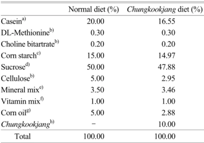
Table 1. Composition of experimental diets Normal diet (%) Chungkookjang diet (%)
Caseina) 20.00 16.55
DL-Methionineb) 0.30 0.30
Choline bitartrateb) 0.20 0.20
Corn starchc) 15.00 14.97
Sucrosed) 50.00 47.88
Celluloseb) 5.00 2.95
Mineral mixe) 3.50 3.46
Vitamin mixf) 1.00 1.00
Corn oilg) 5.00 2.88
Chungkookjangh) - 10.00
Total 100.00 100.00 Sources of ingredients: a) Fonterra Co. (NewZealand), b) Sigma (USA), c) Samyang Genex Co. (Korea), d) Samyang Co. (Korea),
g) Cheiljedang Co. (Korea).
e) AIN-76 mineral AIN-76 mineral mixture (g/kg mixture): calcium phosphate, dibasic 500; sodium chloride 74; potassium citrate monohydrate 220; potassium sulfate 52; magnesium oxide 24;
manganous carbonate 0.3; potassium iodate 0.01; chromium potassium sulfate 0.55; sucrose finely powdered to make 1000.0 g.
f) AIN-76 vitamin mixture (per kg mixture): thiamin HCl 600 mg;
riboflavin 600 mg; pyridoxine HCl 700 mg; nicotinic acid 3 g;
D-calcium pantothenate 1.6 g; folic acid 200 mg; D-biotin 20 mg; vitamin B12 (cyanocobalamin) 1 mg; vitamin A (retinyl palmitate) 4,000 IU; vitiamin E (DL-α-tocopheryl acetate) 5,000 IU; vitamin D3 (cholecalciferol) 2.5 mg; menaquinone 5.0 mg;
sucrose finely powdered to make 1000.0 g.
h) Composition of powder: crude protein 36.3%, ash 0.4%, crude fat 21.2%, crude fiber 20.5% and carbohydrate 1.3%.
citrate buffer (0.4 M, pH 4.5) after an overnight fast. The normal control group animals were injected intraperitoneally with an equivalent amount of buffer (0.4 M citrate buffer, pH 4.5). Diabetic rats were confirmed by measuring the 4-hr fasting blood glucose level from the tail vein at 72-hr after injection with STZ. Animals with a blood glucose level above 300 mg/dl were considered to be diabetic and included in the experiment. Blood glucose levels were determined using the glucose oxidase method with Gluco card IITM (ARKRAY, Japan).
4. Blood analysis
The serum glucose, triglyceride and total cholesterol levels were estimated using a commercial kit (EIKEN Inc., Japan) with Hitachi-7600 Analysis System (Hitachi Ltd., Japan) by the enzymatic methods of Brandstrup et al. (1957), McGrown et al. (1983) and Allain et al. (1974), respectively.
Total lipid was estimated by the colorimetric method using a commercial kit (Advanced Diagnostics Inc., USA) with Spectronic 601 Analysis System (Milton Roy Co., USA).
5. Antioxidant enzyme activities
Kidney tissues were homogenized in 0.25 M sucrose solution using a tissue homogenizer with Teflon pestle at 4℃ to give 20% homogenate (w/v). Homogenates were centrifuged at 600×g for 10 min to remove any cell debris and supernatants were further centrifuged at 10,000×g for 20 min to remove the mitochondria pellets. Finally, the
supernatants were ultracentrifuged at 105,000×g for 60 min to obtain the cytosol supernatant. Protein amounts in the mitochondrial and cytosolic fractions were measured by the method of Lowry et al. (1951) with bovine serum albumin as the standard. The activities of xanthine oxidase (XO), superoxide dismutase (SOD), catalase (CAT) and glutathione S-transferase (GST) were measured by the methods of Stripe and Della (1969), Martin et al. (1987), Aebi (1984) and Habig et al. (1974), respectively.
6. Histological studies
Kidney tissues were routinely fixed in 10% formalin solution and the paraffin-embedded 2 μm sections were stained with periodic acid-Schiff (PAS). Samples were observed under Olympus BX50 light microscope equipped with a Polaroid DMCIe digital camera system (Polaroid, USA). The Armanni Ebstein cells in the cortex and cortico- medullar junction area were quantified by the following method. Four images were taken by 80i microscope (Nikon, Japan) with a ProGres C14 Digital Camera (Jenoptik, Germany) from each animal. Armanni Ebstein cells per tubule were counted at ×400 magnification.
7. Statistical analysis
Values are presented as the means ± SD of ten rats in each group. Data were statistically analyzed by Student's t-test using SPSS-12.0. The limit of statistical significance was set at P<0.05.
RESULTS
1. Water intake, food intake, body weight gain and food efficiency ratio
Water and food intakes in the diabetic groups were signifi- cantly higher (P<0.001) than the NC group by 1,125% and 127%, respectively and they did not differ significantly between the two diabetic groups. Body weight gain and food efficiency ratio in the diabetic groups were significantly lower (P<0.001) than the NC group and they were higher in the DCH group than the DC group by 15% (Table 2).
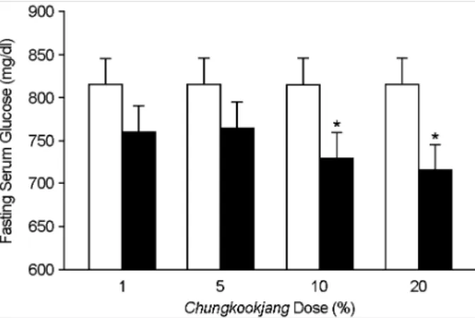
Fig. 1. Effect of different dose of Chungkookjang diet in week 4 on fasting serum glucose in DC and DCH groups. Values are the means ± SD of 10 rats. The value with an asterisk is significantly different from DC group by t-test (*; P<0.05). □: DC, ■: DCH.
2. Blood glucose levels
At 1 and 2 weeks after the experiment, blood glucose levels in the DCH group were significantly lower than the DC group by 29% (P<0.05) and 25% (P<0.05), respectively (Fig. 2).
3. Biochemical measurement
At the end of the 8-week experimented period, a signifi- cant elevation in final fasting serum glucose level was observed in the diabetic groups compared to the NC group (P<0.001) and it was significantly lower in the DCH group than the DC group by 10% (P<0.05). A significant elevation in serum total cholesterol, triglyceride and total lipid were
observed in the DC group compared to the NC group (P<
0.01). Total cholesterol level was lower in the DCH group than the DC group by 7%. Triglyceride and total lipid were Table 2. Water and food intakes, body weight gain and food
efficiency ratio of diabetic rats fed the experimental diets for 8 weeks
Groupsa) NC DC DCH
Water intake
(ml/day) 30.82±3.42 377.53±24.11### 372.07±10.31###
Food intake
(g/day) 19.94±1.50 45.18±2.05### 44.32±3.18###
Body weight
gain (g/day) 2.74±0.42 0.88±0.39### 1.01±0.25###
Food efficiency
ratiob) (%) 13.73±1.71 1.95±0.83### 2.25±0.43###
a) NC: Normal Control, DC: Diabetic Control, DCH: Diabetic Chungkookjang.
b) Food efficiency ratio= (body weight gain/ food intake) ×100.
Values are the means ± SD of 10 rats. The value with a sharp-note is significantly different from NC group by t-test (###; P<0.001).
Table 4. Organ weight in diabetic rats fed the experimental diets in week 8
Organs NC DC DCH 9.57±0.84a) 13.17±1.29### 12.60±1.40###
Liver
2.48±0.08b) 5.05±0.28### 5.00±0.34###
2.25±0.13 3.71±0.40### 3.97±0.57###
Kidney
0.59±0.03 1.42±0.13### 1.46±0.07###
1.25±0.07 1.06±0.11## 0.99±0.13###
Heart
0.32±0.02 0.41±0.04### 0.39±0.02###
a) Absolute organ weight, Unit: g. b) Relative organ weight, Unit:
g/100 g body weight.
Values are the means ± SD of 10 rats. The value with a sharp- note is significantly different from NC group by t-test (##; P<0.01,
###; P<0.001).
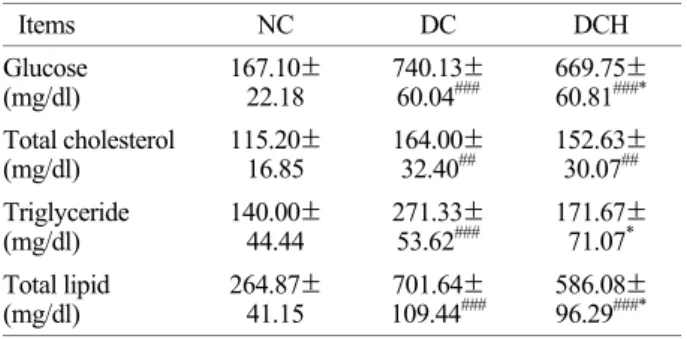
Table 3. Fasting serum glucose and lipid levels of diabetic rats fed the experimental diets in week 8
Items NC DC DCH
Glucose
(mg/dl) 167.10±
22.18 740.13±
60.04### 669.75±
60.81###*
Total cholesterol
(mg/dl) 115.20±
16.85 164.00±
32.40## 152.63±
30.07##
Triglyceride
(mg/dl) 140.00±
44.44 271.33±
53.62### 171.67±
71.07* Total lipid
(mg/dl) 264.87±
41.15 701.64±
109.44### 586.08±
96.29###*
Values are the means ± SD of 10 rats.The value with a sharp- note is significantly different from NC group by t-test (##; P<0.01,
###; P<0.001). The value with an asterisk is significantly different from DC group by t-test (*; P<0.05).
Table 5. Effect of Chungkookjang on kidney XO, SOD, CAT and GST activities in diabetic rats fed the experimental diets in week 8
Enzymes NC DC DCH
XOa) 0.50±0.12 0.71±0.12## 0.61±0.03#*
SODb) 7.63±1.69 8.40±3.84 5.34±1.00##*
CATc) 1.01±0.53 2.09±0.51## 1.70±0.65# GSTd) 20.90±7.58 35.53±4.43### 19.04±4.33***
a) Unit: nmole uric acid formed/mg protein/min.
b) Unit: U (50% inhibition of autoxidation of hematoxylin) mg protein/min.
c) Unit: H2O2 nmole reduced/mg protein/min.
d) Unit: nmole 2, 4-dinitrobenzene-glutathione conjugate/mg protein/
min.
Values are the means ± SD of 10 rats. The value with a sharp- note is significantly different from NC group by t-test (#; P<0.05,
##; P<0.01, ###; P<0.001). The value with an asterisk is significantly different from DC group by t-test (*; P<0.05, ***; P<0.001).
Fig. 2. Changes in blood glucose levels of diabetic rats fed the experimental diets for 8 weeks. Values are the means ± SD of 10 rats. The value with an asterisk is significantly different from DC group by t-test (*; P<0.05, **; P<0.01). ●, NC; ■, DC; ▲, DCH.
significantly lower in the DCH group than the DC group by 36.7% (P<0.05) and 16.5% (P<0.05), respectively (Table 3).
4. Organ weight
Relative weight of liver, kidney and heart were signifi- cantly higher (P<0.001) in the diabetic groups than the NC group. However, they did not differ significantly between the DC and DCH groups (Table 4).
5. Antioxidant enzyme activities
The activities of XO (P<0.01), SOD, CAT (P<0.01) and GST (P<0.001) in the DC group were higher than the NC group. However, they were lowered in the DCH group
compared to the DC group by 14% (P<0.05), 36% (P<0.05), 19% and 46% (P<0.001), respectively (Table 5).
6. Histological analysis
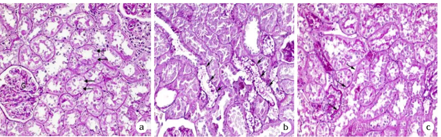
Tubular epithelial cells of the NC group have fine gran- ular cytoplasm and brush board. Some proximal tubules of the DC group are lined by clear epithelial cells (Armanni- Ebstein cells). In the DCH group, this cytoplasmic change was rarely seen and whole structures of kidney are similar to those of the NC group. In an assay via light microscopy, the DC group showed no glomerular enlargement or sclerotic change (Fig. 3A). Fig. 3B shows quantitative analysis of Armanni-Ebstein cells in renal cortex. Armanni-Ebstein cells in the DC group was significantly higher than the NC group (0.134±0.048 vs. 0.009±0.004; P<0.001) by quantitative scoring and it was significantly lower in the DCH group (0.068±0.037; P<0.01) than the DC group.
DISCUSSION
In this study, we investigated the effect of the Chungkookjang on blood glucose, lipid profiles, antioxidant enzyme activities and histological change in kidney of STZ- induced diabetic rats (50 mg/kg body weight i.p.). Although many previous studies (Fujita and Yamagami, 2001; Kim 2008) have shown that fermented soybean improved glucose and lipid metabolism, there were no clear effective evidence of histological changes in kidney of Chungkookjang diet for Fig. 3A. Histological analysis of pathologic renal change in normal and diabetic rats fed Chungkookjang in week 8. Representative photomicrographs of periodic acid Schiff (PAS)-stained renal sections from a non-diabetic control rat (a), an untreated diabetic rat (b) and Chungkookjang treated diabetic rat (c). Tubular epithelial cells have fine granular cytoplasm and brush board (a). Glomerulus (G) shows dispersed capillaries. Clear epithelial cells (Armani-Epstein cells) of the proximal tubules (arrows) are frequently seen in diabetic rat and reduced in Chungkookjang treated diabetic rat. ×200.
a b c
Fig. 3B. Quantitative analysis of Armanni-Epstein cells in renal cortex. Values are the means ± SD. The value with a sharp-note is significantly different from NC group by t-test (###; P<0.001). The value with an asterisk is significantly different from DC group by t-test (**; P<0.01).
diabetic rats up to date. The treatment with Chungkookjang diet (100 g/kg diet) was performed over the 8-week experi- mental period. We chose a 10% Chungkookjang diet based on the dose-response study (Fig. 1).
Diabetic rats showed increased food and water intakes and less body weight gain than the non-diabetic rats, which were attributed to a polyphagic condition and weight loss due to the excessive break down of tissue protein (Chatterjea and Shinde, 2002). The treatment with Chungkookjang diet improved food efficiency ratio and increased body weight gain compared to the diabetic control group, indicating control over polyphagia and muscle wasting caused by the hyperglycemic condition to some extent.
The treatment with Chungkookjang diet also significantly reduced serum glucose level compared to the untreated diabetic group. This may be beneficial in amelioration of the diabetic state and could explain the lowered blood glucose levels in rats fed Chungkookjang diet. The current observation of lipid level in the serum showed that the total cholesterol, triglyceride and total lipid levels in the diabetic control group were significantly higher than the normal group. Serum triglyceride and total lipid levels of the dia- betic group treated with Chungkookjang diet were signifi- cantly lower than the diabetic control group by 36.7% and 16.5%, respectively. Therefore, Chungkookjang diet seems to elicit a positive effect and may be useful in the prevention and treatment of diabetic hypertriglyceridemia and hyper- lipidemia.
Hyperglycemia also generates reactive oxygen species (ROS) that in turn cause lipid peroxidation and membrane damage (Hunt et al., 1988). Previous studies have reported that lipid peroxidation in the liver, kidney, and brain of diabetic rats was increased (Venkateswaran and Pari, 2002;
Latha and Pari, 2003). Antioxidant enzymes are capable of eliminating ROS and lipid peroxidation products, thereby protecting cells and tissues from oxidative damage. Diabetic nephropathy is one of the most important microvascular complications of diabetes mellitus. Recent studies have indicated that ROS plays a key, intermediate role in the development of diabetic nephropathy. High glucose level directly increases hydrogen peroxide production in mesangial cells and lipid peroxidation in glomerular mesangial cells
(Anjaneyulu and Chopra, 2004).
XO has been proposed to be a major source of ROS in diabetes mellitus (Butler et al., 2000). SOD is the first and most important line of antioxidant enzyme defense against oxidative stress, particularly oxygen radicals. SOD acceler- ates dismutation of superoxide radicals to hydrogen per- oxide that in turn is destroyed by CAT and GPx (Deisseroth and Dounce, 1970). Endogenous antioxidant enzymes (SOD, CAT and GST) are responsible for the detoxification of deleterious oxygen radicals (Del Maestro, 1980).
In our study, the activity of XO was increased in the kidneys of diabetic rats, which indicates increase in the generation of ROS. The activities of SOD, CAT and GST were increased in the kidneys of diabetic rats, which could be due to the compensatory reaction against increase in ROS. Data presented in our investigation indicate that the progression of diabetes results in augmentation of oxidative stress accompanied by impaired enzymatic antioxidative defense system in the kidneys of diabetic rats. Diabetic rats fed Chungkookjang showed reversal of these parameters to near normalcy. The treatment with Chungkookjang diet reduced oxidative stress as evidenced by the restoration of the enzymatic antioxidative defense system. These results indicate that treatment with Chungkookjang diet restored the SOD, CAT, GST, and XO activities and reduced oxida- tive stress in diabetic rats.
In this study, the most significant histological changes of the diabetic rats were clear cytoplasm of proximal tubular epithelium, so called Armanni-Ebstein cells (Ritchie and Waugh, 1957). This clearing effect is a result of removal of the accumulated cytoplasmic glycogen particles resulted by the diabetic condition, during the staining procedure (Kang et al., 2005). The accumulation of glycogen occurred mainly in the proximal tubular epithelium. Tubular epithelial changes and the appearance of Armanni-Ebstein cells were reduced in the diabetic group treated with Chungkookjang diet. The glomeruli had no cellular proliferation or sclerotic changes.
These data suggest that treatment with Chungkookjang diet may have protective effect to diabetic changes in kidney though the restoration of enzymatic antioxidative defense system.
In conclusion, these results indicated that Chungkookjang
can prevent or retard the development of diabetic complica- tions via its beneficial effects for correcting the hypergly- cemia, lipid levels, antioxidant enzyme system and protecting against histopathological changes in the kidneys of diabetic rats. Thus present study suggests that Chungkookjang is a potential natural product for the prevention of diabetic complications.
REFERENCES
Aebi H. Catalase in vitro. Methods Enzymol. 1984. 105: 121-126.
Allain CC, Poon LS, Chen CS, Richmond W, Fu PC. Enzymatic determination of total serum cholesterol. Clin Chem. 1974.
20: 470-475.
American Institute of Nutrition. Report of the American Institute of Nutrition ad hoc committee on standard for nutritional studies. J Nutr. 1977. 107: 1340-1348.
Anderson JW, Johnstone BM, Cook-Newell ME. Meta-analysis of effects of soy protein intake on serum lipids. N Engl J Med. 1995. 333: 276-282.
Anjaneyulu M, Chopra K. Nordihydroguairetic acid, a lignin, prevents oxidative stress and the development of diabetic nephropathy in rats. Pharmacology 2004. 72: 42-50.
Bhathena SJ, Velasquez MT. Beneficial role of dietary phyto- estrogens in obesity and diabetes. Am J Clin Nutr. 2002. 76:
1191-1201.
Brandstrup N, Kirk JE, Bruni C. Determination of hexokinase in tissues. J Gerontol. 1957. 12: 166-171.
Butler R, Morris AD, Belch JJF, Hill A, Struthers AD. Allopurinol normalizes endothelial dysfunction in type 2 diabetics with mild hypertension. Hypertension 2000. 35: 746-751.
Chatterjea MN, Shinde R. Text book of medical biochemistry, 5th ed. 2002. pp. 317. Jaypee Brothers, Medical Publishers (P) Ltd. New Delhi, India.
Deisseroth A, Dounce AL. Catalase physical and chemical pro- perties, mechanism of catalysis and physiological role. Physiol Rev. 1970. 50: 3-24.
Del Maestro RF. An approach to free radicals in medicine and biology. Acta Physiol Scand. 1980. 492: 153-168.
Edwads CRW, Baird JD, Toft AD. Davidson's principle and practice of medicine, 17th ed., Endocrine and metabolic diseases (Edwards CRW, Bouchier IAD, Haslett C, Chilvers ER. Eds).
1995. pp. 669-774. Churchill Livingstone, Edinburgh.
Fujita H, Yamagami T. Fermented soybean-derives Touchi-extract
with anti-diabetic effect via α-glucosidase inhibitory action in along-term administration study with KKAy mice. Life Sci.
2001. 70: 219-227.
Habig WH, Pabst MJ, Jakoby WB. Glutathione S-transferase. The first enzymatic step in mercapturic acid formation. J Biol Chem. 1974. 249: 7130-7139.
Hunt JV, Dean RT, Wolff SP. Hydroxyl radical production and autooxidative glycosylation. Glucose autooxidation as the cause of protein damage in the experimental glycation model of diabetes and aging. Biochem J. 1988. 256: 205-212.
Kang J, Dai XS, Yu TB, Wen B, Yang ZW. Glycogen accumulation in renal tubules, a key morphological change in the diabetic rat kidney. Acta Diabetol. 2005. 42: 110-116.
Kim HY, Yoon IS, Kim YC. Antidiabetic, antioxidative and renoprotective effects of Rehmanniae Radix preparata extract in streptozotocin-induced diabetic rats. J Exp Biomed Sci.
2008. 14: 19-26.
Kim NY, Song EJ, Kwon DY, Kim HP, Heo MY. Antioxidant and antigenotoxic activities of Korean fermented soybean. Food Chem Toxicol. 2008. 46: 1184-1189.
Lasaridis AN, Sarafidis PA. Diabetic nephropathy and antihy- pertensive treatment: what are the lessons from clinical trials?
Am J Hypertens. 2003. 16: 689-697.
Latha M, Pari L. Preventive effects of Cassia auriculata L. flowers on brain lipid peroxidation in rats treated with streptozotocin.
Mol Cell Biochem. 2003. 243: 23-28.
Lee JS. Effects of soy protein and genistein on blood glucose, antio- xidant enzyme activities, and lipid profile in streptozotocin- induced diabetic rats. Life Sci. 2006. 79: 1578-1584.
Lowry OH, Rosebrough NJ, Farr AL, Randall RJ. Protein measure- ment with folin phenol reagent. J Biol Chem. 1951. 193: 265 -275.
Maritim AC, Sanders RA, Watkins III JB. Effect of alpha lipoic acid on biomarkers of oxidative stress in streptozotocin-induced diabetic rats. J Nutr Biochem. 2003. 14: 288-294.
Martin JP, Dailey M, Sugarman E. Negative and positive assays of superoxide dismutase based on hematoxylin autoxidation.
Arch Biochem Biophys. 1987. 255: 329-336.
McGrowan MW, Artiss JD, Strandbergh DR, Zak B. A peroxidase- coupled method for the colorimetric determination of serum triglycerides. Clin Chem. 1983. 29: 538-542.
Ritchie S, Waugh D. The pathology of Armanni-Ebstein diabetic nephropathy. Am J Pathol. 1957. 33: 1035-1043.
Stirpe F, Della CE. The regulation of rat liver xanthine oxidase.
Conversion in vitro of the enzyme activity from dehydro-
genase (type D) to oxidase (type O). J Biol Chem. 1969. 244:
3855-3863.
Tomohiro T, Kumai T, Sato T, Takeba Y, Kobayashi S, Kimura K.
Hypertension aggravates glomerular dysfunction with oxida- tive stress in a rat model of diabetic nephropathy. Life Sci.
2007. 80: 1364-1372.
Velasquez MT, Bhathena SJ. Dietary phytoestrogens: A possible role in renal disease protection. Am J Kidney Dis. 2001. 37:
1056-1068.
Venkateswaran S, Pari L. Antioxidant effect of Phaseolus vulgaris in streptozotocin-induced diabetic rats. Asia Pac J Clin Nutr.
2002. 11: 206-209.
Watanabe Y, Hotta N. Tubulointerstitial injury in diabetes mellitus (including Armanni-Ebstein lesion). Ryoikibetsu Shokogun Shirizu. 1997. 17Pt2: 225-228.
Zimmet P, Alberti KG, Shaw J. Global and societal implications of the diabetes epidemic. Nature 2001. 414: 782-787.