J. Exp. Biomed. Sci. 2010, 16(2): 83~90
Anti-inflammatory Effects and its Mechanisms of Hesperidin in an Asthmatic Mouse Model Induced by Ovalbumin
Jeong Hyun Chang†
Department of Clinical Laboratory Science, College of Health & Therapy, Daegu Haany University, Gyeong San 712-715, Korea
Hesperidin, a member of the flavanone group of flavonoids, can be isolated in large amounts from the rinds of some citrus species [e.g., Citrus aurantium L. (bitter orange), Citrus sinensis L. (sweet orange) and Citrus unshiu Marcov.
(satsuma mandarin)], and has been reported to have anticarcinogenic, antihypotensive and antimicrobial properties.
Despite the efficacy of these polyphenolic compounds as immune modulators, the effects of the flavonoids are poorly understood about allergic effect. In this study, we investigated whether hesperidin could influence on Th1 and Th2 balance. Allergic reactions included an increase in the number of eosinophils in bronchoalveolar lavage (BAL) fluid, an increase in inflammatory cell infiltration into the lung tissue around blood vessels and airways, airway luminal narrowing, the development of airway hyper-responsiveness (AHR). The administration of hesperidin before the last airway OVA challenge resulted in a significant inhibition of all asthmatic reactions. Accordingly, this study may provide evidence that hesperidin plays a critical role in the amelioration of the pathogenetic process of asthma in mice. These findings provide new insight into the immunopharmacological role of hesperidin in terms of its effects in a murine model of asthma, and also broaden current perspectives in our understanding of the immunopharmacological functions of hesperidin.
Key Words: Ovalbumin, Hesperidin, AHR, Th1/Th2 balance, IL-4, IL-5, IL-13
INTRODUCTION
Asthma is an inflammatory disease that is characterized by bronchial hyper-responsiveness that can proceed to life-threatening airway obstruction. OVA-induced asthma is characterized by AHR (Airway Hyper Responsiveness) and airway inflammation (Gleich et al., 1997). This inflammation is closely associated with the accumulation of eosinophils, neutrophils, and lymphocytes into the bronchial lumen and lung tissues (Kwak et al., 2003; Hamelmann et al., 1997). These cellular infiltrates release various chemical mediators that can cause AHR (Lee et al., 2002; Tournoy et al., 2000). Recruitment of these inflammatory cells from
the blood to the site of inflammation is regarded as a central event in the development and prolongation of airway inflammation.
The T helper 2 (Th2)-type cytokines interleukins-4 (IL-4), IL-5, and IL-13, expressed by activated CD4+ T cells play a critical role in the pathogenesis of asthma by controlling the key process of immunoglobulin E (IgE) production, growth of mast cells and the differentiation and activation of mast cells and eosinophils (Boschetto et al., 1988; Busse et al., 1993; Bousquet et al., 1990; Kay, 1991; Mappe et al., 1987). In contrast, Th1 cytokines such as interferon-γ (IFN-γ) and IL-12, which down-regulate Th2 responses, inhibit the development of allergic lung inflammation (Iwamoto et al., 1993; Tanaka et al., 2004). Thus, interventions that inhibit Th2 cytokines by enhancing Th1 cytokine production, may be useful in the treatment of allergic asthma (Sur et al., 1996).
The flavonoids comprise a family of common phenolic plant pigments that have been identified as dietary anticarcinogens and antioxidants (Chen et al., 2004).
*Received: May 25, 2010 / Revised: June 8, 2010 Accepted: June 8, 2010
†Corresponding author: Jeong Hyun Chang, Department of Clinical Laboratory Science, College of Health & Therapy, Daegu Haany University, Gyeong San 712-715, Korea.
Tel: 82-53-819-1352, Fax: 82-53-819-1352 e-mail: [email protected]
Hesperidin can be isolated in large amounts from the rinds of some citrus species [e.g., Citrus aurantium L. (bitter orange), Citrus sinensis L. (sweet orange), and Citrus unshiu Marcov. (satsuma mandarin)] and has been reported to have antimicrobial, anticarcinogenic and anti-inflammatory action (Garg et al., 2001). Studies with rats treated with hesperidin have showed a 22% decrease in colon cancer and a 29% reduction in lung cancer, respectively. These effects have been attributed to the fact that juices contain high levels of the flavonoid hesperidin. Satsuma mandarin (C. unshiu) juice enriched with hesperidin has also been shown to significantly reduce the incidence of colon cancer in rats (Tanaka et al., 2004). In a hamster model of atherosclerosis, hesperidin was able to significantly inhibit atherosclerosis and lower cholesterol and triglycerides (Vinson et al., 2002).
In a recent study, treatment with hesperidin inhibited the production of nitrogen dioxide (NO2), and expression of iNOS protein. In the case of COX-2, hesperidin did not affect the protein level in mouse macrophage cells (Sakata et al., 2003). Thus, In vitro, hesperidin inhibited production of IL-8, TNFα, IL-1β, IL-6, IL-12, ICAM-1 and VCAM-1 (Yeh et al., 2007). In this study, we have attempted to characterize the effects of a noncytotoxic concentration of hesperidin in a murine model of asthma. Our findings demonstrated, for the first time, that hesperidin treatment suppressed asthmatic syndrome.
MATERIALS AND METHODS Animals and experimental protocol
Female BALB/c mice, 6~8 weeks of age and free of murine-specific pathogens, were obtained from the Charles River Laboratories (Yokohama, Japan). All experimental animals used in this study were maintained under a protocol approved by the Institutional Animal Care and Use Committee of the Pusan National University Medical School. Mice were immunized intraperitoneally (i.p.) with 20 μg of OVA (Sigma- Aldrich, St. Louis, MO) emulsified in 1 mg of aluminum hydroxide (Pierce Chemical Co., Rockford, IL) on days 1 and 15. Mice were challenged for 30 min via the airway with OVA (5% OVA) each day from
days 21~23 on consecutive days. BAL fluid was obtained at 24 h after the last challenge. At the time of lavage, the mice (10 mice in each group) were killed with an overdose of ether. The chest cavity was exposed to allow for expansion, after which the trachea was carefully incubated and the catheter secured with ligatures. Prewarmed saline solution was slowly infused into the lungs and withdrawn.
The aliquots were pooled and then kept at 4℃. A part of each pool was then centrifuged, and the supernatants were kept at -70℃ until use.
Administration of hesperidin
Mice were injected i.p with 50 or 250 mg/kg/day in 200 μl of hesperidin (Sigma, St. Louis, Mo) each day from days 18 to 20 on consecutive days.
Total cell counting
The total cell numbers were counted with a hemocytometer. Smears of BAL cells prepared with Cytospin II (Shandon, Runcorn, UK) were stained with Diff-Quik solution (Dade Diagnostics of P.R. Inc, Aguada, PR) for differential cell counting. Two independent, blinded investigators counted the cells, using a microscope.
Approximately 200 cells were counted in each of four different random locations.
Measurement of eosinophil peroxide
The suspension of BAL cells and the pulmonary homogenates were frozen/thawed three times using liquid nitrogen and a water bath at 37℃ to obtain the EPO (eosinophil peroxide). The BAL fluid was centrifuged to 4℃ for 10 min and serially diluted in a 96-well plate (75 μl/well) followed by the addition of 150 μl of substrate (1.5 mM o-phenylenediamine and 6.6 mM H2O2 in 0.05 M Tris-HCl, pH 8.0). After 30 min at room temperature, the reaction was stopped by the addition of 75 μl of 30%
H2SO4, and the absorbance of the samples was determined at 492 nm on an ELISA reader.
Immunohistochemistry
At 48 h after the last challenge, lungs were removed from the mice after they had been sacrified. Prior to the
removal of the lungs, the lungs and trachea were filled intratracheally with a fixative (4% paraformaldehyde) using a ligature around the trachea. Lung tissues were fixed with 10% (v/v) paraformaldehyde. The specimens were dehydrated and embedded in paraffin. For histological examination, 4 lm sections of fixed embedded tissues were cut on a Leica model 2165 rotary microtome (Leica, Nussloch, Germany), placed on glass slides, deparaffinized, and sequentially stained with hematoxylin 2 and eosin-Y (Richard-Allan Scientific, Kalamazoo, MI). An inflammation score was graded by three independent investigators who were not associated with this study.
Determination of airway responsiveness to methacholine Airway responsiveness was measured in mice 24 h after the last challenge in an unrestrained conscious state, as described previously (Lee et al., 2008). Mice were placed in a barometric plethysmographic chamber (All Medicus Co., Seoul, Korea) and baseline readings were taken and averaged for 3 min. Aerosolized methacholine in increasing concentrations (2.5 to 50 mg/ml) was nebulized through an inlet of the main chamber for 3 min. Readings were taken and averaged for 3 min after each nebulization. Enhanced pause (Penh), calculated as (expiratory time/relaxation time-1) × (peak expiratory flow/peak inspiratory flow), according to protocol of the manufacturers, is a dimensionless value that represents a function of the proportion of maximal expiratory to maximal inspiratory box pressure signals and a function of the timing of expiration. Penh was used as a measure of airway respons- iveness to methacholine. Results were expressed as the percent increase of Penh following challenge with each concentration of methacholine, where the baseline Penh (after saline challenge) was expressed as 100%. Penh values were averaged for 3 min after each nebulization and evaluated.
Cytokine assay
The levels of cytokines in bronchoalveolar lavage (BAL) fluid were determined by enzyme-linked immunosorbent assay (ELISA). ELISA kits from R&D Systems (Minneapolis, MN) were employed for the measurement of
IL-4, IL-5, and IL-13.
Measurement of OVA-specific serum levels of IgE OVA-specific serum IgE levels were determined by enzyme-linked immunosorbent assay (ELISA) using samples collected 12 h after the last OVA challenge, as described previously (Choi et al., 2009). In brief, a 96 well microtitre plate was coated with OVA (10 mg/ml), and then treated with mouse sera followed by biotin-conjugated rat anti- mouse IgE (pharmingen, San Diego, CA). Then, avidin- horseradish peroxidase (HRP) solution was added to each well. Units are optical density readings at 405 nm.
Densitometric analysis and statistics
Experiments were repeated at least three times with consistent results. Unless otherwise stated, data are expressed as the mean ± S.E.M. ANOVA was used to compare experimental groups to control values while comparisons between multiple groups were performed using Tukey's Multiple Comparison test. Statistical significance was indicated by a P value less than P*** < 0.001, P** < 0.05, P*
< 0.01.
RESULTS
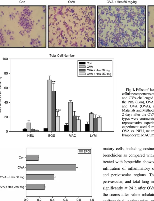
Effect of heperidin on cellular changes in BAL fluids Numbers of total cells, eosinophils, lymphocytes, and macrophages in BAL fluids were increased significantly at 24 h after OVA inhalation compared with the numbers after saline inhalation (Fig. 1). The increased numbers of these cells were significantly reduced by the administration of hesperidin.
Effect of hesperidin on the level of eosinophils in BAL fluids
The levels of eosinophils in BAL fluids were significantly increased at 24 h after OVA inhalation compared with the levels after saline inhalation (Fig. 2). The increased levels of these cells were significantly reduced by about 38.03%
(Hes 50 mg/kg/day) and 51.62% (Hes 250 mg/kg/day) upon administration of ovalbumin.
Effect of hesperidin on pathological changes of OVA- induced asthma
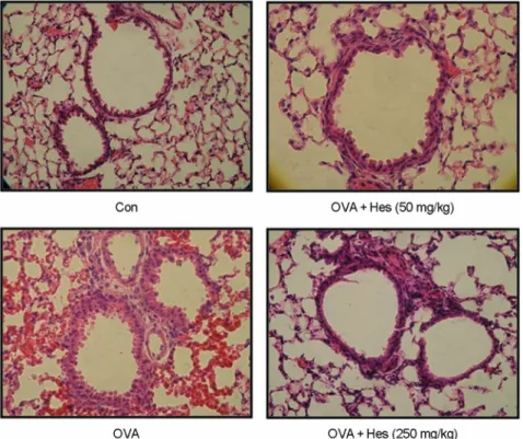
Histological analyses revealed typical pathologic features of asthma in the OVA-exposed mice. Numerous inflam-
matory cells, including eosinophils infiltrated around the bronchioles as compared with the control (Fig. 3). Mice treated with hesperidin showed marked reductions in the infiltration of inflammatory cells in the peribronchiolar and perivascular regions. The scores of peribronchial, perivascular, and total lung inflammation were increased significantly at 24 h after OVA inhalation compared with the scores after saline inhalation (Fig. 3). The increased peribronchial, perivascular, and total lung inflammation were significantly reduced by the administration of hesperidin. These results suggest that hesperidin inhibits antigen-induced inflammation in the lungs, including the influx of eosinophils.
Effect of hesperidin on airway hyper-responsiveness Airway responsiveness was assessed as a percent increase of Penh in response to increasing doses of methacholine. In OVA-sensitized and -challenged mice, the dose-response curve of percent Penh shifted to the left compared with that of control mice (Fig. 4). In addition, the percent Penh Fig. 2. Eosinophil peroxidase (EPO) activity in BAL fluids of
OVA-sensitized and -challenged mice. EPO is an indicator of the numbers of eosinophil levels. The increased levels of EPO were significantly reduced about 38 % (OVA + Hes 50 mg/kg/day), 51% (OVA + Hes 250 mg/kg/day) compared with OVA-treated mice. The results were from one representative experiment out of 5 performed. This experiment used 5 mice (n=5). ***P<0.001 vs.
OVA.
Fig. 1. Effect of hesperidin on total and differential cellular components of BAL fluids of OVA-sensitized and OVA-challenged mice. Mice were treated with the PBS (Con), OVA plus hesperidin (OVA + Hes), and OVA (OVA), respectively, as described in Materials and Methods. The BAL cells were collected 2 days after the OVA challenge. The different cell types were enumerated. The results were from one representative experiment out of 5 performed. This experiment used 5 mice. P*** < 0.001, P** < 0.05 OVA vs. NEU, neutrophil; EOS, eosinophil; LYM, lymphocyte; MAC, macrophages; TOT, total cell.
produced by methacholine administration (at doses from 2.5 mg/ml to 50 mg/ml) increased significantly in the
OVA-sensitized and -challenged mice compared with the controls. OVA-sensitized and -challenged mice treated with hesperidin showed a dose-response curve of percent Penh that shifted to the right compared with that of untreated mice. The shift was dose-dependent. These results indicate that hesperidin treatment reduces OVA-induced airway hyperresponsiveness.
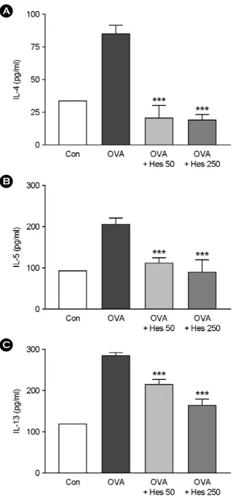
Effect of hesperidin on levels of IL-4, IL-5 and IL-13 in lung tissues of OVA-sensitized and -challenged mice
The regulation of cytokine production is largely mediated by NF-κB activation, and the inhibition of this expression and activation could lead to a reduction in the inflammatory cytokine production. To determine cytokine level in the BAL fluid, IL-4, IL-5 and IL-13 were determined by ELISA. BAL fluids were obtained 4 h after the last airway challenge. The levels of IL-4, IL-5 and IL-13 in the BAL fluids were significantly increased by airway challenge with OVA when compared with that with saline as control (Fig. 5).
Effect of hesperidin on IgE levels in serum
Because Th2 cytokines promote airway inflammation in Fig. 3. Hesperidin inhibits lung inflammation and inflammatory cell infiltration. Mice were sensitized and challenged as described in Materials and Methods. Sections were obtained from the lungs of mice receiving the control (CON), OVA plus hesperidin 50 mg/kg/day (OVA + Hes 50 mg), OVA plus hesperidin 250 mg/kg/day (OVA + Hes 250 mg) and OVA (OVA).
Lungs were removed 2 days after the last airway challenge. Sections were stained by hematoxylin and eosin staining (× 200).
Fig. 4. Effect of hesperidin on airway responsiveness in OVA- sensitized and OVA-challenged mice. Airway responsiveness was measured at 72 h after the last challenge in saline-inhaled mice administered PBS (Con), OVA-sensitized mice administered saline (OVA) and OVA-sensitized mice administered hesperidin (OVA + Hes). Airway responsiveness to aerosolized methacholine was measured in unrestrained, conscious mice. Mice were placed into the main chamber and were nebulized first with PBS, then with increasing doses (2.5 to 50 mg/ml) of methacholine for 3 min for each nebulization. Readings of breathing parameters were taken for 3 min after each nebulization during which Penh values were determined. Data represent mean 7 independent experiments.
asthma through increased IgE levels, we investigated the expression of IgE in serum. IgE favors to Th2 in airway inflammation, we measured how far hesperidin modulated the levels of serum IgE in OVA mice. The levels of serum IgE were found to be significantly increased in OVA mice
compared with those of PBS mice (Con). However, administration of hesperidin significantly decreased the levels of serum IgE. These data indicate that hesperidin modulates Th1/Th2 levels in an OVA-induced asthma model (Fig. 6).
DISCUSSION
This study, for the first time, provides experimental B
A
C
Fig. 5. The effect of IL-4, IL-5 and IL-13 on BAL fluid cytokines. Bronchoalveolar lavage (BAL) was performed 4 h after the last airway challenge as described in the manufacturer. IL-4, IL-5 and IL-13 cytokine levels in the BAL fluids were measured by ELISA Kit. The mice were bled 4 h after the last airway OVA challenge. Data represent mean ± SEM from 4 independent experiments. P*** < 0.001 vs. OVA
Fig. 6. The effect of hesperdin on IgE levels in serum of OVA-sensitized and OVA-challenged. Blood was collected by cardiac puncture to measure serum IgE. All experiments were analyzed using ELISA (n=4).
evidence demonstrating that hesperidin inhibits OVA- induced airway inflammation in a murine model of asthma.
Hesperidin profoundly inhibited asthmatic reactions such as leukocytic recruitment into the airway and lung inflammation.
Based on animal studies, the immunological processes involved in airway inflammation of asthma are characterized by the proliferation and activation of T cells of the subtype Th2 CD4+ (Lu et al., 2010). Ultimately, mediators lead to degranulation of effector /proinflammatory cells with the release of mediators and oxidants, which lead to the injury and inflammation noted in asthma. ROS such as superoxide, hydrogen peroxide, and possibly hydroxyl radicals contribute to inflammatory changes in the asthmatic airway (Park et al., 2009). In support of this concept are the high levels of ROS and oxidatively modified proteins in airways of asthmatics (Heidenfelder et al., 2009). High levels of ROS are produced in the lungs of asthmatic patients by activated inflammatory cells, i.e., eosinophils, alveolar macrophages, and neutrophils.
OVA-induced asthma has been recognized as a disease resulting from chronic airway inflammation character- istically associated with the infiltration of lymphocytes, eosinophils, and neutrophils into the bronchial lumen (Corrigan et al., 1992; Punnonen et al., 1994). There is increasing evidence that cytokine-inducible leukocyte- endothelial adhesion molecules are important in the recruitment and migration of leukocytes to the sites of inflammation (Muller et al., 2003). In this study, hesperidin reduced levels of Inflammatory cells migration in lung tissues of OVA-sensitized and -challenged mice. These effects of hesperidin may explain why the administration of hesperidin significantly reduced the increase in neutrophils, in eosinophils, in lymphocytes, in monocytes, and in the total cells, elicited in the airway lumen 1 days after OVA inhalation (Fig. 1). It has long been postulated that the expression of MMP-9 is regulated by cytokines, especially, TNF-α (Bahar-Shany et al., 2010; Perez-Gracia et al., 2009). Out data demonstrate that hesperidin reduces the increased numbers of inflammatory cells - the source of IL-5 in the airways and then decreases the increased levels of Th2 cytokine in BAL fluids of OVA-sensitized and
OVA-challenged mice (Fig. 5).
Taken together, Our results strongly indicate that hesperidin reduces the pathologic lung damage due to the regulation of Th1/Th2 balances through the inhibition of inflammatory cells migration by suppressing IL-5 pro- duction. This study also supports evidence that hesperidin might offer a new therapeutic approach to allergic airway diseases.
Acknowledgements
This research was supported by a grant from Daegu Haany University Ky·lin Foundation in 2009.
REFERENCES
Bahar-Shany K, Ravid A, Koren R. Upregulation of MMP-9 production by TNFalpha in keratinocytes and its attenuation by vitamin D. J Cell Physiol. 2010. 222: 729-737.
Boschetto P, Fabbri LM, Zocca E, Milani G, Pivirotto F, Dal Vecchio A, Plebani M, Mapp CE. Prednisone inhibits late asthmatic reactions and airway inflammation induced by toluene diisocyanate in sensitized subjects. J Allergy Clin Immunol. 1988. 81: 454.
Bousquet J, Chanez P, Lacoste JY, Barneon G, Ghavanian N, Enander I, Venge P, Ahlstedt S, Simony-Lafontaine J, Godard P. Eosinophilic inflammation in asthma. N Engl J Med. 1990.
323: 1033-1039.
Busse WW, William FC, Sedgwick JD. Mechanism of airway inflammation in asthma. Am Rev Respir Dis. 1993. 147: 20 -24.
Chen CC, Chow MP, Huang WC, Lin YC, Chang YJ. Flavonoids inhibit tumor necrosis factor-alpha-induced up-regulation of intercellular adhesion molecule-1 (ICAM-1) in respiratory epithelial cells through activator protein-1 and nuclear factorkappaB: tructure-activity relationships. Mol Pharmacol.
2004. 66: 683-693.
Choi JR, Lee CM, Jung ID, Lee JS, Jeong YI, Chang JH, Park HJ, Choi IW, Kim JS, Shin YK, Park SN, Park YM. Apigenin protects ovalbumin-induced asthma through the regulation of GATA-3 gene. Int Immunopharmacol. 2009. 9: 918-924.
Corrigan CJ, Kay AB. T cells and eosinophils in the pathogenesis of asthma. Immunol Today 1992. 13: 501-507.
Garg A, Garg S, Zaneveld LJ, Singla AK. Chemistry and
pharmacology of the citrus bioflavonoid hesperidin. Phytother Res. 2001. 15: 655-669.
Gleich GJ, Kita H. Bronchial asthma: lessons from murine models.
Proc Natl Acad Sci U S A. 1997. 94: 2101-2102.
Hamelmann E, Schwarze J, Takeda K, Oshiba A, Larsen GL, Irvin CG, Gelfand EW. Noninvasive measurement of airway responsiveness in allergic mice using barometric plethysmo- graphy. Am J Respir Crit Care Med. 1997. 156: 766-775.
Heidenfelder B, Johnson M, Hudgens E, Inmon J, Hamilton RG, Neas L, Gallagher JE. Increased plasma reactive oxidant levels and their relationship to blood cells, total IgE, and allergen-specific IgE levels in asthmatic children. J Asthma 2009. 46: 687-691.
Iwamoto I, Nakajima H, Endo H, Yoshida S. Interferon gamma regulates antigen induced eosinophil recruitment into the mouse airways by inhibiting the infiltration of CD4+ T cells.
J Exp Med. 1993. 177: 573-576.
Kay AB. Asthma and inflammation. J Allergy Clin Immunol.
1991. 87: 893-910.
Kwak YG, Song CH, Yi HK, Hwang PH, Kim JS, Lee KS, Lee YC. Involvement of PTEN in airway hyper-responsiveness and inflammation in bronchial asthma. J Clin Invest. 2003.
111: 1083-1092.
Lee CM, Chang JH, Moon DO, Choi YH, Choi IW, Park YM, Kim GY. Lycopene suppresses ovalbumin-induced airway inflammation in a murine model of asthma. Biochem Biophys Res Commun. 2008. 374: 248-252.
Lee YC, Kwak YG, Song CH. Contribution of vascular endothelial growth factor to airway hyper-responsiveness and inflammation in a murine model of toluene diisocyanate- induced asthma. J Immunol. 2002. 168: 3595-3600.
Lu Y, Sjöstrand M, Malmhäll C, Rådinger M, Jeurink P, Lötvall J, Bossios A. New production of eosinophils and the corresponding TH1/TH2 balance in the lungs after allergen exposure in BALB/c and C57BL/6 mice. Scand J Immunol.
2010. 71: 176-185.
Mappe CE, Boschetto P, Zocca E, Milani GF, Pivirotto F, Teggazin V, Fabbri LM. Pathogenesis of late asthmatic reactions induced by exposure to isocyanates. Bull Eur Physiopathol Respir. 1987. 23: 583-586.
Muller WA. Leukocyte-endothelial-cell interactions in leukocyte transmigration and the inflammatory response. Trends
Immunol. 2003. 24: 327-334.
Park CS, Kim TB, Lee KY, Moon KA, Bae YJ, Jang MK, Cho YS, Moon HB. Increased oxidative stress in the airway and development of allergic inflammation in a mouse model of asthma. Ann Allergy Asthma Immunol. 2009. 103: 238-247.
Perez-Gracia JL, Prior C, Guillén-Grima F, Segura V, Gonzalez A, Panizo A, Melero I, Grande-Pulido E, Gurpide A, Gil-Bazo I, Calvo A. Identification of TNF-alpha and MMP-9 as potential baseline predictive serum markers of sunitinib activity in patients with renal cell carcinoma using a human cytokine array. Br J Cancer 2009. 101: 1876-1883.
Punnonen J, Aversa G, Cocks BG, de Vries JE. Role of interleukin- 4 and interleukin-13 in synthesis of IgE and expression of CD23 by human B cells. Allergy 1994. 49: 576-586.
Sakata K, Hirose Y, Qiao Z, Tanaka T, Mori H. Inhibition of inducible isoforms of cyclooxygenase and nitric oxide synthase by flavonoid hesperidin in mouse macrophage cell line. Cancer Lett. 2003. 199: 139-145.
Sur S, Lam J, Bouchard P, Sigounas A, Holbert D, Metzger WJ.
Immunomodulatory effects of IL-12 on allergic lung inflammation depend on timing of doses. J Immunol. 1996.
157: 4173-4180.
Tanaka H, Komai M, Nagao K, Ishizaki M, Kajiwara D, Takatsu K. Role of interleukin-5 and eosinophils in allergen-induced airway remodeling in mice. Am J Respir Cell Mol Biol. 2004.
31: 62-68.
Tanaka T, Kohno H, Murakami M, Shimada R, Kagami S, Sumida T, Azuma Y, Ogawa H. Suppression of azoxymethane- induced colon carcinogenesis in male F344 rats by mandarin juices rich in â-cryptoxanthin and hesperidin. Int J Cancer 2000. 8: 146-150.
Tournoy KG, Kips JC, Schou C, Pauwels RA. Airway eosinophilia is not a requirement for allergen-induced airway hyper- responsiveness. Clin Exp Allergy 2000. 30: 7985.
Vinson JA, Liang X, Proch J, Honzt BA, Dancel J, Sandone N.
Polyphenol antioxidants in citrus juices: In Vitro and in Vivo studies relevant to heart disease. Adv Exp Med Biol. 2002.
505: 113-122.
Yeh CC, Kao SJ, Lin CC, Wang SD, Liu CJ, Kao ST. The immunomodulation of endotoxin-induced acute lung injury by hesperidin in vivo and in vitro. Life Sci. 2007. 80: 1821 -1831.