INTRODUCTION
Mylabris, the dried body of the Chinese blister beetle (Myla- bris phalerata Pallas), has been used as Chinese medicine for over 2000 yr. Its active constituent, cantharidin (CA), has anti-tumor properties and causes leukocytosis. However, it has irritant effects on the urinary organs. Norcantharidin (NCTD), the demethylated form of cantharidin (Fig. 1), is easier to be synthesized and is relatively free from side effects.
NCTD inhibits the proliferation of some cancer cells (such as HL60, K562, Bel-7402, MCF-7, Colo205, HT-29, SW480) by interrupting DNA synthesis or upregulating of the CD95 receptor and CD95 ligand on the cell surface and has anti- tumor activity against transplanted hepatoma in mouse model.
These findings suggest that NCTD is a potential antitumor agent (1-3). However, the exact mechanism responsible for the apoptotic effect is not thoroughly elucidated.
Apoptosis, or programmed cell death, is a genetically reg- ulated, self-destructive cellular death process that is impor- tant in development, tissue remodeling, immune regulation, and many diseases (4-7). Cysteine-dependent aspartate-spe- cific proteases (caspases) have been demonstrated to be cru-
cial mediators in apoptotic pathway. Caspases can be divid- ed into two groups: initiator caspases (such as caspase-8 and caspase-9) whose main function is to activate downstream caspases, and executor caspases (such as caspase-3), which mediate apoptosis by proteolysis of specific substrates includ- ing inhibitor of caspase-activated DNase (ICAD) and anti- apoptotic protein, Bcl-2 (8-12). Many Bcl-2 family proteins reside the mitochondrial outer membrane. The balance bet- ween Bax and Bcl-2 (or Bcl-xL) determines the fate of cells in many apoptotic systems. Bcl-2 and Bcl-xLcan be cleaved by caspase-3 and cleavage of these proteins appears to inacti- vate their survival function. In response to the death stimuli, the mitochrondrial membranes are permeabilized, resulting in the release of cytochrome c. In the cytosol, cytochrome c activates apoptosis by binding and activating apoptotic pro- tease activating factor-1 (Apaf-1)-caspase-9 complex, which form an apoptosome acting as a processing/activation center for the downstream caspase-3 (13-17).
In the present study, we demonstrate that caspases activa- tion participated in NCTD-induced apoptosis, and up-reg- ulaton of Bax and down-regulation of Bcl-2 (or Bcl-xL) con- tributed to the NCTD-induced A375-S2 cell apoptosis.
Wei-wei An*�, Min-wei Wang�, Shin-ichi Tashiro�, Satoshi Onodera�, Takashi Ikejima*
*China-Japan Research Institute of Medical and Pharmaceutical Sciences, Shenyang Pharmaceutical University, Shenyang; �Department of Pharmacology, Shenyang Pharmaceutical University, Shenyang, China; �Department of Clinical and Biomedical Sciences, Showa Pharmaceutical University, Tokyo, Japan
Address for correspondence Ikejima Takashi, Ph.D.
China-Japan Research Institute of Medical and Pharmaceutical Sciences, Shenyang Pharmaceutical University, Shenyang 110016, China
Tel, Fax : +86.24-2384-4463 E-mail : [email protected]
560
Norcantharidin Induces Human Melanoma A375-S2 Cell Apoptosis through Mitochondrial and Caspase Pathways
Norcantharidin (NCTD) is the demethylated form of cantharidin, which is the active substance of mylabris. To examine the pathway of NCTD-induced A375-S2 cell death, 3-(4, 5-dimethylthiazol-2-yl)-2, 5-dipheyltetrazolium bromide (MTT) assay, photomicroscopical observation, DNA agarose gel electrophoresis, caspase activi- ty assay and Western blot analysis were carried out. A375-S2 cells treated with NCTD exhibited several typical characteristics of apoptosis. The inhibitory effect of NCTD on human melanoma, A375-S2 cells, was partially reversed by the inhibitors of pan-caspase, caspase-3 and caspase-9. The activities of caspase-3 and -9 were significantly increased after treatment with NCTD at different time. The expression of inhibitor of caspase-activated DNase was decreased in a time-dependent man- ner, simultaneously, the ratio of Bcl-2/Bax or Bcl-xL/Bax was decreased and the expression ratio of proteins could be reversed by caspase-3 inhibitor. The expres- sion of cytochrome c in cytosol was increased after NCTD treatment and caspase- 3 inhibitor had no significant effect on the up-regulation of cytochrom c. These results suggest that NCTD induced A375-S2 cell apoptosis and the activation of caspase and mitochondrial pathway were involved in the process of NCTD-induced A375-S2 cell apoptosis.
Key Words : Cantharidin; Norcantharidin; Cell Line, Tumor; A375-S2 Cells; Apoptosis; Caspase; Mitochon- dria; Proto-Oncogene Proteins c-bcl-2; Cytochromes c
Received : 16 January 2004 Accepted : 20 April 2004
MATERIALS AND METHODS Chemical reagents
NCTD of analytical grade purity was from the Ju-nan Phar- maceutical Works (Junan, China) and dissolved in RPMI- 1640 (HyClone, U.S.A.). Caspase-8 inhibitor (z-IETD-fmk) was from Enzyme Systems (CA, U.S.A.). Caspase-3 inhibitor (z-DEVD-fmk) and pan-caspase inhibitor (z-VAD-fmk) were from Calbiochem (CA, U.S.A.). Caspase-9 inhibitor (Ac- LEHD-CHO), rabbit polyclonal antibodies against ICAD, cytochrome c, Bax and Bcl-xL, mouse polyclonal antibodies against Bcl-2, horseradish peroxidase-conjugated secondary antibody (goat-anti-rabbit or goat-anti-mouse) were from Santa Cruz Biotechnology (Santa Cruz, CA). Caspase-3, -8 and -9 Apoptosis Detection Kits were from Santa Cruz Biotech- nology (Santa Cruz, CA).
Cell culture
A375-S2, melanoma cells, were obtained from American Type Culture Collection (ATCC, #CRL, 1872, MD, U.S.A.) and were cultured in RPMI-1640 medium (HyClone, U.S.A.) supplemented with 10% heat inactivated (56℃, 30 min) fetal calf serum (Beijing Yuanheng Shengma Research Insti- tution of Biotechnology, Beijing, China), 2 M L-glutamin (GIBCO, U.S.A.), 100 kU/L penicillin and 100 g/L strepto- mycin (GIBCO, U.S.A.) at 37℃in 5% CO2.
Cell growth inhibition test
A375-S2 cells (1.0×10⁸cells/L) seeded in 96-well plate (NUNKTM, Roskilde, Denmark) were cultured for 24 hr, then various concentrations of NCTD (60-480 M) were added and cultured for 12, 24, 36, 48 hr further. MTT (thiazolyl blue, Sigma, MO, U.S.A.) test were carried out to detect cell growth using an enzyme-linked immunosorbent assay plate reader (TECAN, Austria) (18). After preincubation with given concentrations of pan-caspase inhibitor (z-VAD-fmk), caspase-8 inhibitor (z-IETD-fmk), caspase-9 inhibitor (Ac- LEHD-CHO), caspase-3 inhibitor (z-DEVD-fmk) for 2 hr,
60 M NCTD were added and cultured for further 24 hr.
Growth inhibition was evaluated by MTT method. The per- centage of cell growth inhibition was calculated as follows:
Relative viability (%)={1-[A492 (control)-A492 (NCTD)]}/
A492 (control)×100
Nuclear damage observed by Hoechst 33258 staining Apoptotic nuclear morphology was assessed using Hoechst 33258 (Sigma, U.S.A.) as previously described (19). A375- S2 cells, containing adherent and floating, were collected by centrifugation at 1,000 g for 5 min, washed two times with PBS. The cells were fixed with 3.7% paraformaldelyde at room temperature for 2 hr, then washed and stained with Hoechst 33258 167 M at 37℃for 30 min. At the end of incubation, the cells were washed and resuspended in PBS for observation of nuclear morphology using fluorescence microscope (Nikon, Osaka, Japan).
Lactate dehydrogenase (LDH) activity-based cytotoxicity assays (20, 21)
The cells were cultured with NCTD for 12, 24 or 36 hr.
Floating dead cells were collected from culture medium by centrifugation (240 g for 10 min at 4℃), and the lactate dehydrogenase (LDH) content from the pellets lysed in 1%
NP-40 for 15 min was used as an index of apoptotic cell death (LDHp). The released LDH in the culture medium (extra- cellular LDH or LDHe) was used as an index of necrotic cell death. The adherent and viable cells were lysed in 1% NP40 for 15 min to release LDH (intracellular LDH or LDHi).
Then the substrate reaction buffer of LDH (L (+)-lactic acid 0.5 mM, indonitrotetrazolium 0.66 mM, phenazine metho- sulfate 0.28 mM, -nicotinamide adenine dinucleotide 1.3 mM in pH 8.2 Tris-HCl) was added. The OD value at 492 nm of reaction for 1 and 5 min were assayed and LDH activi- ties were determined by the average difference between 1 min and 5 min. The percentage of apoptotic and necrotic cell death was calculated as follows:
% apoptosis=LDHp/(LDHp+LDHe+LDHi)×100
% necrosis=LDHe/(LDHp+LDHe+LDHi)×100 Determination of DNA fragmentation by agarose gel electrophoresis
DNA extraction and electrophoresis were performed as described previously (22). In brief, A375-S2 cells, contain- ing adherent and floating, were collected by centrifugation at 1,000 g for 5 min. The cell pellet was suspended in cell lysis buffer [Tris-HCl 10 mM (pH 7.4), EDTA 10 mM (pH 8.0), Triton-100 0.5%) and kept at 4℃for 10 min. The lysate was centrifuged at 25,000 g for 20 min. The supernatant was incubated with RNase A 40 g/L (Sigma) at 37℃for 1 hr, then incubated with proteinase K 40 g/L (Merck) at 37℃for
Fig. 1.Structures of cantharidin (CA) and norcantharidin (NCTD).
O
O O
CH3
CH3
Cantharidin (CA) O
O
O O
Norcantharidin (NCTD) O
1 hr. The supernatant was mixed with NaCl 0.5 M and 50%
2-propanol at -20℃overnight, then centrifuged at 25,000 g for 15 min. After drying, DNA was dissolved in TE buffer [Tris-HCl 10 mM (pH 7.4), EDTA 1 mM (pH 8.0)] and sep- arated by 2% agarose gel electrophoresis at 100 V for 1 hr.
Assay of caspase activities
A375-S2 cells were treated with or without NCTD. Anal- ysis of caspase-3, caspase-8 and caspase-9 activities was per- formed using Caspase Apoptosis Detection Kit (Santa Cruz, CA, U.S.A.) according to the manufacturer’s instruction. In brief, harvested cells at various time points were washed with PBS two times and centrifuged at 150 g for 5 min. The super- natant was aspirated off and 100 L cell lysis buffer (provid- ed) was added to an Eppendorf centrifuge at 500 L per 1×
106cells. Cells in the lysis buffer were incubated on ice for 10 min. Reaction buffer containing 10 L DTT, 10 L of DEVD-AFC, IEVD-AFC or LEHD-AFC substrates and 380 L H2O was added to each aliquot of cell lysate. The reaction mixtures were incubated at 37℃for 1 hr. The fluorescence of the cleaved substrates was determined with a spectrofluo- rometer set at 400 nm excitation wavelength and at 505 nm emission wavelength. The unit of enzyme activity corresponds to the activity that cleaves the respective substrate in 1 min/
mg protein at 37℃. Western blot analysis
A375-S2 cells were treated with 60 M NCTD for 0, 12, 24, 36 hr. Both adherent and floating cells were collected and frozen at -80℃. Western blot analysis was performed as previously described (23) with some modification. Briefly, A375-S2 cells were lysed for 1 hr on ice in lysis buffer [50
mM HEPES (pH 7.4), 1% Triton X-100, 2 mM sodium orthovanadate, 100 mM sodium fluoride, 1 mM EDTA, 1 mM phenylmethanesulfonyl fluoride (PMSF)], supplement- ed with proteinase inhibitors: 100 g/mL aprotinin, 10 g/
mL leupeptin, and 100 g/mL pepstatin. Protein concen- tration was determined by the Bio-Rad DC protein assay (Bio-Rad Laboratories, Hercules, CA). The lysate was cen- trifuged at 16,000 g at 4℃for 10 min. Equivalent amounts of protein lysates were mixted in 2×loading buffer [50 mM Tris-HCl (pH 6.8), 2% SDS, 10% 2-mercaptoethanol, 10%
glycerol, and 0.002% bromphenol blue], heated at 100℃ for 5 min, and then analyzed by electrophoresis in 12% SDS polyacrylamide gel and blotted onto nitrocellulose membrane (Amersham Biosciences, U.K.). After blocked with Tween 20-Tris-buffer saline [50 mM Tris-HCl (pH 7.5), 150 mM NaCl, and 0.02% Tween 20] containing 5% nonfat milk at room temperature with the primary antibodies at 1:500 dilu- tion in blotting buffer. After washed 3 times for 10 min each in Tris-buffered saline, the membrane was incubated with a diluted horseradish peroxidase-labeled secondary antibody (1:500) in blotting buffer at room temperature for 1 hr. After 3 more washes, proteins were detected by chemilumines- cence, according to the manufacturer’s instructions (Bio-Rad Laboratories, Hercules, CA).
Statistical Analysis
All results were confirmed in at least three separate exper- iments. Data are expressed as mean±SD. Data of the repre- sentative were analyzed for statistical significance by Stu- dent’s t test. p-value of less than 0.05 was considered statis- tically significant.
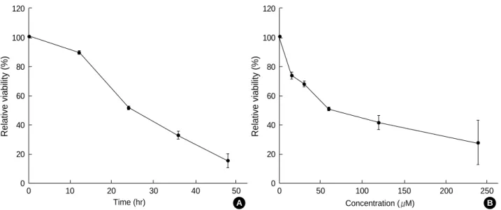
Fig. 2.Cytotoxic effects of NCTD on A375-S2 cell. Cells were treated with various doses of NCTD for 24 hr (A) or 60 M NCTD for various time periods (B). The relative viability of cells was determined by MTT assay. Results are represented as mean±SD, n=3.
Relative viability (%)
120
100
80
60
40
20
00 10 20 30 40 50
Time (hr) A
Relative viability (%)
120
100
80
60
40
20
00 50 100 150 200 250
Concentration ( M) B
RESULTS
Cytotoxicity of NCTD on A375-S2 cells by MTT assay NCTD 15 to 240 M exerted potent inhibitory effect on A375-S2 cell growth. By 24 hr after NCTD 60 M treat- ment, cell death rate reached to almost 50% (Fig. 2).
NCTD-induced morphological changes and DNA fragmentation of A375-S2 cells
In control group, A375-S2 cells were round in shape and
stained homogeneously. After 24 hr treatment with NCTD, blebbing nuclei and granular apoptotic bodies appeared (Fig.
3, arrows).
DNA fragmentation as a hallmark of apoptosis was observed
Fig. 3.Cellular morphology of NCTD-treated A375-S2 cells. Cells were cultured without NCTD (control) or with 60 M NCTD for 24 hr. Morphological change were observed by fluorescent micro- scopy (×200). Arrows indicate condensed nuclei.
Control NCTD
Fig. 4.NCTD-induced DNA fragmentation in A375-S2 cells. The cells were cultured in the presence of NCTD 60 M for 0, 6, 12, 24 and 36 hr. Genomic DNA was extracted and analyzed via elec- trophoresis on 2% agarose gels. Lane M: DNA molecular markers.
M 0 12 24 36 Time (hr)
Cell death ratio (%)
70
60
50
40
30
20
10
0
0 15 30 60 120 240
Concentration ( M) Apoptosis (%)
Necrosis (%)
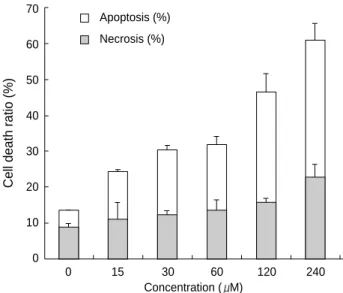
Fig. 5.Ratio of apoptosis and necrosis in A375-S2 cells. Cells were treated with 0, 15, 30, 60,120, 240 M NCTD for 24 hr. The ratios of LDH released from floating dead cells and the culture medium were used to distinguish the proportion of apoptotic and necrotic cells. Results are represented as mean±SD, n=5.
Relative viability (%)
100
80
60
40
20
0
A B C D E F G H I
Fig. 6.Effect of caspase inhibitors on NCTD-induced A375-S2 cells apoptosis. The cells were cultured in the presence or absence of caspase inhibitors. Two hours prior to the addition of 60 M NCTD, pan-caspase inhibitor (z-VAD-fmk, 40 M), caspase-3 inhibitor (z-DEVD-fmk, 20 M), caspase-8 inhibitor (z-IETD-fmk, 20 M), caspase-9 inhibitor (Ac-LEHD-CHO, 20 M) were added, then further incubated for 24 hr. A: NCTD-treated group; B: NCTD- and z-VAD-fmk-treated group; C: NCTD- and z-DEVD-fmk-treated group; D: NCTD- and z-IETD-fmk-treated group; E: NCTD- and Ac-LEHD-CHO-treated group; F: z-VAD-fmk-treated group; G: z- DEVD-fmk-treated group; H: z-IETD-fmk-treated group; I: z-LEHD- fmk-treated group. Results are represented as mean±SD, n=3.
*p<0.05, **p<0.01 vs. group A.
30.44 69.02
** **
**
61.04
37.67 48.50
85.27
94.55 97.52 94.84
in NCTD-treated A375-S2 cells (Fig. 4).
Apoptosis of A375-S2 cell death identified by LDH released assay
The ratio of apoptotic A375-S2 cells increased from 4.6%
at 0 M NCTD to 38.5% at 240 M NCTD, however, that of necrotic cells were still negligible (Fig. 5).
Effect of caspases on NCTD-induced cytotoxicity in A375-S2 cells
A375-S2 cells were treated with 60 M NCTD for 24 hr in the absence or presence of various caspase inhibitors: pan- caspase inhibitor (z-VAD-fmk, 40 M), caspase-3 inhibitor (z-DEVD-fmk, 20 M), caspase-8 inhibitor (z-IETD-fmk, 20 M), caspase-9 inhibitor (Ac-LEHD-CHO, 20 M). Z- VAD-fmk, z-DEVD-fmk and Ac-LEHD-CHO partially
blocked NCTD-induced A375-S2 cell apoptosis. Inhibitory ratio was 30.98%, 38.96%, and 51.50%, respectively. How- ever, caspase-8 inhibitor did not affect the death ratio (Fig. 6).
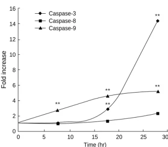
Caspase-3 activity was significantly enhanced after the cells were treated with NCTD 60 M at different time points, however, caspase-9 activity increased moderately and increase of caspase-8 activity was almost negligible (Fig. 7).
The ICAD protein degradation was significant after 24 hr incubation with NCTD (Fig. 8), and this change was blocked by caspase-3 inhibitor.
Involvement of mitochondrial proteins in NCTD-induced A375-S2 cell apoptosis
After incubation with NCTD, protein expression ratio of Bcl-xL/Bax and Bcl-2/Bax was down-regulated, and this change was blocked by caspase-3 inhibitor. At the same time, the protein of cytchrome c was increased in cytosol and caspase- 3 inhibitor had no influence on the increase (Fig. 9).
DISCUSSION
The present study showed that NCTD inhibited A375- S2 cell growth in a time- and dose-dependent manner. At the same time we demonstrated that NCTD induced apop- tosis in A375-S2 cells and the apoptosis was possibly associ- ated with caspases. Caspases are a family of cysteine proteas- es that are activated during the apoptotic processes. Death receptors such as Fas induce caspase-8 activation via Fas-asso- ciated death domain protein (FADD). It was reported that in human colorectal carcinoma cell lines NCTD induced apoptosis by activation of caspase-8, which was prevented by
Fold increase
16
14
12
10
8
6
4
2
0
0 5 10 15 20 25 30
Time (hr) Caspase-3
Caspase-8 Caspase-9
Fig. 7.Effects of NCTD on the activation of caspase-3, -8, -9 in A375-S2 cells. Cells were treated with NCTD 60 M for 0, 8, 18 and 28 hr. Caspase-3, caspase-8 and caspase-9 activities was performed using Caspase Apoptosis Detection Kit according to the manufacturer’s instruction. Results are represented as mean
±SD, n=3. *p<0.05, **p<0.01 vs. 0 hr.
**
**
**
**
**
Fig. 8.Effects of NCTD on ICAD expression in the absence or presence of caspase-3 inhibitor (z-DEVD-fmk). A375-S2 cells were treated with 60 M NCTD only for 0, 12, 24 and 36 hr. Two hours prior to the addition of NCTD, caspase-3 inhibitor (z-DEVD-fmk, 20 M) were added, then further incubated for 36 hr. Cell lysates were separated by 12% SDS-PAGE, and ICAD was detected by Western blot analysis.
ICAD
0 12 24 36 36 Time (hr)
NCTD NCTD+z-DEVD-fmk
45 kD
Fig. 9.Effects of NCTD on Bcl-2, Bcl-xLand Bax expression in the absence or presence of caspase-3 inhibitor (z-DEVD-fmk). A375- S2 cells were treated with 60 M NCTD only for 0, 12, 24 and 36 hr. Two hours prior to the addition of NCTD, caspase-3 inhibitor (z-DEVD-fmk, 20 M) were added, then further incubated for 36 hr. Cell lysates were separated by 12% SDS-PAGE, and Bcl-2, Bcl-xLand Bax proteins were detected by Western blot analysis.
Bcl-xL
Bax
Bcl-2
Cyt c
0 12 24 36 36 Time (hr)
NCTD NCTD+z-DEVD-fmk
32 kD
21 kD
26 kD
15 kD
the pan-caspase inhibitor z-VAD-fmk and caspase-8 inhibitor z-IETD-fmk (1), but the inhibition pathways of NCTD- induced apoptosis remain unclear. Morphological observa- tion, DNA fragmentation and LDH activity assay suggest- ed that NCTD induced A375-S2 cell death involved a mecha- nism of apoptosis. In the present study, NCTD-induced A375-S2 cell death was blocked by pan-caspase inhibitor, indicating that caspase family play a role in the apoptotic process. Caspase-3, and caspase-9 inhibitor (z-DEVD-fmk, Ac-LEHD-CHO, respectively) partially blocked NCTD- induced A375-S2 cell apoptosis, whereas caspase-8 inhibitor (z-IETD-fmk) had no effect on A375-S2 cell death induced by NCTD, indicating that post-mitochondrial caspase-9, but not upper stream caspase-8, activated this apoptotic process.
Chemotherapeutic agents and UV irradiation cause release of mitochondrial cytochrome c, which binds to Apaf-1, and this complex mediates recruitment of procaspase-9 and acti- vates caspase-3. ICAD is expressed as two isoforms, ICAD- L/DFF45 and ICAD-S/DFF35. Once ICAD/DFF45 is cleaved by caspase-3 or caspase-7, CAD is released to the nucleus and induces DNA fragmentation, resulting in the morpho- logical and biochemical features of apoptosis (9, 10, 20, 24, 25). To further confirm the participation of different caspas- es in the cell death, we examined the activities of caspase-3, -8, and -9 and the protein expression of cytochrome c and the substrate for caspase-3, ICAD. The activities of caspase- 9 and caspase-3 were up-regulated at 8 and 18 hr, respec- tively, but capase-8 activity just showed slight change after 28 hr treatment. The protein expression of cytochrome c was up-regulated and the protein expression of ICAD was sig- nificantly down-regulated. Based on these results, we con- cluded that caspase-9-activated apoptotic pathway played a role in the apoptotic pathway of NCTD-treated A375-S2 cells. On the other hand, since NCTD-induced A375-S2 cell apoptosis was only partially reduced by caspase inhibitors, it is possible that other apoptotic pathways might also partic- ipate in this process.
Several pro-apoptotic proteins, such as Bax, Bak, and Bid, translocate to the mitochondrial membrane, and this local- ization is associated with their pro-apoptotic activities. It has been reported that Bcl-2 could exert its action through heterodimerization with Bax. Meanwhile, Bcl-2 and Bcl-xL are cleaved by caspase-3 and are converted to pro-apoptotic proteins similar to Bax. Therefore, the ratio between Bcl-2 and Bax or Bcl-xLand Bax is a decisive factor to activate cell death (9, 15, 26-29). A375-S2 cells treated with NCTD exhibited the elevated ratio between pro-apoptotic Bax and anti-apoptotic Bcl-2 or Bcl-xL. The oligomerization of Bax in the mitochondrial membrane has been shown to induce cytochrome c release, meanwhile pro-apoptotic Bcl-2 cleav- age product was reported to localize on mitochondrial mem- brane and caused release of cytochrome c (9, 30, 31). In our study, the protein expression of cytochrome c was markedly up-regulated followed by the changes of Bcl-2/Bax and Bcl-
xL/Bax ratios at 24 hr in A375-S2 cell treated by NCTD. At the same time, the change of Bcl-2/Bax or Bcl-xL/Bax ratio was blocked by caspase-3 inhibitor, and caspase-3 inhibitor had slight effect on the expression of cytochrome c, indicat- ing a potential positive feedback loop that ensures the death of the cell. These results suggested that the mitochondrial pathway of cell death, including Bcl-2 family and cytochrome c, might be involved in A375-S2 cell death and orchestrate the caspase cascades.
In conclusion, NCTD inhibited A375-S2 cell growth and caspase-9, caspase-3 activation was involved in the apoptotic progression. Simultaneously mitochondrial pathway, includ- ing cytochrome c, Bax and Bcl-2 (or Bcl-xL), contributed to the NCTD-induced A375-S2 cell apoptosis. More detailed mechanism of NCTD-induced A375-S2 cell apoptosis remains to be elucidated.
REFERENCES
1. Peng F, Wei YQ, Tian L, Yang L, Zhao X, Lu Y, Mao YQ, Kan B, Lei S, Wang GS, Jiang Y, Wang QR, Luo F, Zou LQ, Liu JY. Induc- tion of apoptosis by norcantharidin in human colorectal carcinoma cell lines: involvement of the CD95 receptor/ligand. J Cancer Res Clin Oncol 2002; 128: 223-30.
2. Hong CY, Huang SC, Lin SK, Lee JJ, Chueh LL, Lee CH, Lin JH, Hsiao M. Norcantharidin-induced post-G2/M apoptosis is depen- dent on wild-type p53 gene. Biochem Biophys Res Commun 2000;
276: 278-85.
3. Rong Y, Liang FY, Chen L, Du HJ, Liu LY, Sun HL, An W. Nor- cantharidin induces apoptosis in human breast cancer cell line. Can- cer 2000; 19: 1077-81.
4. Kim SO, Han J. Pan-caspase inhibitor zVAD enhances cell death in RAW 246.7 macrophages. J Endotoxin Res 2001; 7: 292-6.
5. Kawazoe N, Watabe M, Masuda Y, Nakajo S, Nakaya K. Tiami1 is involved in the regulation of bufalin-induced apoptosis in human leukemia cells. Oncogene 1999; 18: 2413-21.
6. Hill PA, Tumber A, Meikle MC. Multiple extracellular signals pro- mote osteoblast survival and apoptosis. Endocrinology 1997; 138:
3849-58.
7. Mizukami S, Kikuchi K, Higuchi T, Urano Y, Mashima T, Tsuruo T, Nagano T. Imaging of caspase-3 activation in HeLa cells stimu- lated with etoposide using a novel fluorescent probe. FEBS Lett 1999;
453: 356-60.
8. Zhang Y, Fujita N, Tsuruo T. Caspase-mediated cleavage of p21Waf1/ Cip1 converts cells from growth arrest to undergoing apoptosis. Oncogene 1999; 18: 1131-8.
9. Kirsch DG, Doseff A, Chau BN, Lim DS, de Souza-Pinto NC, Hans- ford R, Kastan MB, Lazebnik YA, Hardwick JM. Caspase-3-depen- dent cleavage of Bcl-2 promotes release of cytochrome c. J Biol Chem 1999; 274: 21155-61.
10. Enari M, Sakahira H, Yokoyama H, Okawa K, Iwamatsu A, Nagata S. A caspase-activated DNase that degrades DNA during apoptosis and its inhibitor ICAD. Nature 1998; 391: 43-50.
11. Doerfler P, Forbush KA, Perlmutter RM. Caspase enzyme activity is not essential for apoptosis during thymocyte development. J Immunol 2000; 164: 4071-9.
12. Cheng EH, Kirsch DG, Clem RJ, Ravi R, Kastan MB, Bedi A, Ueno K, Hardwick JM. Conversion of Bcl-2 to a Bax-like death effector by caspases. Science 1997; 278: 1966-8.
13. Green DR, Reed JC. Mitochondria and apoptosis. Science 1998;
281: 1309-12.
14. Nagata S. Apoptosis by death factor. Cell 1997; 88: 355-65.
15. Wang NS, Unkila MT, Reineks EZ, Distelhorst CW. Transient expres- sion of wild-type or mitochondrially targeted Bcl-2 induces apopto- sis, whereas transient expression of endoplasmic reticulum-targeted Bcl-2 is protective against Bax-induced cell death. J Biol Chem 2001;
276: 44117-28.
16. Klefstrom J, Verschuren EW, Evan G. c-Myc augments the apop- totic activity of cytosolic death receptor signaling proteins by engag- ing the mitochondrial apoptotic pathway. J Biol Chem 2002; 277:
43224-32.
17. Li LY, Luo X, Wang X. Endonuclease G is an apoptotic DNase when released from mitochondria. Nature 2001; 412: 95-9.
18. Gamet-Payraste L, Lumeau S, Cassar G, Dupont MA, Chevolleau S, Gasc N, Tulliez J, Terce F. Sulforaphane, naturally occurring isothiocyanate, induces cell cycle arrest and apoptosis in HT29 human colon cancer cells. Cancer Res 2000; 60: 1426-33.
19. Sarin A, Haddad EK, Henkart PA. Caspase dependence of target cell damage induced by cytotoxic T lymphocytes. J Immunol 1998;
161: 2810-6.
20. Charrier L, Jarry A, Toquet C, Bou-Hanna C, Chedorge M, Denis M, Vallette G, Laboisse CL. Growth phase-dependent expression of ICAD-L/DFF45 modulates the pattern of apoptosis in human colonic cancer cells. Cancer Res 2002; 62: 2169-74.
21. Kim YM, Talanian RV, Billiar TR. Nitric oxide inhibits apoptosis by preventing increases in caspase-3-like activity via two distinct mechanism. J Biol Chem 1997; 272: 31138-48.
22. Herrmann M, Lorenz HM, Voll R, Grunke M, Woith W, Kalden JR. A rapid and simple method for the isolation of apoptotic DNA fragments. Nucleic Acids Res 1994; 22: 5506-7.
23. Sambrook J, Fritsch EF, Maniatis T. Molecular cloning: A labora- tory manual. 2nd ed. Cold Spring Harbor Laboratory Press 1989;
880-98.
24. Villa P, Kaufmann SH, Earnshaw WC. Caspase and caspase inhi- bitors. Trends in Biochem Sci 1997; 22: 388-93.
25. Sakahira H, Enari M, Nagata S. Cleavage of CAD inhibitor in CAD activation and DNA degradation during apoptosis. Nature 1998;
391: 96-9.
26. Yin XM, Oltvai ZN, Korsmeyer SJ. BH1 and BH2 domains of Bcl- 2 are required for inhibition of apoptosis and heterodimerization with Bax. Nature 1994; 369: 321-3.
27. Cheng EH, Levine B, Boise LH, Thompson CB, Hardwick JM. Bax- independent inhibition of apoptosis by Bcl-xL. Nature 1996; 379:
554-6.
28. Adams JM, Cory S. The Bcl-2 protein family: arbiters of cell sur- vival. Science 1998; 281: 1322-6.
29. Reed JC. Double identity for proteins of the Bcl-2 family. Nature 1997; 387: 773-6.
30. Yang J, Liu X, Bhalla K, Kim CN, Ibrado AM, Cai J, Peng TI, Jones DP, Wang X. Prevention of apoptosis by Bcl-2: release of cytochrome c from mitochondria blocked. Science 1997; 275: 1129-32.
31. Kluck RM, Bossy-Wetzel E, Green DR, Newmeyer DD. The release of cytochrome c from mitochondria: a primary site for Bcl-2 regu- lation of apoptosis. Science 1997; 275: 1132-6.