390
©The Korean Society of Food Science and Technology
해양미세조류 에탄올 추출물의 항균활성에 관한 연구
김윤정·하상철
1·김대욱
2·신일식
3,*
한화호텔앤드리조트 식품연구센터, 1대구미래대학교 호텔외식조리과,
2식품의약품안전처 연구기획조정과, 3강릉원주대학교 해양식품공학과
Antibacterial activity of ethanol extracts from marine micro-algae
Yun-Jung Kim, Sang-Chul Ha1, Dae Uk Kim2, and Il-Shik Shin3,* Food Research center, Hanwha Hotel and Resort
1Department of Hotel Culinary Arts, Daegu Future College
2Research Planning and Management Division, Food and Drug Safety Evaluation, Ministry of Food and Drug Safety 3Department of Marine Food Science and Technology, Gangneung-Wonju National University
Abstract The antibacterial activity of ethanol (99.9%) extracts from marine micro-algae, namely, Mixed A (Pavlova sp., Thalassiosira weissflogii, Tetraselmis suecica and Isochrysis galbana were mixed with 1:1:1:1 ratio), Chlorella vulgaris, Nannochloropsis oculata and Chaetoceros calcitrans were estimated against food-borne bacteria, namely, Escherichia coli, Salmonella Typhimurium, Staphylococcus aureus and Bacillus cereus. The extracts from these marine micro-algae showed potent antibacterial activity against all tested bacteria by the paper disk method. The extracts from C. vulgaris showed the strongest antibacterial activity against E. coli with minimum inhibitory concentration (MIC) of 0.62 mg/mL, and minimum bactericidal concentration (MBC) of 2.50 mg/mL. The extract from C. vulgaris contained 2 active compounds, 38.8% linoelaidic acid and 30.0% phytol. These results indicated that the ethanol extract from C. vulgaris may be a putative natural antibacterial agent against food-borne bacteria.
Keywords: marine micro-algae, antibacterial activity, food-borne bacteria, Chlorella vulgaris, natural antibacterial agent
서
론
식품을 안전하게 장기간 저장하기 위한 수단으로 가열살균, 냉 동냉장, 방사선, 초고압, 건조 등 물리적 방법과 과산화수소, 알 코올, 염소제, 보존료 등 화학적 방법이 있다. 식품을 살균하여 보존성을 높이는 방법으로 주로 사용하는 가열 처리 방법은 열 에 의해 제품의 질 저하, 영양가 파괴 등 많은 단점을 가지고 있 다. 이와 같은 단점을 보완하기 위하여 냉장 및 냉동법은 열처리 를 최소화하여 신선함을 유지할 수 있으나 저온유지를 위한 온 도관리와 저장, 유통 비용이 높다. 이러한 제한 요인을 보완하고 부패성 미생물이나 식중독 미생물의 증식을 억제하여 식품을 안 전하게 장기 저장하려는 수단으로 각종 보존료 혹은 살균제를 개 발하여 사용하고 있으며 현재 아질산염(nitrite), 소브산(sorbic acid), 메타아황산소듐(sodium metabisulfite), 벤조산(benzoic acid), 염소 제 등 다양한 합성 보존료가 생산되어 이용되고 있다(1-3). 그러 나 합성보존료는 체내 축적성 등 그 유해성 문제가 지속적으로 대두되고 있고, 물질의 종류, 사용량 등에 따라 잔류독성, 돌연변 이 유발, 기형유발 등 인체에 부정적 영향을 주고 있기 때문에 소비자들의 기피 현상이 두드러지고 있다. 이와 같은 경향으로 인하여 식품산업계에서도 인공 합성보존 제의 사용을 제한하려는 추세이고, 안전성이 확보된 천연 항균성 물질에 대한 연구가 집중적으로 이루어지고 있다(4-6). 일반적으 로 사람들이 오랫동안 식용으로 이용해 온 원료 혹은 자연물로 부터 항균성 성분을 추출하여 보존제로 사용하는 경우, 미국에서 는 이를 generally recognized as safe (GRAS)로 분류하여 관리하 고 있다(7). 따라서 천연 항균물질의 개발과 이용은 인공합성보 존제의 사용으로 인한 부정적인 측면을 해소하고 식품의 저장성 향상과 안전성을 확보할 수 있는 좋은 방안이다(8). 조류(algae)는 육상식물을 제외한 모든 광합성 생물의 통칭으 로, 이들 중 현미경으로 관찰할 수 있는 단세포성 조류를 미세조 류(microalgae)라 하며, 대부분의 식물성 플랑크톤이 이에 속한다. 미세조류는 클로로필(chlorophylls)이나 카로테노이드(carotenoids) 등의 색소를 함유하며 주로 광합성을 통해 세포 성장과 번식을 행하는 식물군으로 약 20,000여종이 있으며, 크기는 1-1,000 µm 로 1차 대사산물인 단백질, 지방질, 탄수화물 등은 물론이고, 2차 대사산물인 다양한 생리활성물질을 생산하다고 알려져 있다(9). 이러한 미세조류에 대한 근년의 연구는 클로렐라(Chlorella sp.) 균체를 이용한 단백질 자원(SCP: single cell protein)으로서의 이 용 방안에 대한 연구에서 시작되었다고 할 수 있는데, 최근에는 미세조류로부터 단백질뿐만 아니라 지질, 당질, 색소와 같은 다 량 물질과 항균성 물질, 항암성 물질, toxin 등과 같은 미량 물질 의 활용 가능성에 대해 연구가 많이 행해지고 있다(10-15). 미세 *Corresponding author: Il-Shik Shin, Department of Marine FoodScience and Technology, Gangneung-Wonju National University, Gangneung, Gangwon 25457, Korea
Tel: +82-33-640-2346 Fax: +82-33-640-2346 E-mail: [email protected]
Received April 17, 2017; revised May 22, 2017; accepted May 22, 2017
조류의 생리 활성 물질 검색에 대한 연구는 과거에도 일부 행해 져 왔으나 최근에 그 존재에 대한 결과가 확인되고 있고(16-17), 산업적 이용 가능성도 있는 것으로 알려져 있는데, 그 중에서도 항균성 물질의 탐색과 그 결과에 대한 것이 보고되고 있다(18). 미세조류 중 Chlorella sp.는 가장 널리 연구되어진 조류이며, 산업적 이용 범위도 넓다. 클로렐라 추출물을 Listeria monocyto-genes에 감염된 마우스에 투여한 결과 항암 및 항균 등 면역증강 을 가져오는 것으로 나타났고, Bacillus sp. 등 장내 유해세균을 현저히 감소시키는 항균활성을 갖고 있는 것으로 알려져 있다 (19). 또한, 미세조류 추출물의 일부는 항산화 활성을 가진다는 보고가 있다(20). 하지만 아직 미세조류의 항균성 물질 탐색은 기초 단계에 있 다고 할 수 있으며, 국내에 분포하고 있는 미세조류에 대한 정확 한 정보가 없는 실정이고 미세조류의 이용성에 대한 연구도 미 미한 실정이다. 이에 본 연구에서는 미세조류를 이용한 식품보존 료의 개발 가능성을 검토하고자 4종의 해양 미세조류로부터 99.9% 에탄올로 추출한 추출물의 식중독균에 대한 항균활성과 그 성분 을 조사하였다.
재료 및 방법
미세조류 본 실험에서 사용된 미세조류는 강릉원주대학교 해양생물연구 교육센터에서 분양받았다. Chlorella vulgaris (Daesang Co., Ltd., Fresh water heterotrophic tank culture) 는 OECD medium (Wako Pure Chemical Industries Ltd., Osaka, Japan)에서 OECD-guideline No. 201에 따라 6일간 배양하였다. Chaetoceros calci-trans KMMCC B-258, Nannochloropsis oculata KMMCC C-16, Pavlova sp. CCMP 459, Thalassiosira weissflogii CCMP-1051, Tetraselmis suecica KMMCC P-9, Isochrysis galbana KMMCC H-2는 염도를 33‰로 조정한 F/2 배지(21)에서 6일간 배양하였다. 세포수 1.0×10⁶ cells/mL의 Chlorella vulgaris, Chaetoceroscalci-trans, Nannochloropsis oculata 각각 400 mL와 세포수 1.0×10⁶ cells/mL의 미세조류 4종(Pavlova sp., Thalassiosira weissflogii, Tetraselmis suecica, Isochrysis galbana)을 각각 100 mL씩 혼합한 mixed microalgae (이하 Mixed A) 400 mL 등 4종류의 미세조류 배양액을 원심분리(4oC, 5,000×g, 20분)하여 세포를 취한 후, 3% 식염수를 넣고 다시 원심분리(4oC, 5,000×g, 20분)하였다. 깨끗한 세포만을 분리하기 위해 위 과정을 2번 반복한 후, 냉동 건조하 여 −80oC의 심온냉동고(deep-freezer)에 보관하여 사용하였다. 미세조류로부터 항균물질의 추출 미세조류로부터 항균물질을 추출하기 위하여 미세조류 세포를 동결건조한 분말 시료에 중량대비 25배에 해당하는 99.9% 에탄 올(Duksan Pure Chemicals, Seoul, Korea)을 가하고 실온에서 3일 동안 추출하여 상층액을 모아 거름종이(Advantec No. 5C, Tokyo, Japan)로 여과한 후, rotary evaporator (Rotavapor R-200, BUCHI Co., New Castle, DE, USA)로 감압 농축하였다. 이 농축한 추출 물에 남아있는 에탄올을 질소생성기로 제거한 후 밀봉하여 4oC
냉장고에 보관하면서 실험에 사용하였다.
공시균주
항균활성 측정에 사용된 균주는 그람음성세균 2종 (Escherichia coli ATCC 11775, Salmonella Typhimurium KCCM 11862)과 그 람양성세균 2종(Staphylococcus aureus ATCC 12692, Bacillus
cereus ATCC 11778)이었다. 각 균주는 American Type Culture Collection (ATCC)과 Korean Culture Center of Microorganisms (KCCM)에서 분양받아 사용하였다. 항균활성 측정에 사용된 모 든 균주는 균배양액 1 mL를 9 mL의 brain heart infusion (BHI) broth (Difco Laboratories, Detroit, MI, USA)에 접종하여 37oC에 서 2회 계대배양 하였고, UV-분광광도계(spectrophotometer, UV 1600 PC, Shimadzu, Tokyo, Japan)를 이용하여 균수를 1.0×10⁵ CFU/mL 농도로 조정한 후 실험에 사용하였다.
항균활성측정
종이원반퍼짐분석(Paper disk diffusion assay)
미세조류 에탄올 추출물의 항균활성 유무를 paper disk diffusion method로 측정하였다(22). 공시균주의 배양액(1.0×10⁵ CFU/mL) 100µL를 멸균한 spreader로 brain heart infusion agar (BHA, Difco Laboratories) plate의 표면에 균질하게 도말한 후, 중앙에 멸 균된 8 mm filter paper disk (Whatman No. 2)를 고정하고 여기 에 Iso mixed, C. vulgaris, N. oculata, C. calcitrans의 추출물 (10 mg/mL) 50µL를 점적하고 37oC에서 24시간 배양하였다. 항균 활성은 disk 주위로 미생물의 생장이 저해되는 inhibition zone의 크기(mm)로 측정하였으며, 미세조류 추출물의 희석에 사용된 에 탄올을 음성 대조군, 항균성이 보고된 allyl isothiocyanate (AIT, >98% purity, Fluka Co., Haan, Germany)를 양성 대조군으로 사용 하였다.
최소저해농도(Minimal inhibitory concentration, MIC) 미세조류 에탄올 추출물의 공시균주에 대한 최소저해농도는 broth dilution method를 사용하여 측정하였다(23). 미세조류 에탄 올 추출물을 membrane filter로 멸균한 다음 BHI broth로 0.02-100 mg/mL 농도로 희석하였다. 멸균한 tube에 BHI broth 100 µL, 미세조류 에탄올 추출물 50 µL, 균 배양액 50 µL를 순차적으로 첨가하여 37oC에서 24시간 배양한 후, tube를 육안(visible)으로 봤
을 때, 균이 증식하지 않은 최소농도를 최소저해농도로 설정하였 다. 각 균주는 균수를 1.0×10⁵ CFU/mL로 조절하였으며 모든 실
험은 3회 반복 수행하였다.
최소살균농도(Minimal bactericidal concentration, MBC) 최소살균농도는 streak plate method를 사용하여 측정하였다(24). 최소저해농도 전후 농도의 배양액을 백금이로 취하여 각각 BHA plate에 streak하고, 37oC에서 48시간 배양한 후, 균이 증식하지 않 는 최소농도를 최소살균농도로 설정하였다. 모든 실험은 3회 반 복 수행하였다.
가스크로마토그래피 질량스펙트럼(Gas Chromatography Mass Spectrum, GC/MS)
C. vulgaris 에탄올 추출물의 성분 분석은 GC/MS (GC-MS-CTC, 7890A/5975C MSD, Agilent Technologies, Santa Clara, CA, USA)를 이용하였다(25).
성분 분석에 사용한 컬럼은 SE-54 (30 m×0.25 mm I.D. 0.25µm film thickness; HP-5MS, Agilent Technologies, Santa Clara, CA, USA)이었으며, 운반기체는 헬륨가스를 사용하였다. 주 입구 온도를 250oC로 하여 splitless mode 주입방법을 사용하였다.
오븐온도는 40oC에서 2분간 유지 시킨 후, 5oC/min의 속도로
150oC까지 올리고 24분간 유지하였으며, 150oC에서 15oC/min의 속도로 300oC까지 올린 후 10분간 유지하였다. 분석한 결과는
결과 및 고찰
항균활성
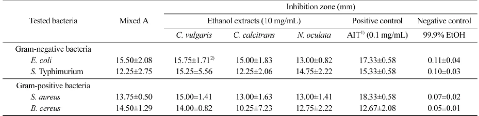
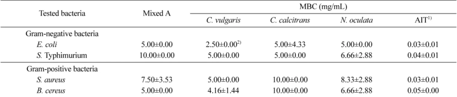
Mixed A, C, vulgaris, N, oculata, C, calcitrans 등 4종류의 미세조류로부터 99.9% 에탄올로 항균물질을 추출하고, 각 추출 물을 10 mg/mL 농도로 조제하여 E. coli, S. Typhimurium, S. aureus, B. cereus에 대한 항균활성의 유무를 disk diffusion method로 측정한 결과는 Table 1과 같다. 4종류의 미세조류 에탄 올 추출물은 공시균주 모두에 대해 항균활성을 나타내었으며, 12.25-15.75 mm의 inhibition zone을 나타내었다. Mixed A, C, vulgaris, C, calcitrans의 추출물은 E. coli에 대하여 보다 강한 항 균활성을 나타내었으며, 그 중 C. vulgaris 추출물의 E. coli에 대 한 inhibition zone이 15.75 mm로 가장 강한 항균활성을 나타내었다. 이러한 결과는 미세조류의 항균물질에 관한 연구에서 유기용 매 추출물이 수용성 추출물보다 항균활성이 높게 나타났다는 보 고(26)와 극성이 다른 3 종류의 유기용매 중 에탄올 추출물의 항 균활성이 가장 강하다고 보고한 연구(25-27)와 유사하였다. 에탄 올은 식용으로서 그 안전성이 높고 최종 제품의 인허가 시에도 유리하므로 미세조류에서 항균활성 물질을 추출할 때에 에탄올 을 사용하는 것이 좋은 것으로 사료된다. 최소저해농도(MIC) 공시균주에 대한 4종류의 미세조류 에탄올 추출물의 최소저해 농도는 Table 2와 같다. 4종류의 미세조류 추출물은 공시균주에 대하여 0.62-1.66 mg/mL의 MIC를 나타내었으며, C. vulgaris 추출 물이 4종의 공시균주에 대한 최소저해농도가 0.62 mg/mL로 가장 강한 항균활성을 나타내었다. 그 다음이 C. calcitrans 추출물로 1.04-1.66 mg/mL의 최소저해농도를 나타내었으며, 그램양성균(S. aureus, 1.66; B. cereus, 1.25 mg/mL)보다 그램음성균(E. coli,
1.04; S. Typhimurium, 1.04 mg/mL)에 대하여 더 강한 항균활성을 나타내었다. MIC의 경우, 통상 육안으로 증식이 인정되지 않는 농도로 결정하는데, 본 실험의 경우, 미세조류 고유의 색으로 인 하여 증식여부를 결정하는 데 있어서 다소 곤란함이 있었다. 이 에 최소살균농도를 측정하여 미세조류 추출물의 항균활성 근거 를 확인하였다. 최소살균농도(MBC) 4종류의 미세조류 에탄올 추출물의 공시균주에 대한 최소살균 농도를 Table 3에 나타내었다. 4종류의 미세조류 추출물은 2.50-10.00 mg/mL의 최소살균농도를 나타내었으며, C. vulgaris 추출물 Table 1. Antibacterial activity of ethanol extracts from marine micro-algae against food-borne bacteria
Tested bacteria Mixed A
Inhibition zone (mm)
Ethanol extracts (10 mg/mL) Positive control Negative control C. vulgaris C. calcitrans N. oculata AIT1) (0.1 mg/mL) 99.9% EtOH Gram-negative bacteria E. coli 15.50±2.08 15.75±1.712) 15.00±1.83 13.00±0.82 17.33±0.58 0.11±0.04 S. Typhimurium 12.25±2.75 15.25±5.56 12.25±2.06 14.75±2.22 15.33±0.58 0.10±0.03 Gram-positive bacteria S. aureus 13.75±0.50 15.00±1.41 13.00±1.63 13.00±1.41 18.33±0.58 0.07±0.02 B. cereus 14.50±1.29 14.00±0.82 10.25±7.23 12.75±2.22 12.67±2.08 0.05±0.01 1)Allyl isothiocyanate
2)Mean±standard deviation of three measurements.
Table 2. MIC of ethanol extracts from marine micro-algae against food-borne bacteria
Tested bacteria Mixed A MIC (mg/mL)
C. vulgaris C. calcitrans N. oculata AIT1) Gram-negative bacteria E. coli 15.50±2.08 0.62±0.832) 1.04±0.36 1.66±0.72 0.02±0.07 S. Typhimurium 12.25±2.75 0.62±0.83 1.04±0.36 1.66±0.72 0.03±0.01 Gram-positive bacteria S. aureus 13.75±0.50 0.62±0.00 1.66±0.72 1.45±0.95 0.02±0.07 B. cereus 14.50±1.29 0.62±0.83 1.25±0.00 1.66±0.72 0.03±0.00 1)Allyl isothiocyanate
2)Mean±standard deviation of three measurements.
Fig. 1. Procedure for extraction of antimicrobial agent from marine micro-algae.
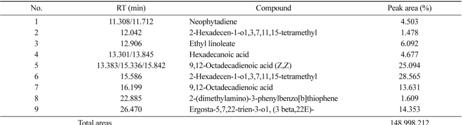
의 항균활성(최소살균농도, 2.50-5.00)이 가장 강한 것으로 나타났 다. C. vulgaris 에탄올 추출물의 최소살균농도는 E. coli에 대해 2.50 mg/mL, S. aureus, B. cereus와 S. Typhimurium에 대해서는 4.16-5.00 mg/mL로 천연항균제 AIT의 최소살균농도(0.03-0.05 µg/ mL)에 비해 활성이 낮았지만, 다른 미세조류 에탄올 추출물보다 강한 항균활성을 나타내었다. C. vulgaris 추출물은 클로렐라 성 장촉진 인자인 CGF (Chrorella Growth Factor)를 비롯한 아미노 산, 당단백질, 비타민 등 기능성 물질을 많이 함유하고 있고(28), C. vulgaris의 추출물이 세균에 대하여 항균활성을 가진다고 보고 된 바 있다(29-31). 이에 GC/MS를 이용하여 C. vulgaris 에탄올 추출물로부터 항균활성을 나타내는 성분의 존재를 확인하였다. C. vulgaris 에탄올 추출물의 항균성분 C. vulgaris 에탄올 추출물의 성분을 GC/MS로 분석하였고, 그 결과를 Fig. 2와 Table 4에 나타내었다. C. vulgaris 에탄올 추출 물의 성분 분석 결과, 동정된 화합물은 모두 9종이었고(Fig. 2), 동정된 성분 중에서 함량이 가장 많은 성분은 9,12-octadecadienoic acid (linoelaidic acid, peak 5, 7)로 38.8%이었으며, 그 다음이 2-Hexadecen-1-o1, 3,7,11,15-tetramethyl (phytol, peak 2,6)로 30.0% 이었다. 미세조류 추출물의 항균활성에 관한 최초의 보고는 Pratt 등(32) 이 Chlorella로부터 추출한 지방산의 중합체인 클로레린(chlorellin) 이 그램양성균과 그램음성균에 대하여 항균활성이 있다고 보고 한 논문이다. 그 이후 미세조류에 의해 생산되는 새로운 항균성 분과 항균 메카니즘을 밝히기 위해 많은 연구가 진행되었으며 (33), 미세조류인 Phaeodactylum tricornutum가 생산하는 불포화지 방산인 에이코사펜타인산(eicosapentaenoic acid)가 multidrug-resis-tant Staphylococcus aureus (MRSA)를 포함한 그램양성균과 그램 음성균에 대하여 항균활성을 나타내었으며, 헥사데카트라이엔산 (hexadecatrienoic acid)는 식중독균인 S. aureus에 대해 항균활성을 나타내었고(34,35). Smith 등(36)은 P. tricornutum로부터 고농도의 팔미톨레인산(palmitoleic acid)와 다른 기능성 지방산이 마이크로 몰 농도에서 그램양성 병원균에 대해 항균활성을 나타내며, 노출 즉시 치명적인 효과를 나타내는 것으로 보고하였으며, Santoyo 등(37)은 미세조류인 Haematococcus pluvialis로부터 추출한 에탄 올 추출물이 E. coli와 S. aureus에 항균활성이 있으며, 그 성분은 짧은 사슬의 지방산인 뷰탄산(butanoic acids)과 락트산메틸(methyl lactic acids)이었다고 보고 한 바 있다. 이러한 보고로 볼 때, 미 세조류가 생산하는 대사산물 중 지방산이 항균활성을 가지는 주 성분이라는 것을 알 수 있다.
미세조류가 생산하는 지방산의 정확한 항균 기작은 아직 밝혀 져 있지 않지만, 지방산은 세균의 여러 기관에 작용하는 데, 그 Table 3. MBC of ethanol extracts from marine micro-algae against food-borne bacteria
Tested bacteria Mixed A MBC (mg/mL)
C. vulgaris C. calcitrans N. oculata AIT1) Gram-negative bacteria E. coli 5.00±0.00 2.50±0.002) 5.00±4.33 5.00±0.00 0.03±0.01 S. Typhimurium 10.00±0.00 5.00±0.00 5.00±0.00 6.66±2.88 0.04±0.01 Gram-positive bacteria S. aureus 7.50±3.53 5.00±0.00 10.00±0.00 8.33±2.88 0.03±0.01 B. cereus 5.00±0.00 4.16±1.44 10.00±0.00 6.66±2.88 0.05±0.00 1)Allyl isothiocyanate
2)Mean±standard deviation of three measurements.
중 세포막이 가장 가능성 높은 표적인 것으로 추측되고 있으며, 지방산의 사슬이 길고 불포화도가 높을수록 항균활성이 강하다 고 보고하고 있다(38). 세균 세포막의 손상은 세포내 물질의 유 실과 영양성분 흡수 저해, 세포호흡의 억제 등을 야기하여 세균 의 증식이 억제되거나 사멸한다. 본 연구에서 C. vulgaris로부터 에탄올로 추출한 추출물에서 가 장 함량이 많은 것으로 동정된 옥타데카다이엔산(octadecadienoic acid)는 일명 리노엘라이드산(linoelaidic acid)로 탄소수 18개의 고 도불포화지방산인 것과 두 번째로 함량이 많은 피톨(Phytol)은 클 로로필을 구성하는 불포화 제1급 알코올의 일종으로서 엽록소의 한 성분이며, 병원성 대장균 및 황색포도상구균에 항균활성을 나 타내는 것으로 보고된(39) 것으로 볼 때, 이 2가지 성분이 항균 활성의 주성분인 것으로 사료된다. 또한 따라서 미세조류인 C. vulgaris의 에탄올 추출물은 항균활성이 강한 고도불포화지방산과 엽록소를 함유하고 있어 천연식품보존료로서의 이용이 가능할 것 으로 사료된다.
요
약
Mixed A, Chlorella vulgaris, Nannochloropsis oculata, Chaeto-ceros calcitrans 등 4종류의 미세조류로부터 99.9% 에탄올로 추 출한 성분의 항균활성을 검증하기 위해 그람음성세균 2종(E. coli, S. Typhimurium)과 그람양성세균 2종(S. aureus, B. cereus)에 대하 여 paper disk diffusion assay, MIC, MBC를 측정하였다. 4종류의 미세조류 추출물은 공시균주에 대하여 0.62-1.66 mg/mL의 MIC를 나타내었으며, C. vulgaris 추출물이 4종의 공시균주에 대한 MIC 가 0.62 mg/mL로 가장 강한 항균활성을 나타내었으며, MBC 또 한 C. vulgaris 에탄올 추출물이 E. coli에 대해 2.50 mg/mL, S. aureus, B. cereus와 S. Typhimurium에 대해서는 4.16-5.00 mg/mL 로 가장 강한 항균활성을 나타내었다.
항균활성이 가장 강한 C. vulgaris 에탄올 추출물의 성분을 분 석한 결과, 함량이 가장 많은 성분은 9,12-octadecadienoic acid (linoelaidic acid, peak 5, 7)로 38.8%이었으며, 그 다음이 2-Hexa-decen-1-o1, 3,7,11,15-tetramethyl (phytol, peak 2,6)로 30.0%이었 다. Octadecadienoic acid는 일명 linoelaidic acid로 탄소수 18개의 고도불포화지방산이며, 두 번째로 함량이 많은 Phytol은 클로로 필을 구성하는 불포화 제1급 알코올의 일종으로서(엽록소의 구성 성분) 병원성 대장균 및 황색포도상구균에 항균활성을 나타낸다 는 보고로 볼 때, 이 2가지 성분이 항균활성의 주성분인 것으로 사료된다.
References
1. Davidson PM, Post LS, Branen AL, Mccurdy AR. Naturally occurring and miscellaneous food antimicrobials. pp. 371419. In: Antimicrobials in Food. Branen AL, Davison PM (eds). Mar-cel Dekker, New York, NY, USA (1983)
2. Lewis RJ. Food additives. pp. 3-27. In: Food additives Hand-book. Dean RW (ed). Nostrand Reinhold, New York, NY, USA (1989)
3. Cherry JP. Improving the safety of fresh produce whit antimicro-bials. Food Technol. 53: 54-59 (1999)
4. Cho SH, Lee SY, Kim JW, Ko GH, Seo IW. Development and application of natural antimicrobial agent isolated from grapefruit seed extract-Antimicrobial activities of grapefruit seed extract. J. Food Hyg. Saf. 10: 33-39 (1995)
5. Lee SH, Lim YS. Antimicrobial effects of Schizandra chinensis extract on pathogenic microorganism. J. Korean Soc. Food Sci. Nutr. 27: 239-243 (1998)
6. Baratta MT, Dorman HJD, Deans SG, Figueiredo AC, Barro JG, Ruberto G. Antimicrobial and antioxidant properties of some commercial essential oils. Flav. Frag. J. 13: 235-244 (1998) 7. Kong YJ, Oh DH. Effect of ethanol extract of Quercus
mongol-ica leaf as natural food preservative. J. Korean Soc. Food Sci. Nutr. 30: 243-963 (2001)
8. Jung JH, Cho SH. Effect of steeping treatment in the natural anti-microbial agent solution on the quality control of processed tofu. Korean J. Food Preserv. 10: 41-46 (2003)
9. Murakami M, Makabe K, Okada S, Yamaguch K, Konosu S. Screening of biologically active compounds in microalgae. Nip-pon Suisan Gakkaishi. 54: 1035-1042 (1988)
10. Aaronson S, Dhawale SW, Patni J, DeAngelis B, Frank O, Baker H. The cell content and secretion of water soluble vitamins in several freshwater algae. Arch. Microbiol. 112: 57-59 (1977) 11. Percival EP, Foyle RAJ. The extracellular polysaccharides of
Por-phyridium cruentum and PorPor-phyridium aerugineum. Carbohyd. Res. 72: 165-176 (1979)
12. Ben-Amotz A, Avron, M. Glycerol, β-carotene and dry algal meal production in commercial cultivation of Dunaliella. pp. 603-610. In: Algae Biomass. Shelef G, Soeder CJ (eds). Elsevier, Amsterdam, Netherlands (1980)
13. Antia NJ, Desai ID, Romily MJ. The tocopherol, vitamin K, and related isoprenoid quinone composition of unicellular red algae. J. Phycol. 6: 305-312 (1970)
14. Kenyon CN, Rippka R, Stanier RY. Fatty acid composition and physiological properties of some filamentous blue-green algae. Arch. Microbiol. 83: 216-236 (1972)
15. Ben-Amotz A, Katz A, Avron M. Accumulation of beta-carotene in halotolerant algae: purification and characterization of beta-car-otene rich globlues from Dunaliella bardawil (Chlorophycea). J. Phycol. 18: 529-537 (1982)
16. Moor RE. Constituents of blue-green algae. pp. 1-49. In: Marine Natural Products. Scheuer PJ (ed). Academic Press, New York,
Table 4. Identification of antibacterial compounds in ethanol extracts from C. vulgaris by GC-MS
No. RT (min) Compound Peak area (%)
1 11.308/11.712 Neophytadiene 4.503 2 12.042 2-Hexadecen-1-o1,3,7,11,15-tetramethyl 1.478 3 12.906 Ethyl linoleate 6.092 4 13.301/13.845 Hexadecanoic acid 4.677 5 13.383/15.336/15.842 9,12-Octadecadienoic acid (Z,Z) 25.094 6 15.586 2-Hexadecen-1-o1,3,7,11,15-tetramethyl 28.565 7 16.199 9,12-Octadecadienoic acid 13.631 8 22.885 2-(dimethylamino)-3-phenylbenzo[b]thiophene 1.609 9 26.470 Ergosta-5,7,22-trien-3-o1, (3 beta,22E)- 14.353 Total areas 148,998,212
NY, USA (1981)
17. Hoppe HA. Marine algae and their products and constituents in pharmacy. pp. 25-119. In: Marine Algae in Pharmaceutical Sci-ence. Hoppe HA, Levring T, Tanaka Y (eds). Walter de Gruyter, Berlin, Germany (1979)
18. Nagai H, Murata M, Torigoe K, Satake M, Yasumoto T. Gam-bieric acids: New potent antifungal substances with unprece-dented polyether structures from a marine dinoflagellate Gambierdiscus toxicus. J. Org. Chem. 57: 54485453 (1992) 19. Hasegawa T, Kimura Y, Hiromatsu K, Kobayashi N, Yamada A,
Makino M, Sano T, Nomoto K, Yoshiko Y. Effect of hot water extract of Chlorella vulgaris on cytokine expresstion patterns in mice with murine acquired immunodeficiency syndrome after infection with Listeria monocytogenes. Immunopharmacology 35: 272-282 (1997)
20. Richmond A. Microalgal biotechnology at the turn of the century. J. Appl. Phycol. 12: 441-451 (2005)
21. Guillard RRL, Ryther JH. Studies of marine planktonic diatoms. I. Cyclotella nana Hudstedt, and Detonula converfacea (Cleve) Gran. Can. J. Microbiol. 8, 229?239 (1962)
22. Bauer AW, Kibby MM, Sherria JC, Turck M. Antibiotic suscepti-bility testing by a standardized single disk method. Am. J. Clin. Pathol. 45: 493-496 (1966)
23. CLSI. Performance Standards for Antimicrobial Susceptibility Testing; Twenty-Fifth Informational Supplement (M100-S25). Clinical Laboratory Standards Institute, Wayne, PA, USA (2015) 24. Bamba H, Kondo Y, Wong RM, Sekine S, Matsuzaki F.
Evalua-tion of assay method of the susceptibility of antimicrobial agents using a 96-well flat-bottom microlpate and a microplate reader. Am. J. Gastroenterol. 92: 659-662 (1997)
25. Plaza M, Santoyo S, Jaime L, Avalo B, Cifuentes A, Reglero G, Garcia-Blairsy Reina G, Senoran FJ, Ibanez E. Comprehensive characterization of the functional activities of pressurized liquid and ultrasound-assisted extraction from Chlorella vulgaris. LWT-Food Sci. Tech. 46: 245-253 (2012)
26. Plaza M, Santoyo S, Jaime L, Garcia-Blairsy Reina G, Herrero M, Senoran FJ, Ibanez E. Screening for bioactive compounds from algae. J. Pharmaceut. Biomed. 51: 450-455 (2010)
27. Santoyo S, Rodriguez-Meizoso I, Cifuentes A, Jaime L, Garcia-Blairsy Reina G, Senoran FJ, Ibanez E. Green processes based on the extraction with pressurized fluids to obtain potent antimicrobi-als from Haematococcus pluvialis microalgae. LWT-Food Sci. Tech. 42: 1213-1218 (2009)
28. Gouveia L, Veloso V, Reis A, Fernandes H, Novais J, Empis J. Evolution of pigment composition in Chlorella vulgaris. Biore-source Technol. 57: 157-163 (1996)
29. Joo DS, Lee EH. Searching of antimicrobial active compounds from microalgae. Kor. J. Life Sci. 8: 173-180 (1998)
30. Eguchi K, Nagase H, Qzawa M, Endoh Y, Goto K, Hirata K, Miyamoto K, Yoshimura H. Evaluation of antimicrobial agents for veterinary use in the ecotoxicity test using microalgae. Chemosphere 57: 1733-1738 (2004)
31. Dantas DC, Kaneno R, Queiroz ML. The effects of Chlorella vulgaris in the protection of mice infected with Listeria monocy-togenes. Role of natural killer cells. Immunopharm. Immunot. 21: 609-619 (1999)
32. Pratt R, Daniels TC, Eiler JB, Gunnison JB, Kumler WD. Chlo-rellin, an antibacterial substance from Chlorella. Science 99: 351-352 (1944)
33. Ghasemi Y, Yazdi MT, Shafiee A, Amini M, Shokravi S, Zarrini G. Parsiguine, a novel antimicrobial substance from Fischerella ambigua. Pharm. Biol. 42: 318-322 (2004)
34. Desbois AP, Mearns-Spragg A, Smith VJ. A fatty acid from the diatom Phaeodactylum tricornutum is antibacterial against diverse bacteria including multi-resistant Staphylococcus aureus (MRSA). Mar. Biotechnol. 11: 45-52 (2009)
35. Smith VJ, Desbois AP, Dyrynda EA. Conventional and uncon-ventional antimicrobials from fish, marine invertebrates and micro-algae. Mar. Drugs 8: 1213-1262 (2010)
36. Smith VJ, Desbois AP, Dyrynda EA. Conventional and uncon-ventional antimicrobials from fish, marine invertebrates and micro-algae. Mar. Drugs 8: 1213-1262 (2010)
37. Santoyo S, Rodrguez-Meizoso I, Cifuentes A, Jaime L, Garca-Blairsy Reina G, Seorans FJ, Ibanez E. Green processes based on the extraction with pressurized fluids to obtain potent antimicrobi-als from Haematococcus pluvialis microalgae. LWTFood Sci. Tech. 42: 1213-1218 (2009)
38. Amaro HM, Guedes C, Malcata FX. Antimicrobial activities of microalgae: an invited review. pp 1272-1280. In: Science against Microbial Pathogens: Communicating Current Research and Tech-nological Advances. Mndez-Vilas A (ed). Formatex Research Center, Badajoz, Spain (2011)
39. Couladis M, Chinou IB, Tzakou O, Loukis A. Composition and antimicrobial activity of the essential oil of Ballota pseudodictam-nus L. Bentham. Phytother. Res. 16: 723-726 (2002)