INTRODUCTION
Collagenous gastritis and collagenous colitis are rare enti- ties, counterparts of each other, characterized by the deposi- tion of a subepithelial collagen band with an inflammatory infiltrate in the gastrointestinal mucosa (1). Collagenous gas- tritis is particularly rare, and less than 20 cases of collagenous gastritis have been reported in the literature since the first description of collagenous gastritis in 1989 by Colletti and Trainer (2). Patients with collagenous gastritis suffer from gastric pain or other accompanying symptoms such as anemia caused by gastric bleeding and watery diarrhea when colitis is present. Some authors suggest that there seems to be two distinctive clinicopathologic subtypes of collagenous gastritis:
1) collagenous gastritis occurring in children or young adults presenting with severe anemia, a nodular pattern on endoscopy, and a disease limited to the gastric mucosa without evidence of colonic involvement, and 2) collagenous gastritis associated with collagenous colitis occurring in adult patient presenting with chronic watery diarrhea (1, 3).
This report describe the first case of collagenous gastritis occurring in a Korean boy who presented with anemia caused by gastric bleeding.
CASE REPORT
An 11-yr-old Korean boy presented with a three-year his-
tory of anemia and intermittent abdominal pain without diar- rhea. The anemia (hemoglobin: 6.8 g/dL) was first detected at the age of 8 yr, when he visited a local clinic due to dizzi- ness and pale appearance. The anemia did not improve with iron replacement, suggesting refractory iron deficiency anemia.
At 9 yr of age, his hemoglobin level had increased up to 11.7 g/dL with iron replacement therapy. At that time, he under- went endoscopy that showed nodular gastritis, hemorrhagic gastritis or angiodysplasia. A screening test for Helicobacter pylori using Campylobacter-like organism (CLO) test was negative.
During his follow-up, he developed intermittent blood-tinged vomiting and epigastric soreness. The vomiting occurred 1 to 4 times a day. He visited Samsung Medical Center for further evaluation and gastrointestinal endoscopic examination. Gas- tric endoscopy revealed a diffuse nodular pattern in the mucosa with easy touch bleeding throughout the stomach (Fig. 1).
There were no remarkable changes in the esophagus, duode- num and colonic mucosa. Physical examination was unremar- kable. Laboratory findings including serologic test for Heli- cobacter pyroli were within normal limits except mild anemia (10.6 g/dL). Endoscopic biopsy from the stomach was per- formed under the impression of hemorrhagic gastritis or an- giodysplasia.
Pathologic findings
Biopsy specimens were obtained from the fundus, body, and antrum of the stomach. Sections of formalin-fixed and
Sanghui Park, Dong Hoon Kim, Youn Ho Choe*, Yeon-Lim Suh
Departments of Pathology and Pediatrics*, Samsung Medical Center, Sungkyunkwan University, School of Medicine, Seoul, Korea
Address for correspondence Yeon-Lim Suh, M.D.
Department of Pathology, Samsung Medical Center, 50 Irwon-dong, Gangnam-gu, Seoul 135-710, Korea Tel : +82.2-3410-2761, Fax : +82.2-3410-0025 E-mail : [email protected]
146 J Korean Med Sci 2005; 20: 146-9
ISSN 1011-8934
Copyright � The Korean Academy of Medical Sciences
Collagenous Gastritis in A Korean Child : A Case Report
Collagenous gastritis, a counterpart of collagenous colitis, is an extremely rare dis- order. The first case of collagenous gastritis in a Korean boy in his pre-teens who had been receiving treatment for refractory iron deficiency anemia has been reported.
The patient had been suffering from intermittent abdominal pain, recurrent blood-tinged vomiting and poor oral intake. The gastric endoscopy revealed diffuse cobble-stone appearance of the mucosa with easy touch bleeding throughout the stomach but no abnormalities in the esophagus, duodenum, and colon. Pathologic examination of the gastric biopsies from the antrum, body and cardia showed a subepithelial col- lagen deposition with entrapped dilated capillaries, moderate infiltrates of lympho- plasma cells and eosinophils of the lamina propria, and marked hypertrophy of the muscularis mucosa. The collagen deposition appeared as discontinuous bands with focally irregular extension into the deeper part of the antral mucosa. It measured up to 150 m. Helicobacter pylori infection was not detected. The biopsies from the duodenum, esophagus and colon revealed no pathologic abnormalities.
Key Words :Gastritis; Gastritis, Collagenous; Child; Endoscopy
Received : 31 December 2003 Accepted : 23 February 2004
Collagenous Gastritis 147
paraffin-embedded specimens were stained with H-E, Mas- son-trichrome, and Congo-red. The collagen band was mea- sured on well-oriented Masson-trichrome sections using an ocular micrometer (4). Optimally oriented tissue from every gastrointestinal endoscopy was measured in at least five sep- arate areas representing the thickest visually determined zones of subepithelial collagen (4). Five points were measured on every fragment of the biopsy specimens and the average size
was calculated.
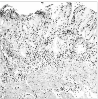
Pathologic examination revealed similar histopathologic changes in the all the gastric specimens. There was a subep- ithelial deposition of thick, homogenous eosinophilic colla- gen in the gastric mucosa. This subepithelial collagen was strongly stained with Masson-trichrome but negative for amy- loid with Congo red. The collagenous deposition appeared as a band-like pattern, which involved the most subepithelial areas of the gastric mucosa. But it was often discontinuous and irregularly extended deeper into proper glands, especially in the antral mucosa (Fig. 2). The thickness of collagenous band measured up to 150 m in the antrum of the stomach but averaged 30-50 m in the fundus and the body. The collagen bands contained entrapped dilated capillaries and mild inflam- matory infiltrates including lymphoplasma cells and eosino- phils (Fig. 3). There were mild to moderate atrophy of the foveolar glands and focal surface epithelial damages in all biop- sies. The surface epithelial changes included partial detach- ment, nuclear stratification, and rare mitosis. There was mod- erate inflammatory infiltrate of the lamina propria without collagen deposits, composed of lymphocytes, plasma cells and many eosinophils. The muscularis mucosa of all specimens showed marked hypertrophy with frequent smooth muscle up-growth into the lamina propria. Intraepithelial lympho- cytic infiltration was rarely seen. Neither Helicobacter pylori nor endocrine cell hyperplasia were found. This histologic finding of the stomach was consistent with collagenous gastritis. The mucosal biopsies from the duodenum, esophagus and colon revealed no remarkable change. On ultrastructural examina- tion of gastric biopsy, subepithelial collagen bands revealed
Fig. 2.Endoscopic biopsy from the antral mucosa shows subepi- thelial collagenous deposition, slough of surface epithelium, inflam- matory cell infiltration, and hypertrophy of muscularis mucosa (H&E,
×100).
Fig. 1.Endoscopy of the stomach shows polypoid mucosal nod- ules around the pyloric ring.
Fig. 3.High power magnification of Fig. 2. The subepithelial collage- nous bands contains entrapped dilated capillaries and inflamma- tory cells (H&E, ×200).
148 S. Park, D.H. Kim, Y.H. Choe, et al.
haphazardly arranged collagen fibrils, and there was focal thick- ening of basement membrane of the proper glands.
DISCUSSION
Collagenous gastritis is defined histologically by the pres- ence of a thickened subepithelial collagen band greater than 10 m thick in association with entrapping dilated capillar- ies and inflammatory cell infiltration of the lamina propria.
Collagenous gastritis was first described in 1989 by Colletti and Trainer (2), since then, less than 20 cases have been report- ed to date. A summary of these cases seems to delineate two major distinct clinicopathologic patterns in patients with col- lagenous gastritis. The first subset is observed in children or young adults with no evidence of extragastric involvement (2, 5), the second subset is associated with collagenous colitis in adult patient (6-8). The first subset is different from adult cases by the severity of the presentation with severe anemia probably due to gastrointestinal bleeding. The gastrointesti- nal bleeding may be caused by entrapped dilated capillaries in the collagenous bands together with superficial epithelial damage.
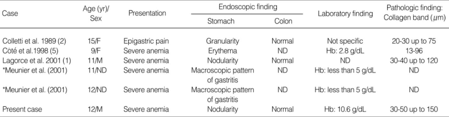
The clinicopathologic and endoscopic features of our patient are similar to the pediatric cases of collagenous gastritis report- ed by Lagorce-Pages et al. (1) and Cote et al. (5). There have been six pediatric cases of collagenous gastritis including this case in the literature (Table 1). The average age at detection was 11.7 yr old. The patients presented with severe anemia or epigastric pain. The gastric endoscopy revealed nodular pattern of the mucosa in three children, erythema in a child and macroscopic pattern of gastritis in two children. The le- sions were limited to the gastric mucosa without evidence of colonic involvement. On the other hand, adult patients of collagenous gastritis showed a variety of clinical and endo- scopic features and simultaneous occurrence of collagenous colitis, collagenous duodenitis, lymphocytic colitis, ulcerative colitis, and celiac disease (1-4, 6-10). There have been 13 adult cases of collagenous gastritis. The incidence of adult cases was more frequent in women than in men and the average age at
detection was 46 yr old (1-4, 6-11). Their main clinical symp- toms were chronic watery diarrhea, although there were excep- tional cases that showed symptoms of the pediatric patients such as anemia, epigastric pain or abdominal distension. There were three cases of collagenous gastritis occurring in young adults aged 20 and 22 yr. The presenting symptoms were epi- gastric pain in 2 patients and severe anemia in one. Of these three patients, a 20 yr old woman had no pathologic findings in the colonic biopsy and no association with atopic dermatitis and bronchial asthma (11), and a 20-yr-old man showed ini- tial presentations of features with collagenous gastritis and was subsequently found to have collagenous colitis (7). The remaining patient showed severe anemia at presentation but colonoscopic finding was not available. In young adult cases of collagenous gastritis except a case associated with collage- nous colitis, the clinical manifestations were similar to those of pediatric patients.
The cause and pathogenesis of collagenous gastritis remain unknown. Three major pathogenic hypothesis have been pro- posed for increased deposition of subepithelial collagen in col- lagenous colitis: 1) chronic inflammation (12-16) and autoim- munity (17), 2) abnormality of the pericryptal fibroblast sheath (18-21), and 3) leakage of plasma proteins and fibrinogen with subsequent replacement with collagen. The most popular theory is that the collagen deposition represents a reparative process secondary to initial injuries caused by drug, toxic or infectious agents. This hypothesis is supported by morpho- logic similarity of collagenous gastritis to collagenous colitis and demonstration of types I and II collagen, a repair-type collagen in collagen deposition of collagenous colitis. How- ever, collagen deposition of collagenous gastritis could not be explained only by reparative process because other gastropa- thies including H. pylori or nonspecific gastritis did not show a thick band-like collagen deposition seen in collagenous gas- tritis. The mechanism of collagen deposition may be complex.
An immune mediated process has been suggested as a mech- anism for collagen deposition of collagenous gastritis because of frequent association with immune related disorders such as collagenous colitis and lymphocytic colitis, and constant signs of immune activation in gastric biopsy from the patients
Case Age (yr)/
Sex Presentation Endoscopic finding
Stomach Colon
Laboratory finding Pathologic finding:
Collagen band ( m)
Colletti et al. 1989 (2) 15/F Epigastric pain Granularity Normal Not specific 20-30 up to 75
Cote et al.1998 (5) 9/F Severe anemia Erythema ND Hb: 2.8 g/dL 13-96
Lagorce et al. 2001 (1) 11/M Severe anemia Nodularity Normal ND 30-40 up to 120
*Meunier et al. (2001) 11/ND Severe anemia Macroscopic pattern ND Hb: less than 5 g/dL ND of gastritis
*Meunier et al. (2001) 12/ND Severe anemia Macroscopic pattern ND Hb: less than 5 g/dL ND of gastritis
Present case 12/M Severe anemia Nodularity Normal Hb: 10.6 g/dL 30-50 up to 150
Table 1.Clinicopathologic features of 6 previously reported pediatric patients with collagenous gastritis (ages less than 18 yr)
ND, not described; Hb, hemoglobin. *This article was written in French and abstract was only available for review.
∨ ′
∨ ′
Collagenous Gastritis 149
with collagenous gastritis. The signs of immune activation include overexpression of HLA-DR by epithelial cells and CD25 positive cells in the lamina propria. The collagen depo- sition may have resulted from activated immune cells produc- ing cytokines and growth factors and thus stimulating the production or reducing the turnover of extacellular matrix.
The gastric mucosa of our patient showed prominent eosino- philic infiltrate of lamina propria that may play a role in im- mune activation. Our patient had no history of drug or spe- cific infection.
In summary, we described typical clinicopathologic find- ings of pediatric collagenous gastritis. This is the first patho- logically proven case of collagenous gastritis in Korea and the sixth pediatric case in the literature. This patient is being followed up at regular intervals with oral antacid and iron supplementation and needs to be checked up for the devel- opment of lower gastrointestinal symptoms and association with immune mediated disorders. More cases will shed light on the pathogenesis of this rare disease entity.
REFERENCES
1. Lagorce-Pages C, Fabiani B, Bouvier R, Scoazec JY, Durand L, Flejou JF. Collagenous gastritis: a report of six cases. Am J Surg Pathol 2001; 25: 1174-9.
2. Colletti RB, Trainer TD. Collagenous gastritis. Gastroenterology 1989; 97: 1552-5.
3. Stancu M, De Petris G, Palumbo TP, Lev R. Collagenous gastritis associated with lymphocytic gastritis and celiac disease. Arch Pathol Lab Med 2001; 125: 1579-84.
4. Vesoulis Z, Lozanski G, Ravichandran P, Esber E. Collagenous gas- tritis: a case report, morphologic evaluation, and review. Mod Pathol 2000; 13: 591-6.
5. Cote JF, Hankard GF, Faure C, Mougenot JF, Holvoet L, Cezard JP, Navarro J, Peuchmaur M. Collagenous gastritis revealed by severe anemia in a child. Hum Pathol 1998; 29: 883-6.
6. Castellano VM, Munoz MT, Colina F, Nevado M, Casis B, Solis-Her- ruzo JA. Collagenous gastrobulbitis and collagenous colitis: case
report and review of the literature. Scand J Gastroenterol 1999; 34:
632-8.
7. Pulimood AB, Ramakrishna BS, Mathan MM. Collagenous gastri- tis and collagenous colitis: a report with sequential histological and ultrastructural findings. Gut 1999; 44: 881-5.
8. Stolte M, Ritter M, Borchard F, Koch-Scherrer G. Collagenous gas- troduodenitis on collagenous colitis. Endoscopy 1990; 22: 186-7.
9. Groisman GM, Meyers S, Harpaz N. Collagenous gastritis associat- ed with lymphocytic colitis. J Clin Gastroenterol 1996; 22: 134-7.
10. Borchard F, Niederau C. Collagenous gastroduodenitis. Dtsch Med Wschr 1989; 114: 1345.
11. Kajino Y, Kushima R, Koyama S, Fujiyama Y, Okabe H. Collagenous gastritis in a young Japanese woman. Pathol Int 2003; 53: 174-8.
12. Jawhary AJ, Talbot IC. Microscopic lymphocytic and collagenous colitis. Histopathology 1996; 29: 101-10.
13. Teglbjaerg PS, Thaysen EH, Jansen HH. Development of collagenous colitis in sequential biopsy specimens. Gastroenterology 1984; 87:
703-9.
14. Jessurun J, Yardley JH, Lee EL, Vendrell DD, Schiller LR, Fordtran JS. Different names for the same condition? Gastroenterology 1986;
91: 1583-4.
15. Rams H, Rogers AI, Ghandur-Mnaymneh L. Collagenous colitis.
Ann Intern Med 1987; 106: 108-13.
16. Lawson JM, Wolosin J, Mottet MD, Brower RA. Collagenous coli- tis: an association with fecal leukocytes. J Clin Gastroenterol 1988;
10: 672-5.
17. Jawhari A, Talbot IC. Microscopic, lymphocytic and collagenous col- itis. Histopathology 1996; 29: 101-10.
18. Widgren S, Jlidi R, Cox JN. Collagenous colitis: histologic, morpho- metric immunohistochemical and ultrastructural studies. Report of 21 cases. Virchow Arch A Pathol Anat Histopathol 1988; 413: 287-96.
19. Kingham JG, Levison DA, Morson BC, Dawson AM. Collagenous colitis. Gut 1986; 27: 570-77.
20. Colina F, Solis-Herruzo JA, Munoz-Yague MT, Vazquez G, Perez- Barrios A. Collagenous colitis: the clinical and morphological fea- tures. Postgrad Med J 1982; 58: 390-95.
21. Gledhill A, Cole FM. Significance of basement membrane thicken- ing in the human colon. Gut 1984; 25: 1085-8.
∨ ′ ′