183 Received:July 22, 2015, Revised:August 17, 2015, Accepted:August 24, 2015
Corresponding to:Mi Il Kang, Department of Internal Medicine, Dankook University Medical College, 119 Dandae-ro, Dongnam-gu, Cheonan 31116, Korea. E-mail:satisfe00@naver.com
pISSN: 2093-940X, eISSN: 2233-4718
Copyright ⓒ 2016 by The Korean College of Rheumatology. All rights reserved.
This is a Free Access article, which permits unrestricted non-commerical use, distribution, and reproduction in any medium, provided the original work is properly cited.
Case Report
Journal of Rheumatic Diseases Vol. 23, No. 3, June, 2016 http://dx.doi.org/10.4078/jrd.2016.23.3.183
Dermatomyositis: A Rare Extra-intestinal Manifestation of Ulcerative Colitis
Chang Hyun Park1, Na Hye Myong2, Hyun Don Joo1, Mi Il Kang1
Departments of 1Internal Medicine and 2Pathology, Dankook University Medical College, Cheonan, Korea
Inflammatory myositis as an extra-intestinal manifestation of inflammatory bowel disease (IBD) is rare. Coexistence of im- muno-mediated diseases in patients with IBD and myositis suggests a common etiopathogenic mechanism underlying these conditions. The current report refers to a rare case of a 45-year-old Korean female with ulcerative colitis (UC) who developed dermatomyositis. She presented with skin rash and proximal muscle weakness, and her disease activity of UC was in remission state. Electromyography, magnetic resonance imaging, and deltoid muscle biopsy were performed. She was diagnosed with dermatomyositis associated with UC and treatment with glucocorticoids and azathioprine resulted in improvement in muscle power and skin rash. Clinicians should be aware of this unusual extra-intestinal manifestation. (J Rheum Dis 2016;23:183-186) Key Words. Inflammatory bowel diseases, Ulcerative colitis, Myositis, Polymyositis
INTRODUCTION
Ulcerative colitis (UC), an inflammatory bowel disease (IBD) of unknown etiology, is frequently associated with extra-intestinal manifestations [1]. Common sites in- clude the joints (arthritis/arthralgia), skin (erythema no- dosum, pyoderma gangrenosum), and eyes (iritis, uvei- tis) [2], while muscular diseases such as myositis have rarely been reported. Only a few cases of myositis asso- ciated with UC have been described in the literature [3].
We report on a case of a middle-aged woman with quies- cent UC who developed dermatomyositis responsive to combined treatment with glucocorticoid and immuno- suppressive agent. To the best of our knowledge, this is the first reported case of dermatomyositis associated with UC in Korea.
CASE REPORT
We report on a case of a 45-year-old woman with UC since 2012, diagnosed based on colonoscopic appearance
and biopsy and maintained on mesalazine 1 g supposito- ries daily. The patient had been in remission for 1 year when she presented in January of 2014 with skin erup- tions and both upper and lower limb proximal muscle weakness. She had not taken any medication associated with myopathy for 1 year. Colonoscopic findings five months prior to this event were normal. Two months be- fore admission, she had developed an erythematous rash on her proximal extremities, neck, and eyelid, and eyelid swelling. She complained of significant pruritus, and mild myalgia on both upper arms and thighs. Three weeks be- fore admission, she had begun to have difficulty climbing stairs, and lifting packages. The patient did not complain of arthralgia or inflammatory back pain.
On physical examination, she had Medical Research Council (MRC) scale grade 4 symmetric weakness of the proximal upper and lower extremities, and full strength in distal limbs.
No sensory disturbance was detected. And there was no evidence to suggest spondyloarthropathy.
Widespread macular erythematous patches of both up-
Chang Hyun Park et al.
184 J Rheum Dis Vol. 23, No. 3, June, 2016
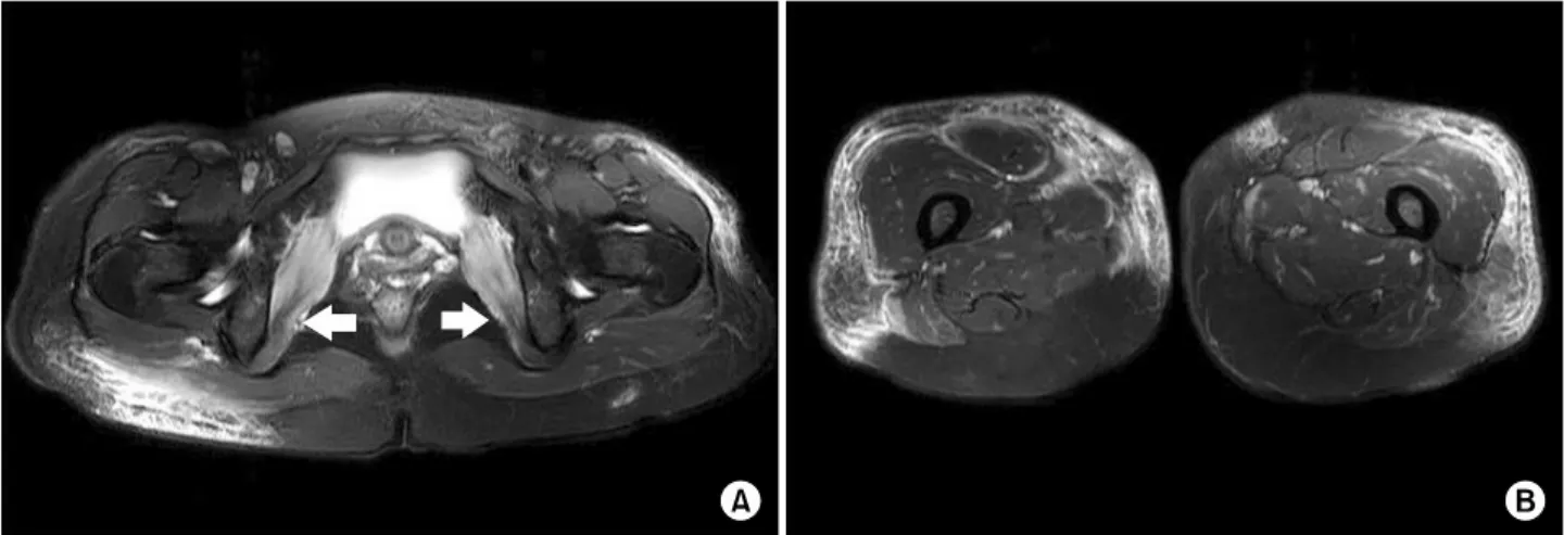
Figure 1. (A) Axial T2 weighted image with fat saturation showed bilateral symmetrical diffuse increased signal intensity in both obturator internus muscles (arrows). (B) Axial T2 weighted image with fat saturation showed epimysial accentuation of both vastus muscles with diffuse interstitial edema in the subcutaneous fat layer on both thighs.
Figure 2. Biopsy from the left deltoid muscle revealed a size variation of myofibers and multifocal lymphocyte infiltration in the perimyseal area and perivascular area. Some degenerat- ing and regenerating myofibers are shown. There is no evi- dence of vasculitis (H&E, ×100).
per arms and thighs were noted, with no nail findings. A V-sign on the neck and heliotroph erythema and swelling of eyelid were observed. Physical examination was other- wise unremarkable, with normal vital signs.
Laboratory tests showed the following: white blood cell count was 4,110/mm3, hemoglobin was 12.6 g/dL, and pla- telet count was 272,000/mm3. Creatine phosphokinase (CPK) was increased to 434 IU/L (26 to 174 IU/L) and lactate dehydrogenase (LDH) was increased to 624 IU/L (240 to 480 IU/L), while serum aldolase level was within normal range (<7.5 U/L). Erythrocyte sedimentation rate value was elevated at 66 mm/h (0 to 30 mm/h) and C-reactive protein was slightly increased to 0.56 mg/dL (0 to 0.5 mg/dL). The level of antinuclear antibodies was 1:40 positive and both anti-Jo-1 antibody and anti-neu- trophil cytoplasmic antibody were negative. Thyroid- stimulating hormone, thyroxine was within normal range. Computed tomography of the abdomen, pelvis, and chest was normal.
On electromyography (EMG), only the presence of short potentials of small amplitude at the level of the right del- toid muscle was observed. These findings were sugges- tive, but not conclusive, of a diagnosis of inflammatory myositis. Nerve conduction studies were normal. Magnetic resonance imaging (MRI) of both thighs showed sym- metrical diffuse increased signal intensity in T2 weighted images with fat saturation in both obturator internus muscles (Figure 1A) and epimysial accentuation of both vastus muscles with diffuse interstitial edema in the sub- cutaneous fat layer on both thighs consistent with in- flammatory myositis (Figure 1B). Biopsy from the left
deltoid muscle showed multifocal lymphocytic peri- vascular infiltration within perimyseal spaces, and peri- myseal lymphocytic infiltration had occurred. Multiple degenerated and regenerated myofibers were also observed. In conclusion, muscle biopsy results were con- sistent with inflammatory myositis (Figure 2). No other extra-intestinal manifestations were observed.
Once the diagnosis of dermatomyositis according to cri- teria of Bohan and Peter [4] was established, the patient was initially treated with oral methylprednisolone of 40 mg daily. Thus, azathioprine 50 mg daily was added, with stepwise reduction of methylprednisolone. Symptoms of myopathy and skin manifestations showed a gradual de-
Dermatomyositis Associated with Ulcerative Colitis
www.jrd.or.kr 185
crease until complete disappearance, while muscle strength had improved considerably 3 months after start- ing therapy. Serum levels of CPK and LDH returned to normal values. Treatment resulted in complete recovery of skin manifestation and return of muscle power to normal. Disease activity of UC was also remission state for more than 1 year.
DISCUSSION
Inflammatory myopathy is a rare form of extra-intestinal manifestation (EIM) in IBD. UC appears to be less com- monly associated with an inflammatory myopathy than Crohn’s disease [3]. Dermatomyositis is a particularly rare EIM of UC. The earliest case of myositis associated with UC was reported by Oshitani et al. [5], who de- scribed dermatomyositis and mononeuritis multiplex in UC and Basedow’s disease. Two cases have been reported [5,6], and this is the first case of dermatomyositis with UC reported in Korea.
The relationship between EIMs and IBD was recognized several years ago, but pathologic factors remain unclear, warranting further investigation. There are some hypoth- eses on the pathogenetic autoimmune mechanisms of EIM in IBD patients. According to a common im- mune-mediated mechanism hypothesized by some au- thors, bowel inflammation and mucosal damage can lead to the release of antigens stimulating an immune response.
The release of intestinal antigens and subsequent pro- duction of antibody-forming immune complexes are re- sponsible for muscle injury [7,8].
Other mechanisms suggested by some authors include an immune response initiated by an infectious agent, sec- ondarily driven against body components such as large bowel and striated muscles. Various infectious agents, in- cluding persistent infection with measles virus, para- myxovirus, Mycobacterium paratuberculosis, herpes virus, and Escherichia coli, have been implicated in the patho- genesis of IBD or myositis. It can probably initiate antigen driven production of autoantibodies (anti-Jo-1), result- ing in generation of myositis [9,10]. Therapy for IBD with glucocorticoids, or azathioprine could also result in mus- cle damage [9]. However, this does not explain myositis that develops before IBD and the same was not true for this case [10]. Regarding disease activity, in some re- ported patients, myositis is associated with an acute ex- acerbation of IBD, but activity of bowel disease does not appear to be mandatory for the onset of myositis. In some
IBD cases, myopathy was diagnosed in a quiescent phase [3], and the current patient was also in clinical remission of UC when symptoms of myositis appeared. However, symptoms associated with myositis, such as fever, myal- gia, muscle fatigue, and weakness may be considered as a part of the clinical manifestations of IBD in patients with active disease. Thus, misinterpretation can occur in diag- nosis of myopathy in association with disease activity of IBD and caution is required [3].
Patients with IBD are often under treatment with sys- temic steroids; therefore, inflammatory myopathy must be differentiated from steroid myopathy, which may also induce an increase in serum CPK levels. These conditions may be differentiated by muscle biopsy. In the absence of histological evidence, if the myopathy is secondary to ste- roids, dose reduction of steroids should be followed by a rapid drop in serum CPK levels, while in the case of my- ositis complicating IBD, myopathy can either progress or remain unchanged [3].
Inflammatory myopathy is considered a rare EIM of IBD, but in fact may be a more common occurrence than re- ported in patients with IBD. As already mentioned, symp- toms of inflammatory myopathy may overlap and be con- cealed with clinical manifestations of active IBD. In addi- tion, diagnosis of subclinical myositis in IBD patients has also been reported with the finding of elevated serum CPK levels and obvious pathological changes in biopsy specimens from striated muscles in the absence of typical symptoms [11]. To put this in perspective, serum CPK should be measured in patients with IBD who show symptoms of myalgia and weakness.
Treatment of inflammatory myopathy is usually based on oral administration of glucocorticoids, either alone or in combination of steroids with immunosuppressive agents, such as azathioprine, methotrexate, or cyclo- sporine [7,12,13]. The efficacy of these immunosuppressive agents in the treatment of inflammatory myopathy sup- ports the hypothesis of a common mechanism of myositis and IBD.
SUMMARY
This case report shows that the diagnosis of in- flammatory myositis should be considered in IBD pa- tients complaining of myalgia or muscular weakness. It can be speculated that the true prevalence of myositis in of IBD is underestimated because nonspecific symptoms are not directly correlated with myositis.
Chang Hyun Park et al.
186 J Rheum Dis Vol. 23, No. 3, June, 2016
Inflammatory myositis should be strongly suspected in UC patients with muscle weakness and elevated serum CPK levels, which may be helpful in the early diagnosis and appropriate treatment of UC. Immune mediated my- ositis should be considered as an important differential diagnosis for myopathy occurring in IBD secondary to glucocorticoid treatment [14]. Early recognition of these extra-intestinal manifestations should be helpful in guid- ing therapy that will reduce overall morbidity in affected patients. Hence, an appropriate approach to muscu- loskeletal manifestations in IBD requires close coopera- tion between gastroenterologists and rheumatologists.
ACKNOWLEDGMENTS
We thank Dr. Ji young Lee for the preparation and inter- pretation of MRI images.
CONFLICT OF INTEREST
No potential conflict of interest relevant to this article was reported.
REFERENCES
1. Bernstein CN, Blanchard JF, Rawsthorne P, Yu N. The prev- alence of extraintestinal diseases in inflammatory bowel disease: a population-based study. Am J Gastroenterol 2001;96:1116-22.
2. Orchard TR, Wordsworth BP, Jewell DP. Peripheral arthro- pathies in inflammatory bowel disease: their articular dis- tribution and natural history. Gut 1998;42:387-91.
3. Paoluzi OA, Crispino P, Rivera M, Iacopini F, Palladini D, Consolazio A, et al. Skeletal muscle disorders associated
with inflammatory bowel diseases: occurrence of myositis in a patient with ulcerative colitis and Hashimoto's thyroi- ditis−case report and review of the literature. Int J Colorec- tal Dis 2006;21:473-7.
4. Bohan A, Peter JB. Polymyositis and dermatomyositis (first of two parts). N Engl J Med 1975;292:344-7.
5. Oshitani H, Wakabayashi Y, Sawaguchi Y, Koike H, Yoshino Y. A case of dermatomyositis followed by multiple mono- neuritis, with the history of ulcerative colitis and Basedow's disease. J Kyorin Med Soc 1981;12:47-53.
6. Hayashi T, Nakamura T, Kurachi K, Asai Y, Nakajima A, Suzuki S, et al. Ulcerative colitis accompanied with sarcoi- dosis and dermatomyositis: report of a case. Dis Colon Rectum 2008;51:474-6.
7. Leibowitz G, Eliakim R, Amir G, Rachmilewitz D. Dermato- myositis associated with Crohn's disease. J Clin Gastroen- terol 1994;18:48-52.
8. Hodgson HJ, Potter BJ, Jewell DP. Immune complexes in ul- cerative colitis and Crohn's disease. Clin Exp Immunol 1977;29:187-96.
9. Braun-Moscovici Y, Schapira D, Balbir-Gurman A, Nahir AM. Inflammatory bowel disease and myositis. Clin Rheumatol 1999;18:261-3.
10. Shimoyama T, Tamura Y, Sakamoto T, Inoue K. Immune- mediated myositis in Crohn's disease. Muscle Nerve 2009;39:101-5.
11. Hayashi K, Kurisu Y, Ohshiba S, Kawamura H, Furutama D, Takada K, et al. Report of a case of Crohn's disease asso- ciated with hyper-creatine phosphokinase-emia. Jpn J Med 1991;30:441-5.
12. Danieli MG, Malcangi G, Palmieri C, Logullo F, Salvi A, Piani M, et al. Cyclosporin A and intravenous immunoglob- ulin treatment in polymyositis/dermatomyositis. Ann Rheum Dis 2002;61:37-41.
13. Metzger AL, Bohan A, Goldberg LS, Bluestone R, Pearson CM. Polymyositis and dermatomyositis: combined metho- trexate and corticosteroid therapy. Ann Intern Med 1974;
81:182-9.
14. Podolsky DK. Inflammatory bowel disease. N Engl J Med 2002;347:417-29.