www.jpis.org
pISSN 2093-2278 eISSN 2093-2286 Copyright © 2012 Korean Academy of PeriodontologyThis is an Open Access article distributed under the terms of the Creative Commons Attribution Non-Commercial License (http://creativecommons.org/licenses/by-nc/3.0/).
Evaluation of calcium sulphate barrier to collagen membrane in intrabony defects
Shilpa Budhiraja1, Neeta Bhavsar2, Santosh Kumar1,*, Khushboo Desai1, Sareen Duseja3
1Department of Periodontology, Karnavati School of Dentistry, Gandhinagar, India
2Department of Periodontology, Government Dental College, Asarwa, India
3Department of Prosthodontics, Karnavati School of Dentistry, Gandhinagar, India
Purpose: The aim of this study was to clinically and radiographically evaluate and compare treatment of intrabony defects with the use of decalcified freeze-dried bone allograft in combination with a calcium sulphate barrier to collagen membrane.
Methods: Twelve patients having chronic periodontal disease aged 20 to 50 years and with a probing depth >6 mm were se- lected. Classification of patient defects into experimental and control groups was made randomly. In the test group, a calcium sulphate barrier membrane, and in control group, a collagen membrane, was used in conjunction with decalcified freeze- dried bone graft in both sides. Ancillary parameters as well as soft tissue parameters along with radiographs were taken at baseline and after 6 months of surgery. Parameters assessed were plaque index, modified gingival index, probing depth, rela- tive attachment level, and location of the gingival margin. A Student’s t-test was done for intragroup and a paired t-test for in- tergroup analysis.
Results: Intragroup analysis revealed statistically significant improvement in all the ancillary parameters and soft tissue pa- rameters with no statistically significant difference in intergroup analysis.
Conclusions: The study concluded that a calcium sulphate barrier was comparable to collagen membrane in achieving clini- cal benefits and hence it can be used as an economical alternative to collagen membrane.
Keywords: Bone substitute, Calcium sulphate, Collagen, Guided tissue regeneration.
INTRODUCTION
Humans have always struggled directly or indirectly with various oral diseases. Gingival and periodontal diseases, in their various forms, have afflicted humans since the dawn of history. In the 21st century, the goals for periodontal therapy are continually being refined not only to achieve a healthy periodontium but also to recreate a normal anatomic rela- tionship and function. Periodontal therapy has the principle objective of the morphological and functional reconstruction of lost periodontal supporting tissues. However, conventional periodontal therapy has been found to result in repair rather than regeneration, and this process is often characterized by
the establishment of long junctional epithelium between the root surface and gingival connective tissue.
The first stage of development of periodontal regeneration methods focused on reconstruction of osseous lesions utiliz- ing a variety of bone replacement grafts. Different types of grafts have been used. Allografts like decalcified freeze-dried bone allograft (DFDBA) showed some promising results, but the ability to regenerate the cementum, periodontal ligament (PDL), and alveolar bone was found to be limited. The era of grafts thus made way for the era of the pioneering technique of guided tissue regeneration (GTR). The placement of the occlusive membrane guides the progenitor cells residing in the PDL to repopulate the osseous defects in order to form
Received: Oct. 29, 2012; Accepted: Nov. 28, 2012
*Correspondence: Santosh Kumar
Department of Periodontology, Karnavati School of Dentistry, 907/A, Uvarsad 382422, Gandhinagar, Gujarat, India E-mail: [email protected], Tel: +91-960-1800-379, Fax: +91-79-23970296
Collagen is the primary structural protein of connective tis- sue [1]. Resorbable collagen membranes have advantages over traditional materials in achieving clot stabilization, wound stability, space provision, and epithelial cell exclusion.
When placed in human periodontal defects, calcium sulphate was found to be biocompatible [2]. It is porous and resorbable [3] in nature and it eliminates the need for suturing.
Thus the aim of the present study is to clinically and radio- graphically evaluate and compare intrabony defect treatment results using DFDBA in combination with a calcium sulphate barrier to collagen membrane.
MATERIALS AND METHODS
Twelve patients with chronic periodontal disease aged 20 to 50 years and having three-wall defects with probing depth
>6 mm were selected from the patient pool of the Depart- ment of Periodontology, Government Dental College and Hospital, Ahmedabad, Gujarat, India. Informed consent was obtained from the patients after explaining to them the na- ture of the study. The inclusion criteria of the study were all of the following: 1) clinical and radiographic evidence of at least one pair of intrabony defects in two different quadrants, 2) periodontal defects with probing depths greater than or equal to 6 mm, 3) endodontically asymptomatic teeth, 4) pa- tients having adequate gingival coverage in selected teeth, and 5) a defect depth of at least 3 mm as detected on the ra- diographs.
After reevaluation of the phase I therapy, patients with any of the following were excluded from the study: 1) patients who were medically compromised or on any drug therapy, which may influence the healing of tissues, 2) smoking or use of other tobacco products, 3) unacceptable oral hygiene habits, 4) history of previous treatment by open debridement and/or regenerative therapy at the same site.
Presurgical therapy
At phase I therapy, thorough scaling and root planing was done and oral hygiene instructions were given. Customized acrylic occlusal stents with vertical grooves were prepared for each patient on their respective study cast to record the direc- tion of insertion of the periodontal probe interproximally and to standardize it for future measurements. Classification of defects in patients as experimental or control was done at this time. Sites were classified randomly as test (t) or control (c).
Clinical and radiographic measurements
Parameters were recorded at baseline, 3 months, and 6 months postoperatively to evaluate the soft tissue response
were plaque index [4], modified gingival index [5], probing depth, relative attachment level (RAL), and location of the gingival margin. Radiographic evaluation [6] was done preop- eratively and 6 months postoperatively by using the long cone paralleling technique in the field of interest. An indige- nously developed standardized X-ray grid was placed in front of the intraoral periapical film on a film holder and an X-ray was taken of the selected teeth.
Surgical procedure
All patients were prescribed to take 100 mg doxycycline a day in capsule form before surgery and continue to take the same medicine postoperatively. The surgical area was anes- thetized using 2% lidocaine hydrochloride with adrenaline (1:80,000). Sulcular incisions were made aimed at preserving as much of the interproximal tissue as possible by using a no.
15 Bard-Parker blade and mucoperiosteal flaps were raised at the buccal and lingual aspects to expose the 3 to 4 mm alveo- lar process. The inner surfaces of the flaps were curetted, tak- ing care not to thin them excessively and hence compromise the blood supply to the healing wound. A thorough debride- ment of the defects was done and the exposed root surfaces were thoroughly planed to a smooth hard surface. After hy- dration of the graft (DFDBA, Dembone, Pacific Coast Tissue Bank, Los Angeles, CA, USA) particles in physiologic saline, they were incrementally condensed into the defect. Overfill- ing of the defect was avoided. Interrupted interdental presu- turing was done with 3.0 Mersilk suture material.
Following graft placement, a bioresorbable collagen mem- brane (Progide, equinox, Zeist, Holland) was trimmed and adapted over the entire defect on the control site so as to cover 2 to 3 mm of surrounding alveolar bone and to ensure stability of the graft material. On the experimental site, a calcium sul- phate (DentoGen-Calcium Sulphate Hemihydrate, Biodenix Technologies Inc., Richmond, CA, USA) barrier was applied.
The barrier material was prepared by mixing the calcium sul- phate powder in the sterile box with the liquid provided by the company. The ideal mix was dry enough to clump together but wet enough to spread with an instrument when posi- tioned on the bone graft surface. A dry surgical field was very important at this stage. The mix was then placed over the graft.
The barrier should be 1.5- to 2-mm thick. It should cover all of the bone graft particles and extend on to the surrounding bone for an additional 2 to 3 mm. A total soft tissue primary closure was obtained by using 3-0 sutures in interrupted fash- ion. A periodontal dressing was used to cover the treated sites.
Postoperative care
Postoperatively subjects were prescribed 100 mg doxycy-
completion of surgery, and 400 mg analgesic ibuprofen three times a day for 5 days. Rinsing with 0.2% chlorhexidine diglu- conate solution twice daily for 4 weeks was also mandatory.
They were advised to avoid hard chewing on the surgical area for 10 days. Pack and suture removal was done on the 10th day.
Statistical analysis
The data obtained from the present study was suitably tab- ulated in appropriate tables. The Student’s t-test and paired t-test were applied to achieve the level of significance, when- ever indicated. The mean and standard deviation were calcu- lated at 3 months only to show the trends over time. For be- tween-group comparison from baseline to 3 months, the Student’s t-test was used. These t-test values were compared
RESULTS
Wound healing was uneventful for all treated cases. Soft tissues healed within normal limits and no significant visual differences were noted between the treatment groups. A sta- tistically significant reduction was noted in the plaque score and gingival inflammation in both test and control groups (Table 1). Between the groups, there is no statistically signifi- cant (P>0.05) difference in probable pocket depth (PPD) re- duction, as both the groups show similar results. There is no statistically significant (P>0.05) difference in mean RAL gain (Table 2) and mean gingival margin level (Table 3) as both the groups show similar results. There is a statistically significant (P<0.01) increase in defect fill from before surgery to 6 months Table 1. Comparison of mean probable pocket depth reduction at baseline to 6 months postoperatively between the groups (mm).
Group Baseline 6-month Probable probing
depth reduction Mean difference
(0–6 months ) T-value Significance
Calcium sulphate/DFDBA 6.92±0.90 3.25±0.75 3.66±0.65 -0.167 -0.432 0.67 (P>0.05)
Collagen membrane/DFDBA 7.08±1.60 3.33±0.98 3.75±0.86 -0.167 -0.364 0.72 (P>0.05)
Values are presented as mean±standard deviation.
DFDBA: decalcified freeze-dried bone allograft.
Table 2. Comparison of mean relative attachment level at baseline to 6 months post operatively between the groups (mm).
Group Baseline 6-month Relative attachment
level gain Mean difference
(0–6 months) T-value Significance
Calcium sulphate/DFDBA 9.42±1.08 6.25±1.35 3.16±0.57 -0.333 -0.67 0.517 (P>0.05)
Collagen membrane/DFDBA 9.75±1.91 6.08±1.10 3.66±1.07 -0.167 -0.432 0.674 (P>0.05)
Values are presented as mean±standard deviation.
DFDBA: decalcified freeze-dried bone allograft.
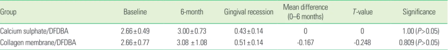
Table 3. Comparison of mean gingival marginal level at baseline to 6 months post operatively between the groups (mm).
Group Baseline 6-month Gingival recession Mean difference
(0–6 months) T-value Significance
Calcium sulphate/DFDBA 2.66±0.49 3.00±0.73 0.43±0.14 0 0 1.00 (P>0.05)
Collagen membrane/DFDBA 2.66±0.77 3.08 ±1.08 0.51±0.14 -0.167 -0.248 0.809 (P>0.05)
Values are presented as mean±standard deviation.
DFDBA: decalcified freeze-dried bone allograft.
Table 4. Comparison of mean change in defect fill (mm and %) at baseline and 6 months within and between the groups.
Group Preoperative 6-month Defect fill
T-value Significance
mm %
Calcium sulphate/DFDBA 7.0±1.04 2.83±1.02 4.16±0.57 65.87 15.43 P<0.01
Collagen membrane/DFDBA 7.33±1.15 2.83±1.11 4.50±0.67 67.69 13.45 P<0.01
Values are presented as mean±standard deviation.
DFDBA: decalcified freeze-dried bone allograft.
in both the treatment groups (Table 4) (Figs. 1–4).
DISCUSSION
For many years, periodontology has been taken as a branch dealing with treatment strategies for attempting to save den- tition suffering from periodontitis, and history has shown that our profession has been successful, to some extent.
The evidence of previous literature has shown that osseous grafting techniques represent one of the modes of therapy for managing the combination of pocket and osseous de- fects. Also, the biological and histological aspects of guided tissue regeneration (GTR) have been established through plenty of animal and human studies. Collagen membrane is one of the most commonly used resorbable membranes, as it is the basic structural unit of connective tissue. It has also the ability to aggregate platelets. Collagen membranes have
been found to have the property of clot stabilization, wound stability, space provision [7], and epithelial cell exclusion which are important factors determining tissue regeneration when a barrier technique is used.
In 1975, Frame [3] listed the following properties of an ideal biodegradable bone implant material: 1) acceptance by the tissues with no untoward reactions, 2) porosity to allow some bone in-growth, 3) biodegradability to preclude areas of weak- ness or possible infection after the new bone has beenformed, 4) ability to be sterilized without a change in its properties, 5) availability and minimal cost. He identified calcium sulphate as such a material.
The use of calcium sulphate as a barrier membrane for GTR has been established by various clinical reports. Calcium sul- phate retards epithelial and connective tissue in-growth to produce a predictable regenerative response [7]. It shows a lack of infection if the material becomes exposed. The mate- Figure 1. Preoperative radiograph showing defect site prior to
treatment with calcium sulphate barrier and decalcified freeze-dried bone allograft.
Figure 3. Preoperative radiograph showing defect site prior to treatment with collagen and decalcified freeze-dried bone allograft.
Figure 2. Postoperative radiograph showing defect fill after under- going treatment with calcium sulphate barrier and decalcified freeze- dried bone allograft.
Figure 4. Postoperative radiograph showing defect fill after under- going treatment with collagen membrane and decalcified freeze- dried bone allograft.
ing root concavities [8]. Above all, it is readily available, easily sterilized, inexpensive, completely resorbable, and biocom- patible, and in the presence of bone and periosteum, it be- comes osteogenic. Also, it has been found that the compres- sive strength of calcium sulphate is greater than that of can- cellous bone with a resorbable capacity of 5 to 7 weeks [9].
Opinions on the effectiveness of DFDBA as a bone implant to augment GTR is varied [10,11]. DFDBA has been used dur- ing GTR procedures because it is thought to be a space main- tainer and to enhance periodontal tissue regeneration by its osteoinductive properties. Urist and Strates [11] reported that the addition of bone grafting materials resulted in signifi- cantly greater pocket depth reduction than in the membrane group only.
Thus, in the present study we have used calcium sulphate as a barrier membrane along with DFDBA as a test group in comparison to collagen membrane along with DFDBA as a control group.
Ancillary parameters were employed to monitor periodon- tal condition throughout the study namely, the Plaque Index [4] and Modified Gingival Index [5].
All soft tissue parameters were measured from the lower border of a customized acrylic stent covering the occlusal surface of the teeth. The acrylic stent was shown to provide a stable, fixed reference point [12] for measurements as com- pared to the cemento-enamel junction, which is often ob- scured by calculus or soft tissues and difficult to locate clini- cally. Moreover, the occlusoapical groove helped to standard- ize the direction of probe insertion.
In the present study, the initial mean probing depths be- tween the groups were not significantly different (Table 1).
After 6 months, the results showed a statistically significant (P<0.01) reduction in probing depth in both treatment groups because of resolution of inflammation. This result showed a statistically significant PPD reduction from baseline to 6 months in the two groups. The findings of this study are in accordance with similar studies conducted by Orsini et al. [13]
and Paolantonio et al. [14]. Similar results were also observed in studies conducted by Trejo et al. [15], Aichelmann-Reidy et al. [16], and Vouros et al. [17].
RAL assessments, made from a fixed reference point (lower border of stent) have the advantage of being a more realistic portrayal of attachment gain or loss than PPD, as they are not influenced by gingival margin changes. The RAL is different from the clinical attachment level, but is recommended for clinical studies due to its ease and reproducibility [18]. In the present study, the RAL gain was statistically significant (Table 2) in both treatment groups after 6 months. The results were analogous to a study by Orsini et al. [13], Paolantonio et al. [14],
[17], Zybutz et al. [19].
The gingival recession (Table 3) noted in the present study was 0.43±0.14 mm for the test site and 0.51±0.14 mm for the control site. Recession at the test site was not significantly dif- ferent from that of the control site (P>0.05). The reduction in the size of the gingiva was attributed to resolution of inflam- mation (shrinkage) after healing. The results were in accor- dance with the results of studies done by Paolantonio et al. [14], Trejo et al. [15], Aichelman-Reidy et al. [16], and Vouros et al. [17].
Radiographic changes in bone were assessed by taking serial radiographs exposed with a standardized millimetre grid. Us- ing such a grid provides an accurate way of assessing changes in bone height less than 1 mm. Measurements were made from the crest of the defect to the base of the defect so as to calculate the defect depth (difference of two measurements).
The radiographic picture (Table 4) in the present study shows gain or defect fill at the test and control site (P<0.01). These results were analogous to those of Trejo et al. [15], Aichelmann- Reidy et al. [16], Zybutz et al. [19], and Zitzmann et al. [20] .
The radiographic findings confirm the clinical findings of the soft tissue response. Despite a lack of concrete evidence of regeneration, the radiographic parameters provided a noninvasive means of evaluating the effect of calcium sul- phate/DFDBA and collagen membrane/DFDBA, as compared to histologic analysis, which is rarely possible in human sub- jects due to ethical constraints. The improvements in param- eters show that both the groups, calcium sulphate/DFDBA and collagen membrane/DFDBA, experienced significant benefits with the periodontal grafting materials.
The intergroup statistical analysis showed no statistically significant differences (P>0.05) in the results between the groups, as the calcium sulphate/DFDBA group showed almost the same results as that of the collagen membrane/ DFDBA group after 6 months in terms of all of the parameters.
All of the above-mentioned comparable results achieved by calcium sulphate/DFDBA compared to collagen membrane/
DFDBA indicated that calcium sulphateis as efficacious as that of collagen membrane as a barrier material. It holds a great promise for improved clinical performance and can be used as a cost-effective replacement for collagen membrane.
CONFLICT OF INTEREST
No potential conflict of interest relevant to this article was reported.
REFERENCES
1. Bunyaratavej P, Wang HL. Collagen membranes: a review.
2. Shaffer CD, App GR. The use of plaster of paris in treating infrabony periodontal defects in humans. J Periodontol 1971;42:685-90.
3. Frame JW. Porous calcium sulphate dihydrate as a biode- gradable implant in bone. J Dent 1975;3:177-87.
4. Turesky S, Gilmore ND, Glickman I. Reduced plaque for- mation by the chloromethyl analogue of victamine C. J Periodontol 1970;41:41-3.
5. Lobene RR, Weatherford T, Ross NM, Lamm RA, Menaker L. A modified gingival index for use in clinical trials. Clin Prev Dent 1986;8:3-6.
6. Reddy MS. Radiographic methods in the evaluation of periodontal therapy. J Periodontol 1992;63(12 Suppl):1078- 84.
7. Sottosanti J. Calcium sulfate: a biodegradable and bio- compatible barrier for guided tissue regeneration. Com- pendium 1992;13:226-8, 230, 232-4.
8. Anson D. Calcium sulfate: a 4-year observation of its use as a resorbable barrier in guided tissue regeneration of periodontal defects. Compend Contin Educ Dent 1996;17:
895-9.
9. Sukumar S, Drizhal I, Paulusova V, Bukac J. Surgical treat- ment of periodontal intrabony defects with calcium sul- phate in combination with beta-tricalcium phosphate:
clinical observations two years post-surgery. Acta Medica (Hradec Kralove) 2011;54:13-20.
10. Blumenthal N, Steinberg J. The use of collagen membrane barriers in conjunction with combined demineralized bone-collagen gel implants in human infrabony defects. J Periodontol 1990;61:319-27.
11. Urist MR, Strates BS. Bone formation in implants of par- tially and wholly demineralized bone matrix. Including observations on acetone-fixed intra and extracellular pro- teins. Clin Orthop Relat Res 1970;71:271-8.
12. Clark DC, Chin Quee T, Bergeron MJ, Chan EC, Lautar- Lemay C, de Gruchy K. Reliability of attachment level
plastic stent. J Periodontol 1987;58:115-8.
13. Orsini M, Orsini G, Benlloch D, Aranda JJ, Lazaro P, Sanz M, et al. Comparison of calcium sulfate and autogenous bone graft to bioabsorbable membranes plus autogenous bone graft in the treatment of intrabony periodontal de- fects: a split-mouth study. J Periodontol 2001;72:296-302.
14. Paolantonio M, Perinetti G, Dolci M, Perfetti G, Tete S, Sammartino G, et al. Surgical treatment of periodontal in- trabony defects with calcium sulfate implant and barrier versus collagen barrier or open flap debridement alone: a 12-month randomized controlled clinical trial. J Periodon- tol 2008;79:1886-93.
15. Trejo PM, Weltman R, Caffesse R. Treatment of intraosse- ous defects with bioabsorbable barriers alone or in com- bination with decalcified freeze-dried bone allograft: a randomized clinical trial. J Periodontol 2000;71:1852-61.
16. Aichelmann-Reidy ME, Heath CD, Reynolds MA. Clinical evaluation of calcium sulfate in combination with demin- eralized freeze-dried bone allograft for the treatment of human intraosseous defects. J Periodontol 2004;75:340-7.
17. Vouros I, Aristodimou E, Konstantinidis A. Guided tissue regeneration in intrabony periodontal defects following treatment with two bioabsorbable membranes in combi- nation with bovine bone mineral graft. A clinical and ra- diographic study. J Clin Periodontol 2004;31:908-17.
18. Caton JG, Greenstein G. Factors related to periodontal re- generation. Periodontol 2000 1993;1:9-15.
19. Zybutz MD, Laurell L, Rapoport DA, Persson GR. Treatment of intrabony defects with resorbable materials, non-re- sorbable materials and flap debridement. J Clin Periodon- tol 2000;27:169-78.
20. Zitzmann NU, Rateitschak-Pluss E, Marinello CP. Treat- ment of angular bone defects with a composite bone grafting material in combination with a collagen mem- brane. J Periodontol 2003;74:687-94.