人힘Utd‘tt~ 협젠썩 It~. 짜 23 샌 까 2 빠 pp. 263 - 267, 1987 Journal 01 Korean Radiological Society, 23(2) 263-267 1987
§후藏의 固形송L 頭狀上皮睡傷
연세 대 학교 의과대 학 방사선과학교실
김 기 황
•정 우 희 * 이 미 경 * 민 진 식 **
- Abstract -
Solid and Papillary Epithelial Neoplasm of the Pancreas
Ki Whang Kim, M.D., Woo Hee
’
ong, M.D. 기 Mikyung Lee, M.D.', Jin Sik Mon, M.D."Department of Radiology, Yonsei University College of Medicine
During last 2 years, we have experienced 3 cases 01 solid and papillary epithelial neoplasm 01 the pancreas AII occured in young women (21, 27, and 29 years) who present with abdominal pain and mass Grossly the tumors all appeared well circumscribed with local or extensive cystic change and hemorrhage Microscopically, the tumors were characterized by distinctive solid and papillary pattern.
(T and sonography 01 the tumor show a well-demarcated mass that can be solid, mixed cystic and solid
。 rmainly cystic
ERCP was done in one case which showed tapered obstruction 01 main duct with round swing 01 side branches
1. 서 트료 」
취장의 고형유두상상피종양 (solid and papillary Epithelial Neoplasm of the Pancreas)는 대 단허 희귀한 질환으로 주로 젊은 여자에서 발생하는 저등급
(low grade) 악성 암-0 로 管願提과 달리 피 막으로 잘 싸여져 있고 국소 임파절이나 원격 전이를하지 않아섣 제에 의해 근치할 수 있는 종양이다1-3) 그러므로 수숭 전에 이 질환의 가능성을 예견할 수 있마연 수술 방침
*
연세대학교 의과대학 병리학교실‘ Department of Pathology, Yonsei University College of Medicine
**
연세대학교 의과대학 외과학교실•• Department of Surgery, Yonsei University College of
ι1edicine
이 논운은 1987 년 3 월 l3 일에 접수하여 1987 년 3 월 27 일에 채택되었음.
을 결정하는데 중요한 일이라 할 수 있다.
저자블은 과거 2 년간 연세대학교 의과대학 방사선과 학교실에서 경험하였던 3 예의 취장의 고형유두상 상파 종양의 방사선학적 소견을 분석 하여 운헌 고찰과 함께 보고하는 바이다.
2. 증례 보고 증 례 l
21 세 여자 환자로 평소 건강하였으나 1 년선부터 상 복부에 종괴가 촉지되었고 2 개월선부터 싱와부 불쾌감 이 생기어 내원하였다 이학석 소견상 십요벼 및 좌 상 복벼 I 4 X 3 cm 크기의 난원형외 양동이 있는 단단한 종괴가 촉지되는 외에는 특이한 소견이 없었다.
렬액 검사는 이상이 없었고 상부 위장판 조영술상 심 이지장파 위선정 -부우|가 외적인 종괴에 외한 압박 소견 이 있었마. 초응파장 고형과 낭성의 혼함 에코를 보이 는 10 cm 크기의 종괴가 취장 우부에서 판찰되었고,선
- 263-
- 大웰 I.iJc M綠뽑짱용;웰 : 第23卷 第2 號 1987 -
산화단층촬영승에 종괴는 주위 조직고}는 경계가 비교적 분명했으며 내부에는 괴사 및 냥성 변화를 볼 수 있었 다. 주위 임파절 전이 냐 간선이의 소견은 없었고 담도 의 확장 역시 볼 수 없었다 (Fig.1).
수술시 취장 두부에 피막이 찰 싸여진 10 cm 크기의 종괴을 제거하였고 국소 임파절 비대는 있었£나 선。l 는없었다.
증례 2
29 세 여자 환자로 만 2 년전 임신 풍 복부 동통」즈로 초음파 검사중 취장 말만부에 낭성 종괴를 발견하였다
(Fig. 2-a). 취장의 假짧뼈 의심하어l 추적 관찰하던 중 크기가 약간 증대되어 내원하였다. 가족력과 과거력상 이상 소견이 없었고 이학적 검시장 초음쿄에서 보이는 종괴는 촉지되지 않았다. 혈액검사상 이상소견이 없었다.
초음파검사상 2 년전 6.5cm 크기의 취장 말단부 낭성 종괴는 좌우 직경이 8cm로 약간 커졌고 후벽에 적은
excrescence 는 여전히 판찰되 었다 (Fig.2-b). 종괴
A
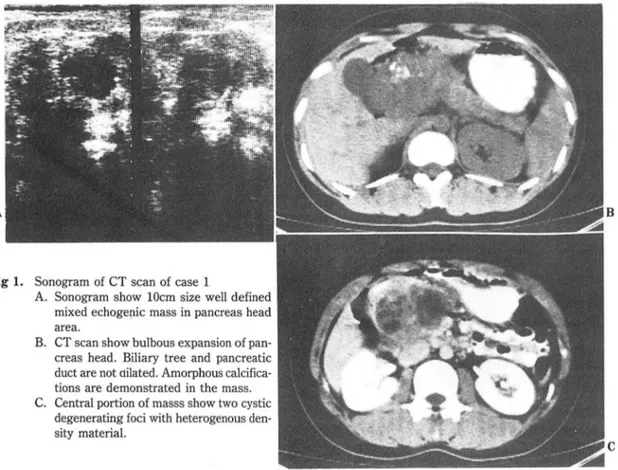
Fig 1. Sonogram of CT scan of case 1
A. Sonogram show 10cm size well defined mixed echogenic mass in pancreas head area.
B. CT scan show bulbous expansion of pan- creas head. Biliary tree and pancreatic duct are not oilated. Amorphous calcifica- tions are demonstrated in the mass C. Central portion of masss show two cystic
degenerating foci with heterogenous den- sity material.
의 한펀에서 中隔을 볼 수 있었다. 수술상 취장의 염증 은 없었고 난형의 단단한 8cm 크거의 종괴를제거하였 다. 종괴는 펴액1] 의해 잘 싸여져 있었으며 비장 정액 과 유착이 있었으냐 국소 임파절이나 간전이는 없었다.
증례 3
27 세 여자 환자로 펑소 건강하였으며 2 일천 심한 상 복~부 동통을 주소로 업원하였다. 급성 뱅색을 보였 o 며 이학적 소견상 좌상훈부 압통응 있었 A냐 종괴는 촉지 되지 않았다. 혈액 검사상 이상소견은 없었다. 초응파 검시장좌상북부에 7 x 5 cm 크기의 경계가잘지워지 며 내부 에코가 不均質인 종괴을 판찰하였고 비장 정액 은 종괴의 후땅에 위치하였다 (Fig. 3-a). 조영제 투여 후 복부 전산화단층촬영상 주위와 경계가 분명하며 벽 이 앓은 저 밀도 불균질의 종괴가 취장 말단부에 있고 내 부 밀도는 44-83 HU 을 보였다. 주위 임파선 전이나 간 선이는 없었다 (Fig. 3-b). 역행성 취담판 조영숭상 취 판 말만부가 tapered obstruction 을 보였고 때分技는
B
C
깅 기 황 외 , 뼈8훌의 lιl形~UWj* 上 æ!lli:짜
A B
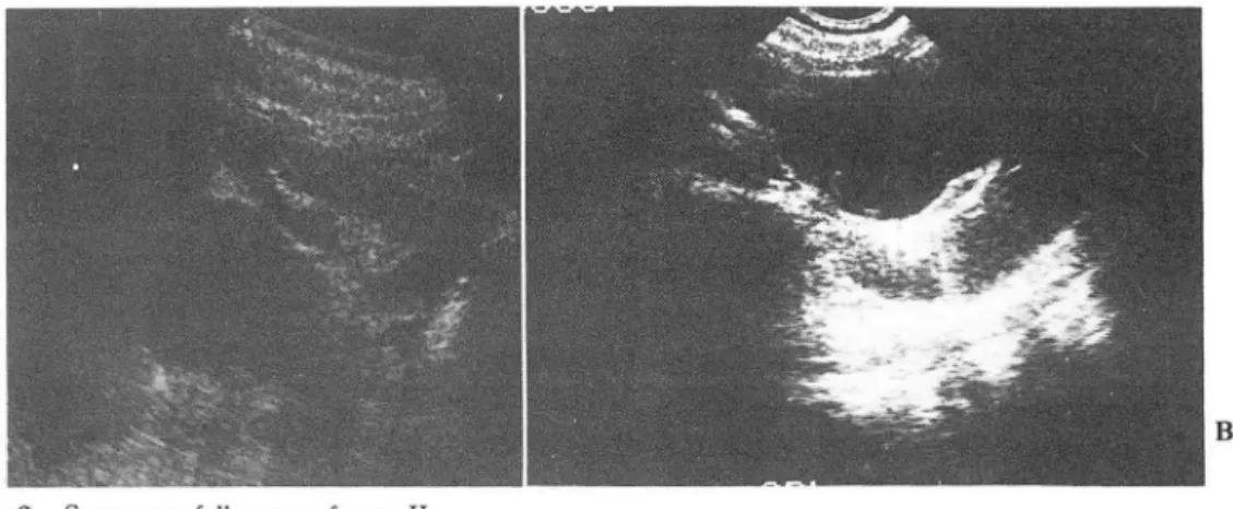
Fig 2. Sonogram follow up of case II
A. There show 6cm size cystic mass in left upper abdomen with excrescence in cyst wall.
B. Two year fol1ow sonogram show increas size of cystic lesion (measured 6 x 8cm). There show in- complete septation and excrescence in medial and posterior aspect of cystic mass.
Sonogram and CT scan of Case III.
A. Sonogram show wel1 defined mass in tail of pancreas with heterogenous echogenicity
B. CT scan also show well defined low den- sity mass with enhancing wal1, mimick- ing cystic lesion.
C. ERCP show tapered obstruction of pan- creatic duct with round swing of side branches
C
round swing을 보여주었다 CFig. 3-c). 수숭상 피막 취장의 고형유두성 상피종양은 Frantz4) 가 1959 년 이 잘 형성된 종괴를 적출하였고 주우l 냐 간 선이는 없 “papi11ary tumor of the pancreas - benign or 었다 malignant ?"라고 키술한 이후 1985 년까지 세계적£
로 약 80 예가 보고되었마1) 국내에서는 검등석 배등 6)
3. 고 찰 김등 7)에 의한 7 예와 저자들의 3 예를 합하여 10 예가
大합放射級짧學會끓‘ : 第 23卷 第2 號 1987 -
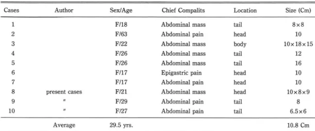
Table 1. Reported Cases in Korea
SexJAge Chief Compalits Location Size (Cm)
F/18 Abdominal mass tail 8x8
F/63 Abdominal pain head 10
F/22 Abdominal mass body 10x 18x 15
F/26 Abdominal mass tail 12
F/26 Abdominal mass tail 16
F/17 Epigastric pain head 10
F/17 Abdominal pain head 10
F/21 Abdominal mass head 10x8x9
F/29 Abdominal pain tail 8
F/27 Abdominal pain tail 6.5x6
29.5 yrs. 10.8 Cm
Cases Author
14
?“
。이
A%
FD
”b
δI
8 present cases 9
10
Average
보고되었t:-j-(Table 1). 임상적인 특정은 Campagno 풍 8)에 의한 52 예 분석에 의하연 주로 젊은 여자 (평균 연령이 24 세)에 딸생하며 임상적으로 북부 종괴냐 동 통을 주소혹 하며 종괴는 대부분이 적지 않으며 취장尾 部에 흔하게 발생하며 평균 lO cm 라고 한다. 국내 문 헌보고를 보면 10 예 천예에서 여자에 발생하였고 평균 연령이 29.5 세며 발생부위는 취장 尾部가 5 예,頭部가 4 예, 體部가 l 예였다. 크기는 형균 10.8 cm로 외국 문헌 보교와 일치했다.
종양 자체가 뱅려학적으로 특정적인 소견이 있어 여 러 명칭£로 불리우고 있다. 예를 들어 solid and pa.
pil1ary epithelial neoplasm1,9-Jll, papil1ary low grade carcinoma 석 papil1ary cystic neoplasm of the pancreas1석 papil1ary tumor of the pancre.
as13~ papillary cystic carcinoma of the pancre.
as14) 등으로 불리고 있 다.
영리 육안소견으로 종양은 피믿L에 찰 싹여 있고 피악 의 침습이냐 주위 임파선 전이가 없고 종양내 낭성 변 호입+ 출혈 부위를 판찰할 수 있다인
조직학적으로 두가지 특칭적인 소견으로 a) 다발성의 걸고 가는 유두상 구조와 b) 다양한 크기의 출혈 공간 이 산재되어 있는 세포의 sheets 로 쿠성된 固形부위를 가지고 있다 11 , 1한 세포학적으혹 多形態 (pleomorphi.
sm) 이나 異型態 (atypism) 가 거의 없고 有絲分짧 (mi.
tosis) 이 척고 15-17) 피막으로 싸여 있어 양성 종양을 의심케 하냐 multiple section 을 해보연 파 악을 칭 습 한 곳을 볼 수 있고 아주 드물게 원격 장기로의 천이를
볼 수 있어 low grade 악성 종양이라 할 수 있다9) 방사선학적 소견은 특칭적이라 할 수 없£냐 절환을 암시할 수 있다고 한다 1 , 7~ 초음파 검사냐 천산화단층촬 영상에 피막이 잘 싸여진 커다란 종패 내부에 낭성변화 나 출혈 정도에 따라 고형의 軟組織종피, 고형파 낭성의 혼합종괴 혹은 낭성 모양을 냐타낼 수 있다 II
또한 종괴가 대만히 큰데 바해 그 행동 양상이 정적 이어 주변 장거의 침송이 없고 임파절이냐 원격 장기의 전이가 없다. 내부 석회화는 흔하지 않요며 II 출혈의 期 에 따라 초음파상 종괴내 에코가 증가할 수도 없을수도 있다. 이 부위는 전산화단층촬영상 물보다 높은 혈액의 밀도로 보여지며 출혈을 의심케 할 수 있다. 동액혈판 검사 소견으로는 이 종양은 대부분 처혈판을 갖는- 종괴 로 혈판의 천위(displacement)를 보여준다고 한다 1 ,
역행성 취담판죠영술 소견은 잘 알려져 있지 않으며 김등 5)의 보고에 의한 1 예는 취판의 전위얀 볼 수 있 었다.
저자의 경우 취장관 말단 부위가 tapered obstruc.
tion 을 보였고 測部가지 가 종괴에 의해 round swing 을보였마.
강벌해야 할 질환은 저자의 3 예에 의거해 보연 증례 1 과 같이 종괴가 냉성과 고형부위가 혼합될 경우 cy.
stic neoplasm 이나 희귀하게 궤사를 일£컨 판세포암 을 강옐해야 하며 cystic neoplasm의 경우 발영 연령 이 형균 50 대이며 15 , 18) 종양내에 출혈부위가 없음이 감 멸정이며 18) 판세포암의 경우 낭성변화자체가 희귀하며
-깅 기 황 외 : 뼈않의 固形¥L될없t 上皮睡흙-
연령군도 높고 종%에 의한 주위의 2 차적 변화가 많다.
증례 2 의 경우는 주로 낭성 변화로 單房낭선종( unilo- cular cystadenoma) 이냐 假훌뼈( pseudocyst) 를 강 벨해야 하며 낭선종의 경우는 연령외에는 감별점이 없 다. 가낭포는 中隔이 냐 tumor excrescence 블 볼 수 없는 점과 18) 임상적으로 취장염이나 외상의 t영력이 도 움을 줄 수 있다. 중례 3 의 경우 초음파상 고형 종괴의 소견은 小島細뼈睡이냐 microcystic adenoma 와감멸 해야 하는데 小島細뼈睡은 혈관조영검사장 Hypervasc-
ular 한 정이 강벌에 도움을 줄 수 있고 1) microcyst- ic adenoma 는 연령이 높은 점과 중심부에 방사장 반 혼이 있으연 도움이 된다 18)
4.
결 료르 」1. 3 예의 취장의 고형유두상상파종%능은 모두 30 세
미만의 젊윤 여성에서 발생하였다.
2. 종%뇨은 주위와 경계가 잘 지어지는 큰 종괴로 입 파선 전이냐 간선이는 없었다.
3. 종양내부는 낭성 변화와 출혈의 정도에 따라 각 기 고형, 고형과 낭성의 혼합형 그러고 낭성의 소견을 보여주었다.
4. 1 예의 ERCP 소견은 취장 미부판이 tapered obstruction 과 뻐部가지 의 round swing 을 보여 주었 다.
REFERENCES
1. Friedman AC, Lichtenstein JE, Fishman EK: 50lid and Papillary Epithelial Neoplasm of the Pancreas. Radiology 154: 333-337, 1985.
2. Oeπel JE, Mendelsohn G. Compagno J: Solid and papillary epithelial neoplasms of the pancreas. In: Humphrey GB, editor. Pancreatic tumor in children. The Haque: Martinus
Nijhoπ: 167-171, 1982
3. Komorn 비 Zirkin RM, Nathan LE: Papillary cystic n∞'plasm of the pancreas: Repoπ of two cases of a surgically curable tumor. Surgery 99: 110-113, 1986
4. Hamoudi AB, Misugi K, Grosfeld JL: Papillary epithelial -
neoplasm of pancreas in a child: Report of a case with electron microsocpy. Cancer 26: 1126-1134, 1970 5. 김석주, 여향순, 박홍배 : 2 case of pap illary low
grade carcinoma of pancreas and ERCP Fi- ndings (학술대회초록). 대한내과학회잡지 : 26: 1164. 1983
6 배한익, 서인수, 문세광 등 :취장의 유두상낭선암 3 예 보고. 대한영 리학회지 18 : 409-415. 1984 7. Kim SY, Lim JH, Lee JD: Papillary Carcinoma of the Pan-
creas: Findings of US and CT Radiology 154, 338, 1</85 8. Compagno J, Oertel JE, Kremzar M: Solid and papillary epithelial neoplasm of the pancreas, probably of small duct or,땅in: a clinicopathologic study of 52 cases (a이: Lab In- vest 40: 248-249, 1979
9. Sanfey H, Mendelsohn G, Cameron JL: Solid and papillary Neoplasm of the Pancreaε A potential curable surgical lesion. Ann 5urg 197, 272-275, 1983
10. Kuo TI, Su 1), chien CH: Solid and Papillary Neoplasm of the Pancreas: Report of three cases from taiwan. Cancer 54: 1469-1474, 1984
11. Balthazar 티, Subramanyum BR, Lefleur RS: Solid and Papillary Epithelial Neoplasm of the Pancreas. Radiology 150: 39-40, 1984
12. Boor PJ, Swanson MR: Papillary~ystic neoplasm of the pan- creas Am J Surg Pathol 3:69-75, 1979
13. Hamoudi AB, Misugik, Grosfeld JL: Papillary Epithelial Neoplasm of pancreas in a child: Report of a case with electron microscopy. Cancer 26: 1126-1134, 1970 14. Dales RL. Garcia J
c.
Davies RS: Papillary Cystic Carcinomaof the pancreas. J 5urg Oncol 22: 115-117, 1983 15. Morrison DM, Laurence D, Jewell MD: Papillary Cystic
Tumor of the pancreas. Arch pathol Lab Med. 108’
723-727, 1984
16. Cubilla AL, Fitzgerald PJ: Classification of Pancreas Cancer (Nonendocrin밍 Mayo Clin Proc 54:449-458, 1979 17. Kloppel G, Heitz PU: Pancreatic Pathology, London, Chur-
chill Livingstone, 1984 p. 101-102.
18. Friedman AC, Lichtenstein Jε Dachman AH: Cystic Neoplasms of the Pancreas, Radiology-Pathologic Correla- tion. Radiology 149: 45-50, 1983.
- 267-