서론
관류전산화단층촬영(CT perfusion; 이하 CTP)은 빠르고 정 확하며 뛰어난 접근성을 보이는 효과적인 신경계 진단도구로 응급상황에서 천막상부의 급성뇌경색의 진단에 널리 사용되어 왔다(1).
하지만, 천막상부의 뇌경색에 비해 천막하부의 뇌경색의 진단에서 CTP의 유용성에 대한 연구는 아직 적은 실정이다 (2).
이에 본 저자들은 다중채널 CTP가 기존의 고식적 CT (con- ventional CT; 이하 CT)에 비해 천막하부 특히, 급성교뇌경색 의 진단에 있어서 진단적 유용성에 대해 알아보고자 하였으며 기존 고식적 방법의 CT와 결과를 비교하고자 하였다.
대상과 방법
대상환자
본 연구는 2006년 2월부터 2010년 3월까지 응급실을 내원 한 급성교뇌경색이 의심되는 환자 즉, 운동 및 감각의 이상, 두 통, 구음장애, 저린감, 안구운동장애, 어지럼, 현훈, 운동실조 등 전정기관의 이상증상을 보이는 178명의 환자 중, 뇌관류전 산화단층촬영을 시행한 후 확진검사로 확산강조자기공명영상 (diffusion weighted MR imaging; 이하 DWMRI)을 시행한 74 명을 대상으로 하였다.
연구에 포함시킨 조건은 1) 급성교뇌경색의 환자로 2) 증상 발생 후 24시간 이내에 내원하여 3) 관류영상을 포함한 CT를 시행한 후, 4) 24시간 이내에 DWMRI로 확진된 환자를 대상
J Korean Soc Radiol 2013;68(1):9-16
Received October 12, 2012; Accepted October 23, 2012 Corresponding author: Myung Soon Kim, MD Department of Radiology, Wonju Christian Hospital, Wonju College of Medicine, Yonsei University, 20 Ilsan-ro, Wonju 220-701, Korea.
Tel. 82-33-741-1462 Fax. 82-33-732-8281 E-mail: [email protected]
Copyrights © 2013 The Korean Society of Radiology
Purpose:
CT perfusion (CTP) is an important modality in the diagnosis of acute stroke, and the range of its use is gradually expanding from supratentorial to whole brain stroke. We assessed the diagnostic value of multichannel CTP in comparison with conventional CT (CT) in acute pontine infarct.Materials and Methods: CTP and follow-up diffusion weighted magnetic reso-
nance imaging were performed in 74 patients diagnosed with acute pontine infarct among 178 suspicious ones. Diagnostic accuracy of CTP and CT was evaluated and quantitative analysis was performed to define the factors that may influence the detection rate.Results: In the diagnosis of acute pontine infarct, the sensitivity, specificity, and ac-
curacy of CTP was 56.8%, 91.4%, and 77.0% and of conventional CT scan was 47.3%, 93.3%, and 74.2%, respectively. There was no statistically significant differ- ence. Receiver operation characteristic curve revealed both types of imaging to have diagnostic usefulness (p < 0.01) in acute pontine infarct. Among the factors that may affect the detection rate, infarct volume was found to be statistically sig- nificant (CTP: p < 0.01, CT: p = 0.01).Conclusion: This is the first study that analyzed the difference between CTP and CT in
the diagnostic accuracy of acute pontine infarction. Both CTP and CT are useful diag- nostic tools although CTP seems to have a slightly higher detection rate than CT.Index terms
Acute Pontine Infarction Multidetector CT Perfusion Conventional CT
Diffusion Weighted Magnetic Resonance Imaging
Comparison of Diagnostic Usefulness of Multi-Channel CT
Perfusion and Conventional Brain CT in the Acute Pontine Infarct 급성 허혈성 교뇌경색에서 다중채널 관류전산화단층촬영과 고식적
전산화단층촬영의 진단적 유용성의 비교
Sung A Kim, MD, Myeong Sub Lee, MD, Myung Soon Kim, MD, Young Ju Kim, MD
Department of Radiology, Wonju Christian Hospital, Wonju College of Medicine, Yonsei University, Wonju, Korea
색조차이에 의해 병변을 인지할 수 있는지를 두 명의 영상의학 과 전문의가 평가하였고, DWMRI에서 급성교뇌경색이 확진된 예를 분석하였다.
병변의 분석
두 명의 영상의학과 전문의가 각각 74명의 CTP 지도를 육안 적으로 관찰하여 색조변화에 따른 병변의 유무를 판독하였다.
본 저자들은 CTP에서 CBF, CBV, MTT에서 하나 이상의 지 도에서 색조변화가 있는 것을 모두 의미가 있다고 판단하였고, 특히, MTT에서만 색조변화가 있는 4예는 허혈성 변화에서 경 색으로의 진행 가능성이 있어, 의미있는 소견으로 보았다.
CTP 및 CT의 진단율에 영향을 줄 수 있는 요인들로 병변의 용적, DWMRI 신호강도는 정량적으로 분석하였다.
확산강조영상에서 진단이 확정된 예를, 1) CTP 및 CT 모두 에서 병변을 발견할 수 있었던 경우, 2) CTP에서는 발견이 안 되었으나, CT에서는 발견할 수 있었던 경우(Fig. 2), 3) CTP 에서는 발견할 수 있었으나, CT에서는 발견할 수 없었던 경우 (Fig. 3), 4) DWMRI에서는 발견할 수 있었으나, CTP 및 CT 모두에서 발견할 수 없었던 경우로 나누어 분석하였다.
병변의 크기 측정
병변의 크기는 체적으로 나타냈으며, DWMRI에서 고신호강 도를 보이는 병변의 부피는 TeraRecon/iNtuition (Foster City, CA, USA)을 사용하여 병변의 관심영역(region of interest)을 추적하였고, 단면과 간격 두께에 따라 자동으로 계산하는 방법 으로 구하였다(Fig. 4). 병변의 경계가 모호한 부분은 DWI의 대조도를 높여 병변의 고신호강도가 더 드러나도록 조정하였다.
통계분석
각각의 CTP 지도에서 음성을 보이는 그룹과 양성을 보이는 그 룹 간의 변수들의 관계는 receiver operation characteristic 곡선으 으로 하였고, 1) 신부전, 2) 조영제의 과민반응, 3) 뇌내출혈 또
는 뇌종양을 가진 환자는 제외하였다.
대상환자의 평균연령은 71.2세(44세~90세)였고, 성별은 남 자 46명, 여자 28명이었다.
CT 및 MR 검사방법
CT는 64-채널 다중검출방식 관류전산화단층촬영(64-chan- nel multidetector CT: Brilliance 64, Philips Medical Systems, Best, Netherlands)으로 40 mm wide detector와 toggling ta- ble technique을 이용하여 고식적인 축상면 CT 영상을 얻었다.
고식적인 축상면 CT는 절편두께 5 mm, 튜브전압 80 kVp, 튜 브전류 200 mA, 32 × 1.25 검출기 조준(detector collima- tion)과 회전당 0.42초의 스캔(scan)속도로 안와 이개선부터 두정골부위까지 촬영하였다.
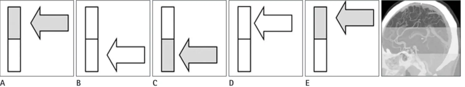
Z축을 따라 toggling table technique을 이용하여 스캔하였다 (40 mm × 2 locations = 80 mm)(2, 3). 총 3.8초 간격으로 16 절편의 연속된 15 스캔을 얻을 수 있었다(Fig. 1).
관류 정도를 정량적으로 평가하는 데는 대체로 상대적 뇌혈류 량(relative cerebral bloodflow; 이하 rCBF), 뇌혈용적(relative cerebral blood volume; 이하 rCBV) 및 평균통과시간(mean transit time; 이하 MTT) 지도를 이용하였고 지도상 경색진행부 위와 생존부위의 관류량을 측정하여 경계치를 찾았다(4).
관류 CT에서 얻어진 데이터영상들은 Extended Brilliance Workstation (Philips Medical Systems, Best, Netherlands)을 통해, 후처리 작업을 거쳐서 CBV, CBF, MTT 지도 등 후처리 CTP 영상을 얻었다(5).
확진검사로서의 MR 영상은 3T DWI와 고식적 T2 강조(in- cluding fluid-attenuated inversion recovery) 자기공명영상장 치(3T MR imaging scans: Achieva, Philips Medical Systems, Best, Netherlands)를 사용하였다.
CTP의 각 CBV, CBF, MTT 지도에서 정상 교뇌와 비교하여
A B C D E
Fig. 1. Toggling table technique.
A. The scanner obtains images during a single rotation at location upper half location of the object.
B. Table moves upward to locate the lower half of the object under the X-ray tube.
C. Scanner obtains images during a single rotation at location.
D. Table moves 4 cm in the opposite direction to the original position.
E. Second cycle of scanning is repeated.
수록 CTP 및 CT에서의 진단율이 p < 0.01 및 p = 0.01로 통 계적 의의가 있었다(Table 2).
하지만, 다른 인자 DWI의 신호강도(signal intensity:mean) 는 CTP 및 CT에서 p = 0.86, p = 0.49로 모두 진단적 의의 는 없었다(Table 2).
또한, 각 CTP의 컬러지도(rCBV, rCBF and MTT) 간의 진 단율은 통계적 의의가 없었고, 이처럼 CTP의 급성교뇌경색의 진단율이 CT보다 약간 높기는 하였으나, 통계적 의의는 없었다 (p > 0.05)(Table 1).
고찰
CTP는 빠르고 정확하며 뛰어난 접근성을 보이는 효과적인 신 경계 진단도구로 응급상황에서 천막상부의 급성뇌경색의 진단 로 분석하였고, 또한, CTP 및 CT 간 진단율의 차이는 McNemar
검정으로 분석하였으며, p value < 0.05를 통계적으로 유의한 값으로 설정하였다(Table 1).
결과
DWMRI에서 급성교뇌경색으로 확진된 78예 중 CTP 및 CT 에서 각각 42예(56.8%)와 35예(47.3%)에서 뇌경색에 해당 되는 소견을 보였으며, 발견되지 못한 예는 CTP 및 CT에서 각 각 32예(43.2%)와 39예(52.7%)였다.
급성교뇌경색의 진단은 CTP가 민감도, 특이도, 정확도에서 각 각 56.8%, 91.4%, 그리고 77.0%였고, CT의 민감도, 특이도, 정 확도는 각각 47.3%, 93.3%, 그리고 74.2%였다(Table 1).
각 진단율에 영향을 미친 인자로는 경색부피(volume)가 클
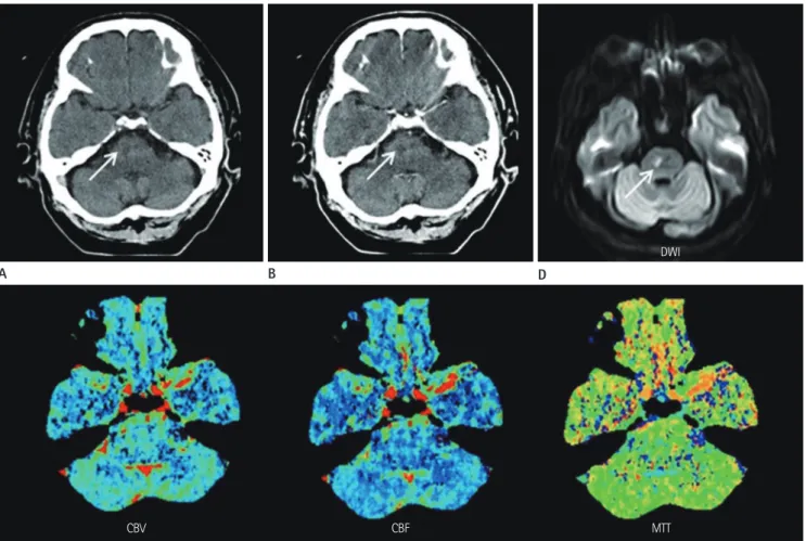
Fig. 2. A case of abnormal finding on conventional CT, but not identified by CTP in right pontine infarction. Three days before examination, this 66-year-old man experienced dysarthria and left side weakness.
A, B. Early conventional CT scans show abnormal hypodensity (arrow) in the right pons (A: precontrast CT scan, B: enhanced CT scan).
C. All CTP maps confirm the absence of perfusion abnormality in the right pons.
D. Follow-up DWI of MRI, obtained 1 day after stroke, shows infarction (arrow) in the former ischemic portion of the right anteromedial pons.
infarct volume: 0.501 mL, diffusion restriction value: 1893.3 (mean).
Note.-CBF = cerebral bloodflow, CBV = cerebral blood volume, CTP = CT perfusion, DWI = diffusion weighted imaging, MTT = mean transit time
A B
C
D
DWI
CBV CBF MTT
정도의 좁은 공간 범위를 갖으며(2, 3, 6, 7), 이러한 제한된 범위의 CTP는 천막상부의 급성 허혈성 뇌경색을 진단하는 데 에 널리 사용되어 왔다(1). 초기 모델인 4- 또는 16-채널 다중
검출방식 CT (multidetector CT; 이하 MDCT)는 20~24 mm
Fig. 4. Volumetric measurement of the infarct lesion. Boundary of the lesion was drawn and then the volume of the area was automatically cal- culated.
Fig. 3. A case of positive abnormal finding on CTP and but not identified by conventional CT in right pontine infarction. One day before examina- tion, this 51-year-old man experienced dysarthria and left side weakness.
A, B. Early conventional CT scans appear normal in the right pons (A: precontrast CT scan, B: enhanced CT scan).
C. All CTP maps show perfusion abnormality (arrow) in the right pons.
D. Follow-up DWI of MRI, obtained 1 day after stroke, shows infarction (arrow) in the former ischemic portion of the right anteromedial pons.
infarct volume: 0.976 mL, diffusion restriction value: 669.5 (mean).
Note.-CBF = cerebral blood flow, CBV = cerebral blood volume, CTP = CT perfusion, DWI = diffusion weighted imaging, MTT = mean transit time
A B
C
D
DWI
CBV CBF MTT
Table 1. Comparison of Sensitivity, Specificity, and Accuracy between CTP and CT (n = 178)
TP TN FP FN Sensitivity Specificity Accuracy p Value
CTP 42 95 9 32 56.8 91.4 77.0 0.093
CT 35 97 7 39 47.3 93.3 74.2
The McNemar test showed significant differences at p < 0.05.
Note.-CT = conventional CT, CTP = CT perfusion, FN = false negative, FP = false positive, TN = true negative, TP = true positive Table 2. Relative Factors of Abnormal Positive Findings of CTP
Gender Age CT Positive Map on CTP Signal (Mean) on DWMRI Infarct Vol. (cm3)
1 M 70 P CBV, CBF, MTT 2004.7 1.77
2 M 50 P CBV, CBF, MTT 2716.5 2.02
3 F 69 P CBV, CBF, MTT 1222.5 1.8
4 F 67 P CBV, CBF, MTT 1710 3.34
5 M 66 P CBV, CBF, MTT 640 1.15
6 M 81 P CBV, CBF, MTT 2183.3 2.46
7 M 71 P CBV, CBF, MTT 3576.0 0.649
8 F 47 P CBV, CBF, MTT 1990.7 2.47
9 M 73 P CBV, CBF, MTT 1983.3 0.754
10 F 70 P CBV, CBF, MTT 1883.9 0.875
11 M 61 P CBV, CBF, MTT 1866.9 2.17
12 F 74 P CBV, CBF, MTT 2653.4 1.29
13 F 72 P CBV, CBF, MTT 1748 1.287
14 M 61 P CBV, CBF, MTT 3318.4 1.23
15 M 67 P CBV, CBF, MTT 3864.9 1.48
16 F 79 P CBV, CBF, MTT 2419.4 0.464
17 M 74 P CBV, CBF, MTT 2502.7 0.87
18 F 84 P CBV, CBF, MTT 2313.6 0.373
19 M 53 P CBV, CBF, MTT 2020 1.287
20 M 68 P CBV, CBF, MTT 983 1.255
21 F 64 P CBV, CBF, MTT 3217 2.65
22 M 77 P CBV, CBF, MTT 2548 1.35
23 M 69 P CBV, CBF, MTT 3575.8 2.07
24 M 61 P CBV, CBF, MTT 832.6 1.15
25 M 55 P CBV, CBF, MTT 1865 0.374
26 F 70 P CBV, CBF, MTT 1303 0.896
27 F 59 N CBV, CBF, MTT 1159.3 1.34
28 M 76 N CBV, CBF, MTT 1911.8 1.64
29 M 55 N CBV, CBF, MTT 1894.2 0.891
30 F 76 N CBV, CBF, MTT 1214.4 0.844
31 F 82 N CBV, CBF, MTT 2943.2 1.41
32 M 69 N CBV, CBF, MTT 1524 1.608
33 F 52 N CBV, CBF, MTT 669.5 0.976
34 M 56 N CBV, CBF, MTT 2005.0 1.07
35 M 68 N CBV, CBF, MTT 2219.6 1.1
36 F 66 N MTT 1928.0 1.55
37 M 80 N MTT 1477.0 0.322
38 F 70 N MTT 723 1.31
39 M 47 P MTT 1315 1.59
40 F 79 N CBF, MTT 3278.4 1.63
41 M 63 N CBF, MTT 3567.0 0.717
42 F 77 P CBV, CBF 886.1 0.422
기술의 발달과 더불어 MDCT의 개발로 인해 스캔속도가 빨라 지고 해상력이 높아졌으며 후처리 컴퓨터 소프트웨어의 지속적 인 발달로 인하여 뇌경색의 진단에 있어 CTP의 유용성이 점차 증대되고 있다(10). 하지만 천막상부의 상대적으로 큰 뇌경색 진단과 달리 상대적으로 크기가 작은 천막하부의 병변의 진단 에 있어서는 CTP의 진단정확도에 대한 연구가 적었다(2).
본 연구에서 저자들은 뇌간에 발생한 뇌경색에서 CTP의 유 용성이 있는지 알고자 하였으며 CT보다 급성교뇌경색의 진단 정확도에 있어 진단적 우위가 있는지 여부를 알아보고자 했다.
는 유용하나, 천막하부를 포함한 전체 범위의 뇌병변을 진단하 는 데는 제한점이 많았다(2, 4, 8, 9). 하지만, 저자들이 사용 한 64-채널 MDCT는 toggling table technique (Jog mode)을 도입한 다중채널 CT 기술을 사용하여 한 번의 조영제 주입으로 80 mm의 스캔범위를 포함할 수 있어, 급성 허혈성 뇌경색 환 자의 천막상부 및 천막하부의 병변을 포함한 전체 뇌를 검사할 수 있다(2, 7). 64채널 이상의 다중채널 CTP는 천막상부의 뇌 경색뿐 아니라, 천막하부를 포함하는 전체 뇌경색의 진단에 점 차 확대되어 사용되고 있다(2, 7). 최근 CT 기기의 튜브제조 Table 2. Continued
Gender Age CT Positive Map on CTP Signal (Mean) on DWMRI Infarct Vol. (cm3)
43 M 70 N N 2692.8 0.374
44 M 48 N N 1231.3 0.443
45 F 82 N N 3371.2 1.28
46 M 70 N N 2552.2 2.79
47 M 45 N N 3566.2 0.576
48 M 82 N N 1987.4 0.706
49 F 72 N N 1573.7 0.353
50 M 70 N N 1800 0.163
51 M 68 N N 2166.3 0.448
52 M 75 N N 2190.0 0.232
53 M 44 N N 2278.9 0.514
54 F 61 N N 1722.0 0.338
55 F 77 N N 2021.1 0.514
56 M 77 N N 1857.8 0.441
57 F 71 N N 2089.6 0.424
58 M 66 N N 1393.0 0.3849
59 F 64 N N 2405 0.316
60 M 90 N N 785 0.269
61 M 75 N N 3518 2.16
62 F 77 N N 710 0.654
63 F 66 N N 2124 1.02
64 M 67 N N 1126 0.533
65 M 53 N N 3392.3 1.51
66 M 60 N N 1974.4 1.19
67 F 67 N N 600 0.48
68 M 80 P N 2397 1.1
69 M 67 P N 1893.3 0.501
70 M 52 P N 1602.5 0.559
71 M 68 P N 1402 0.601
72 F 65 P N 2886.0 1.34
73 M 65 P N 3244.6 1.32
74 M 69 P N 1772.3 0.369
CT p < 0.01 p < 0.01 p = 0.86 p = 0.01
CTP p = 0.49 p < 0.01
The ROC curve showed significant differences at p < 0.05.
Note.-CBF = map of cerebral blood flow, CBV = map of cerebral blood volume, CT = conventional CT, CTP = CT perfusion, DWMRI = diffusion weighted MR imaging, MTT = mean transit time, N = negative finding, P = positive finding
cerebral ischemia: the “toggling-table” technique. AJNR Am J Neuroradiol 2001;22:1077-1080
4. Koenig M, Klotz E, Luka B, Venderink DJ, Spittler JF, Heuser L. Perfusion CT of the brain: diagnostic approach for early detection of ischemic stroke. Radiology 1998;209:85-93 5. Axel L. Tissue mean transit time from dynamic computed
tomography by a simple deconvolution technique. Invest Radiol 1983;18:94-99
6. Wintermark M, Fischbein NJ, Smith WS, Ko NU, Quist M, Dillon WP. Accuracy of dynamic perfusion CT with decon- volution in detecting acute hemispheric stroke. AJNR Am J Neuroradiol 2005;26:104-112
7. Youn SW, Kim JH, Weon YC, Kim SH, Han MK, Bae HJ. Per- fusion CT of the brain using 40-mm-wide detector and toggling table technique for initial imaging of acute stroke. AJR Am J Roentgenol 2008;191:W120-W126 8. Röther J, Jonetz-Mentzel L, Fiala A, Reichenbach JR, Her-
zau M, Kaiser WA, et al. Hemodynamic assessment of acute stroke using dynamic single-slice computed tomo- graphic perfusion imaging. Arch Neurol 2000;57:1161- 1166
9. Nabavi DG, Cenic A, Craen RA, Gelb AW, Bennett JD, Ko- zak R, et al. CT assessment of cerebral perfusion: experi- mental validation and initial clinical experience. Radiology 1999;213:141-149
10. Choi JH, Seo JJ, Kim JK, Chung TW, Jeong YY, Park JG, et al. The usefulness of perfusion CT in acute cerebral isch- emic infarction. J Korean Radiol Soc 2003;49:7-14 11. Mayer TE, Hamann GF, Baranczyk J, Rosengarten B, Klotz E,
Wiesmann M, et al. Dynamic CT perfusion imaging of acute stroke. AJNR Am J Neuroradiol 2000;21:1441-1449 결과적으로 CTP가 CT에 비해 급성교뇌경색의 진단에서보다
높은 진단율을 보였으나, 통계적으로 의의는 없었다. 저자들은 CTP 지도 사이에서도 진단율의 차이가 있는지 보았고 MTT 지 도가 더 많은 경우에서 뇌경색을 진단할 수 있었다.
천막상부의 넓은 영역의 경색일 경우 CBF 지도를 이용하여 민 감도와 특이도는 거의 100%까지 보고되었으며 천막상부의 뇌 혈류 상태를 파악하는 데 도움이 되었다(11). 하지만 본 연구에 서는 CTP가 천막하부 특히, 교뇌경색에서 CT보다 높은 진단율 을 보이지는 않았으며, CTP의 각 지도에서도 각각의 진단율의 차이는 보이지 않았는데 이는 천막상부의 병변보다 천막하부 특 히, 교뇌 병변의 크기가 상대적으로 매우 작고 천막하부의 혈류 가 천막상부와 다르기 때문으로 생각된다.
본 연구의 제한점은 첫째, 대상군이 비교적 적다는 점이며, 둘째, 후두와에서의 빔 경화 허상(beam hardening artifact)으 로 인한 영상 균질도 및 정량적 측정에서 부정확한 결과가 있을 수 있다는 점이다. 앞으로 CT 기술의 발달과 다중채널 CT 기 술의 발달로 이러한 부분들이 개선될 수 있을 것으로 생각된다.
본 연구는 급성교뇌경색의 진단에 있어서 CTP와 CT를 비교 한 첫 번째 연구이며, CTP와 CT 모두 급성교뇌경색의 진단에 유용하였고 CTP가 CT에 비해 통계적 우위는 보이지 않았다.
참고문헌
1. Ledezma CJ, Wintermark M. Multimodal CT in stroke im- aging: new concepts. Radiol Clin North Am 2009;47:109- 116
2. Lee IH, You JH, Lee JY, Whang K, Kim MS, Kim YJ, et al. Ac- curacy of the detection of infratentorial stroke lesions us- ing perfusion CT: an experimenter-blinded study. Neuro- radiology 2010;52:1095-1100
3. Roberts HC, Roberts TP, Smith WS, Lee TJ, Fischbein NJ, Dillon WP. Multisection dynamic CT perfusion for acute
급성 허혈성 교뇌경색에서 다중채널 관류전산화단층촬영과 고식적 전산화단층촬영의 진단적 유용성의 비교
김성아 · 이명섭 · 김명순 · 김영주
목적: 급성뇌경색 환자에서 관류전산화단층촬영(CT perfusion; 이하 CTP)은 중요한 진단방법으로 천막상부뿐 아니라 천막하부의 뇌경색까지 그 이용 범위가 점차 넓어지고 있다. 본 저자들은 급성교뇌경색에서 CTP와 고식적 CT (conven- tional CT; 이하 CT)의 진단율을 비교해 보고자 하였고 각각의 진단방법에 영향을 줄 수 있는 요인을 알아보고자 하였다.
대상과 방법: 급성교뇌경색이 의심되는 증상을 보이는 환자를 대상으로 CT와 다중채널을 사용한 CTP 검사를 하였고 확 진을 위하여 diffusion weighted imaging을 포함한 MRI를 시행하였으며 총 178명 중 경색으로 진단된 74명을 대상으로 하 였다. CTP와 CT의 진단율을 비교하여 보았고 영향을 줄 수 있는 요소를 알아보기 위해 정량적 분석을 하였다.
결과: 급성교뇌경색에서 CTP의 진단적 민감도, 특이도, 정확도는 각각 56.8%, 91.4%, 77.0%였고, CT는 47.3%, 93.3%, 74.2%로 통계적으로 유의한 차이는 없었다. Receiver operation characteristic 곡선에서 두 방법이 모두 진단적 유 용성이 있는 것으로 나타났다(p < 0.01). 진단율에 영향을 미치는 인자로는 경색용적(CTP: p < 0.01, CT : p = 0.01)이 통계적으로 의의가 있었다.
결론: 본 연구는 급성교뇌경색의 진단에 있어서 CTP와 CT를 비교한 첫 번째 연구이며, CTP와 CT 모두 급성교뇌경색의 진단에 유용하였고 CTP가 CT에 비해 통계적 우위는 보이지 않았다.
연세대학교 원주의과대학 원주기독병원 영상의학과