서 론
전산화 단층촬영술 스캔(computed tomography scan) 발달로 치명적인 신경학적 장애를 유발할 수 있는 수술 후
CLINICAL ARTICLE
Korean J Neurotrauma 2012;8:79-86 ISSN 2234-8999
경막외 출혈의 조기 발견이 가능해졌다.11) 이런 영상기법이 발 전하여도, 두개강내 수술 후 경막외 출혈은 신경외과 수술의 중요하고 치명적일 수 있는 합병증이다. 경막외 출혈의 발생 빈도는 연구자에 따라 다르게 보고되나, 보통 1% 내외로 알 Received: June 20, 2012 / Revised: August 4, 2012 / Accepted: August 9, 2012
Address for correspondence: Won Il Joo, MD
Department of Neurosurgery, Yeouido St. Mary’s Hospital Neuroscience Center, College of Medicine, The Catholic University of Korea, 62 Yeouido-dong, Yeongdeungpo-gu, Seoul 150-713, Korea
Tel: +82-2-3779-2248, Fax: +82-2-786-5809, E-mail: [email protected]
The statistical consultation was supported by Catholic Research Coordinating Center of the Korea Health 21 R&D Project (A070001), Minis- try of Health & Welfare, Republic of Korea.
두개강내 수술 후 발생할 수 있는 경막외 출혈의 위험요인의 분석
가톨릭대학교 의과대학 여의도성모병원 신경외과학교실
구원모 .주원일 .나형근 .박해관 .조정기 .이경진
Analysis of Risk Factors for the Development of Post-Operative Epidural Hematoma after Intracranial Surgery
Won Mo Gu, MD, Won Il Joo, MD, Hyoung Kyun Rha, MD,
Hae Kwan Park, MD, Chung Kee Chough, MD and Kyung Jin Lee, MD
Department of Neurosurgery, Yeouido St. Mary’s Hospital Neuroscience Center, College of Medicine, The Catholic University of Korea, Seoul, Korea
Objective: Patients undergoing intracranial operations often suffer from post-operative epidural hematoma (EDH). The incidence and risk factors for with the occurrence of EDH after intracranial operations are not well described previously.
The objective of this study was to identify the risk factors and the incidence of post-operative EDH adjacent and regional to the craniotomy.
Methods: This was a retrospective study of 23 (2.4%) patients, between January 2005 and December 2011, who under- went epidural hematoma evacuation after primary intracranial during this period, 941 intracranial operations were per- formed. The control group (46 patients) and hematoma group (23 patients) were categorized on the basis of having under- gone the same pre-operative diagnosis and treatment within 3 months of their operations. The ages of the hematoma and control group were individually matched to similar ages within 10 years of each other to minimize bias of age.
Results: Univariate analysis showed that the significant pre-operative and intra-operative factors associated with post- operative EDH were a pre-operative Glasgow Coma Scale (GCS) scored <8 (crude odds ratio 8.295), prothrombin ratio
>1.0 (p=0.014), prothrombin time (PT) >11.3 sec (p=0.008), intra-operative blood loss >650 mL (p=0.003) and crani- otomy size >7,420 mm2 (p=0.023). In multivariate analysis, intra-operative blood loss exceeding 650 mL (median of to- tal patients) placed a patient at significantly increased risk for post-operative EDH.
Conclusion: Recognizing the limitations of the study, large intra-operative blood loss, wide craniotomy area, prolonged PT and a pre-operative GCS <8 are presented implicated with an increased risk of post-operative EDH after intracranial surgery.
(Korean J Neurotrauma 2012;8:79-86) KEY WORDS: Intracranial epidural hematoma ㆍPost-operative hematoma ㆍCraniotomy ㆍRisk factor.
려지며 발생 위험 요소는 부분적으로 밝혀져 있다.4) 본 연구에서의 분석한 환자군은 두개강내 수술 후 영상검 사에서 보일 수 있는 임상적 증상이 보이지 않는 적은 양의 경 막외 혈종은 제외하고, 뇌실질에 대한 직접적 압박을 유발해 신경학적 증상이 발생하여 이차적 수술을 받은 환자 중, 수 술부위(regional)와 인접부위(adjacent) 경막외 혈종에 대한 환자군을 분석하였다. 지금까지 발표된 경막외 혈종에 대한 문헌은 대부분 척추수술 후 발생하는 경막외 출혈에 대한 연 구였으며, 신경외과 영역의 기본 술기인 개두술(craniotomy) 후 발생하는 출혈의 위험인자 분석연구는 일부에 불과하였
다.13,17,24) 따라서 본 저자는 두개강내 수술 후 발생하는 경막
외 출혈의 위험 요소를 밝히기 위해, 본원에서 두개강내 수술 후 재수술이 필요했던 수술 후 경막외 출혈 환자들을 분석 하였다. 일부 연구에서는 수술 후 경막외 출혈의 병리학적 기전으로 수술 중 뇌척수액 과다배액으로 인한 뇌실질의 수 축으로 혈관파열이나, 경막 분리로 인해 경막외 출혈이 유발 가능하다고 주장한다.13)
저자는 물리학적 기전으로 인한 경막의 분리로 인한 요인 뿐만 아니라, 수술 전 혈액학적 문제나 기저질환, 수술 중 발생 할 수 있는 변수들을 분석하였다. 본 연구는 본원에서 지난 7년간 개두술을 시행받은 환자를 대상으로 수술 후 경막외 출혈의 발생정도를 입증하고, 수술 후 경막외 출혈을 예측하 고 예방할 수 있는 위험요소를 평가하도록 설계되었다.
대상 및 방법
2005년 1월부터 2011년 12월까지 7년동안 본원에서 시행 한 두개강내 천막상부(supratentorial)수술 941건 (뇌실 외 배 액 및 천공 배액술 등 비개두술은 제외)에 대한 후향적 연구
를 진행하였다. 941예 중에서, 우리는 수술 후 경막외 출혈에 의한 뇌실질 압박 및 신경학적 증상 발생으로 인해 본과적 감압 수술을 받았던 23명의 환자 (2.4%)를 분석하였다 (Fig- ure 1). 임상적 증상을 보이지 않는 출혈은 환자군에서 배제 하였으며 뇌종양, 두개강내 동맥류, 동정맥 기형, 자발성 두 개내 출혈, 두개골성형술, 외상성 뇌손상이 환자군에 포함되 었다. 수술 후 경막외 출혈이 생긴 환자군에서 위험요소 분석 시 병리학적 소견이나 나이에 따른 선택 오류를 최소화하기 위해, 환자군과 동일한 진단명으로 3개월내 수술한 환자 중 환자군과 10살 이내로 연령 분포를 보이는 환자 2배수의 대 조군을 추출하였으며, 각 집단의 임상자료, 수술기록, 영상학 적 소견의 결과를 검토하여 가능한 위험요인을 후향적으로 분석하였다. 이전 연구에서 밝혀진 수술 후 경막외 출혈에 대 한 가능한 위험요소로, 혈액응고 인자의 진단검사학적 이상 소견, 항응고제 및 항혈소판제 복용여부, 음주, 흡연 같은 과 거력 등을 위험인자의 독립변수로 설정하였다.17)
또한 저자는 수술 후 경막외 혈종의 발생률이 골편 제거 유무, 경막외 배액관 삽입유무와 같은 수술 술기에 따라 차 이가 생길 것으로 가정하여 위험요소를 설정하였다. 설정된 위험요인에 대한 상세한 검토는 수술 전(pre-operative)과 수술 중(intra-operative)으로 분류하였고, 수술 전 변수에 는 나이, 성별, 동반질환, 수술 전 의식수준, 항혈소판제 항응 고제의 수술 전 사용, 헤모글로빈, 혈소판수, prothrombin time (PT) 및 activated partial thromboplastin (aPTT) 결 과가 포함되었다. 수술 중 변수에는 수술 중 실혈량, 수술시 간, 경막 절개(durotomy)여부, 경막외 배액의 사용 여부, 개 두술 된 골편의 크기 (Figure 2)를 가능한 위험인자로 설정 하였다. 진단검사적 수치와 같은 연속변수들(continuous variables)은 임상적 정상범위 내 값이나 전체 환자에서 중
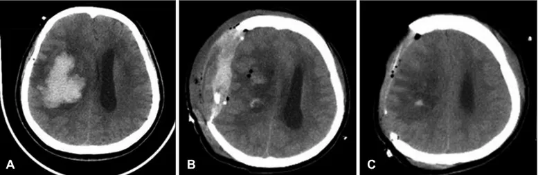
FIGURE 1. Results of computed tomography (CT) examinations. A: CT scan showing intracranial hematoma at right basal ganglia with midline shifting. B: Post-operative immediate brain CT scan showed almost completely removed intracerebral hemorrhage, and also showed large amount of epidural hematoma (EDH) with aggravated mass effect. C: After reoperation was performed to remove the EDH beneath the previous craniectomy site, brain CT scan showed no residual epidural hematoma. A marginal amount of intra- cerebral hemorrhage remained.
A B C
앙값(median value)을 기준값(cut-off value)으로 설정하 여 조건부 다중 회기 분석(conditional logistic regression analysis)을 수행하였다. 모든 통계 분석은 단일변량 및 다 변수 분석 수행을 위해 통계 분석 시스템, 버전 9.2. (SAS Institute, Cary, NC)를 사용하였다.12) 비연속 변수는 chi- square test 및 Fisher’s exact test를 이용하였으며 연속 변수 는 Student’s t-test 및 Mann-Whitney U-test 방법을 사용 하여 통계 분석하였으며, 확률값(p-value) <0.05를 통계적 으로 유의한 것으로 간주하였으며, 단일변량(univariable) 및 다변량(multivariable)에 대한 분석은 수술 후 경막외 출혈 에 대한 위험 요소를 밝히기 위한 개별적 위험 요소로 조 건부 다중 회귀 분석을 통해 이루어졌다.
결 과
환자군 23명 중, 13명은 남성이었고 10명은 여성이었고, 연 령 중앙값은 50.0세 (범위, 20~77세)였다. 대조군 환자의 연령 중앙값은 51.0세 (범위, 25~73세)였다. 환자군 중 17명 (73.9%) 과 대조군 중 34명 (73.9%)은 정규수술을 받았으며, 그 외에는 응급수술을 시행하였다. 전체 941명의 환자는 동일한 수술 후 치료(postoperative care)를 받았으며, 개두술(cranioto- my)을 받은 811명 (86.1%)의 환자 중, 16명 (1.9%)의 환자가 경 막외 출혈로 인해 재수술이 필요했다. 130명 (13.9%)의 두개절 제술(craniectomy)을 받은 환자 중에서, 7명 (5.1%)의 환자는 경막외 출혈이 생겼다. Chi-square test에 의한 유의성을 따져 볼 때 두개절제술의 유무에 따른 수술 후 경막외 출혈 (p=0.05)
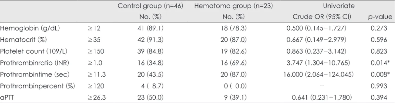
발생에는 통계학적 유의한 차이가 있었다. 69명의 환자 (환자 군 23명, 대조군 46명)의 임상적, 진단검사학적, 수술 중 변 수에 대한 위험요인 분석비교는 Table 1에 요약되어 있다. 두 그룹 간 PT test, 개두술의 골편 크기, 수술 중 혈액 손실량이 두 군 간에 통계적 유의한 차이를 보였고, 두 군 간 수술 전 확 인 가능한 위험 요소에 대한 통계적 유의성 (p<0.05)을 알아 내기 위해, 단일변수에 대한 조건부 다중 회귀분석(unpaired conditional logistic regression analysis)을 시행하였다 (Ta- ble 2, 3). 전체 환자의 중앙값을 기준으로 11.3초 이상의 PT (p=0,008), 1.0 이상의 PT international normalized ratio (INR)(p=0.014) 그리고 GCS 8점 이하에서, 수술 후 경막외 출 혈 환자군에서 유의하게 높게 나타났다. 환자군과 대조군 사 이에 통계학적 단일변수 비교분석상 연장된 PT수치 (>11.3초) 가 그렇지 않은 경우에 비해 발생률이 16배 높게 나타나며, 통 계적으로 유의성을 보였다.
또한 환자군에서 PT (INR)>1.0일 경우 그렇지 않은 경우 에 비해 3.747배 높은 발생률을 보이며 통계적으로 유의하게 나 타났다. 수술 전 의식 상태의 경중에 따라 A (GCS 13-15), B (GCS 9-12), C (GCS 3-8)로 세 group으로 나누었고 group C와 group A에서는 통계적으로 유의한 수준으로 group C 에서 8.295배 높게 경막외 출혈이 발생하였다. 환자군과 대 조군 간 고혈압, 당뇨병, 알코올 섭취 과거력, 흡연, 항응고제 또는 항혈소판제의 사용 및 혈소판 감소증에 관해서는 통계 적으로 유의한 차이는 없었다. 수술 중 발생할 수 있는 위험 요소에 대한 단일변량분석의 결과는 Table 4에 나와있다. 전 체 수술 환자의 수술 중 실혈량은 중앙값 650 mL로 나타났 고, 650 mL 이상의 수술 중 혈액 손실을 입은 환자군에서 9.862배로 경막외 출혈 발생률을 보였고 (p=0.003), 중앙값 7,420 mm2를 초과하는 넓은 골편을 가지는 개두술 환자군 에서 3.845배 높게 나타냈다 (p=0.023). Table 5는 단일변수 분석에서 유의한 차이를 보였던 독립변수로 연장된 PT수 치 (PT>11.3초) 수술 전 의식수준 (GSC<8), 수술 중 출혈량 (mL) 및 개두술시 골편 크기 (mm2)를 사용하여 조건부 다 중 다변수 회귀분석(conditional logistic multivariate re- gression analysis)의 결과를 보여준다. 위 변수들 중, 650 mL 를 초과하는 수술 중 출혈은 다른 독립변수와 관계없이 수술 후 경막외 출혈의 위험을 유의하게 증가시켰다 (p=0.02).
PT, PT INR, 수술 전 의식수준, 개두술시 골편크기는 다변량 분석에서는 통계적 유의성이 없었다.
고 찰
경막외 출혈은 대부분 외상의 결과로 발생하며 중년이나 젊 FIGURE 2. Data concerning craniotomy extent. Craniotomy ex-
tent was measured from plain lateral skull X-ray that is, from max- imal perpendicular diameter (radius a and b) of the craniotomy.
The resulting size of craniotomy was then approximated using the formular for a circle (size=π×ab).
TABLE 1. Clinical characteristics of the control and hematoma groups
Control group [n=46 (%, 66.7%)]
Hematoma group [n=23 (%, 33.3%)] p-value
Gender Male 30 (65.2) 13 (56.5) 0.482
Female 16 (34.8) 10 (43.5)
Age Mean±SD 50.2±12.5 50.4±14.0 0.948
Diabetes mellitus No 44 (95.6) 21 (91.3) 0.596
Yes 2 ( 4.4) 2 ( 8.7)
Hypertension No 34 (73.9) 16 (69.6) 0.703
Yes 12 (26.1) 7 (30.4)
Alcohol use No 32 (69.6) 16 (69.6) 0.999
Yes 14 (30.4) 7 (30.4)
Tobacco use No 37 (80.4) 19 (82.6) 0.999
Yes 9 (19.6) 4 (17.4)
Hemoglobin (g/dL) Mean±SD 13.7±1.5 13.7±1.8 0.836
Hematocrit (%) Mean±SD 40.7±4.4 40.6±5.4 0.904
Platelet count (×109/L) Mean±SD 215.5±55.5 227.6±76.1 0.458
Prothrombinratio (INR) Mean±SD 1.0±0.1 1.0±0.1 0.290
Prothrombin time (sec) Median (minimum-maximum) 11.1 (9.4-17.7) 11.6 (10.6-13.3) 0.002*
Prothrombin percent (%) Median (minimum-maximum) 99.9 (10.5-142.3) 97.9 (80.8-116.6) 0.390
aPTT Mean±SD 25.7±3.8 25.3±3.7 0.657
Anti platelet and anticoagulant use
No Yes
42 (91.3) 4 ( 8.7)
23 (100.0) 0 (0.0)
0.293
GCS (pre-op) Median (minimum-maximum) 15.0 (3.0-15.0) 12.0 (3.0-15.0) 0.095
GCS Category A (13-15) 34 (74.0) 11 (47.8) 0.085
B ( 9-12) 6 (13.0) 6 (26.1)
C ( 3- 8) 6 (13.0) 6 (26.1)
Operation time (min) Median (minimum-maximum) 222.5 (90.0-670.0) 240.0 (100.0-1,020.0) 0.259 Intra-op EBL (mL) Median (minimum-maximum) 500.0 (10.0-2,000.0) 1,000.0 (70.0-8,000.0) <0.0001*
Epidural drain No 32 (69.6) 15 (65.2) 0.715
Yes 14 (30.4) 8 (34.8)
Durotomy No 9 (19.6) 3 (13.0) 0.738
Yes 37 (80.4) 20 (87.0)
Craniotomy area (mm2) Median (minimum-maximum) 8,265.1 (2,642.1-15,848.8) 5,729.6 (1,209.6-25,717.4) 0.018*
*probability values <0.05 were considered statistically significant. INR: international normalized ratio, aPTT: activated partial thromboplastin, GCS: Glasgow Outcome Scale, pre-op: pre-operative, intra-op: intra-operative, EBL: estimated blood loss, SD: standard deviation
TABLE 2. Univariate conditional logistic regression analysis (pre-operative patient details) Control group (n=46)
No. (%) Hematoma group (n=23) No. (%)
Univariate
Crude OR (95% CI) p-value
Gender Female 16 (34.8) 10 (43.5) 2.303 (0.433-12.236) 0.328
Age ≥65 years 5 (10.9) 5 (21.7) 2.195 (0.576- 8.362) 0.250
Diabetes mellitus Yes 2 ( 4.3) 2 (8.7) 2.732 (0.226-33.004) 0.429
Hypertension Yes 12 (26.1) 7 (30.4) 1.356 (0.356- 5.171) 0.656
Alcohol use Yes 14 (30.4) 7 (30.4) 1.000 (0.329- 3.038) 0.999
Tobacco use Yes 9 (19.6) 4 (17.4) 0.871 (0.243- 3.114) 0.831
Antiplatelet
anticoagulant use Yes 4 ( 8.7) 0 (0.0) - 0.993
GCS category A 34 (73.9) 11 (47.8) 1.000 0.082
B 6 (13.0) 6 (26.1) 5.107 (0.981-26.591)
C 6 (13.0) 6 (26.1) 8.295 (1.027-67.032)*
*95% Confidence interval were excluded 1.0. GCS: Glasgow Outcome Scale, OR: odds ratio
은 성인에게 많이 발생한다. 경막외 출혈은 두개관(calvaria) 과 경막 사이에 혈액이 축적된 것을 말하며 전두엽과 측두엽 에서 호발한다. 외상후 경막외 출혈 중 만성 경막외 출혈은 약 10%에서 발생된다고 보고되며, 자발성 경막외 출혈은 흔 하지 않다.15) 저자는 두개강내 수술 후 발생한 경막외 출혈 의 가능한 위험요인을 분석해 보았다.
개두술시 골편 제거 후 두개골(bone edge)과 경막 사이 봉 합술, 제거된 골편 삽입시 골편과 경막 사이 천막봉합(tent- ing suture)을 통해 수술 후 경막외 출혈 유병률을 줄일 수 있 다고 보고된다.4) 본 연구에 해당되는 모든 예에서 골편 삽입 시 상기 수술 술기 적용하였으며 육안적으로 보이는 모든 출 혈 부위 지혈 후, 근육 및 연부조직 피부 봉합하였고, 모든 증
례에서 경막외 배액관이나 모상건막하(subgaleal) 배액관을 삽입하였다.
수술 후 경막외 출혈은 치명적인 신경학적 후유증을 남길 수 있기 때문에 발생 가능한 위험인자에 대한 분석이 필요 하다. 사전에 제거 및 교정 가능한 위험인자를 밝힘으로써 수술 후 경막외 출혈의 발생 빈도를 의미 있게 줄일 수 있을 것 이다. 경막외 출혈의 위험요인 분석에 대한 연구는 주로 척추 수술 후 합병증 분석 연구로 많이 보고되었으나,2,22) 개두술 후 경막외 출혈의 유발률이나, 위험요소에 대한 연구는 소수 에서 이뤄졌다.19,20) 일반적으로 수술시 불충분한 지혈로 인해 경막외 출혈이 발생하는 것으로 생각하여, 개두술 부위(re- gional)나 골편제거 인접부(adjacent) 두개내 경막외 출 TABLE 3. Univariate conditional logistic regression analysis (pre-operative laboratory data)
Control group (n=46) Hematoma group (n=23) Univariate
No. (%) No. (%) Crude OR (95% CI) p-value
Hemoglobin (g/dL) ≥12 41 (89.1) 18 (78.3) 0.500 (0.145-1.727) 0.273
Hematocrit (%) ≥35 42 (91.3) 20 (87.0) 0.667 (0.149-2.979) 0.596
Platelet count (109/L) ≥150 39 (84.8) 19 (82.6) 0.863 (0.237-3.142) 0.823
Prothrombinratio (INR) ≥1.0 16 (34.8) 16 (69.6) 3.747 (1.304-10.765) 0.014*
Prothrombintime (sec) ≥11.3 20 (43.5) 20 (87.0) 16.000 (2.064-124.045) 0.008*
Prothrombinpercent (%) ≥120 4 ( 8.7) 0 ( 0.0) - 0.993
aPTT ≥26.3 23 (50.0) 9 (39.1) 0.641 (0.231-1.780) 0.394
*probability values <0.05 were considered statistically significant. INR: international normalized ratio, aPTT: activated partial thromboplastin, OR: odds ratio
TABLE 4. Univariate conditional logistic regression analysis (intra-operative data)
Control group (n=46) Hematoma group (n=23) Univariate
No. (%) No. (%) Crude OR (95% CI) p-value
Operation time (mins) ≥230 22 (47.8) 14 (60.9) 2.171 (0.614-7.673) 0.229
Intra-op EBL (mL) ≥650 16 (34.8) 19 (82.6) 9.862 (2.214-43.937) 0.003*
Epidural drain Yes 14 (30.4) 8 (34.8) 1.250 (0.409-3.821) 0.696
Durotomy Yes 37 (80.4) 20 (87.0) 2.171 (0.364-12.944) 0.395
Craniomy size (mm2) ≥7,420 18 (39.1) 16 (69.6) 3.845 (1.202-12.302) 0.023*
*probability values <0.05 were considered statistically significant. intra-op: intra-operative, EBL: estimated blood loss, OR: odds ratio
TABLE 5. Multivariate conditional logistic regression analysis
Control group (n=46) Hematoma group (n=23) Multivariate
No. (%) No. (%) Adjusted OR (95% CI)* Adj. p-value*
Prothrombinratio (INR) ≥1.0 16 (34.8) 16 (69.6) 0.807 (0.091-7.181) 0.847
Prothrombintime ≥11.3 20 (43.5) 20 (87.0) 26.128 (0.727-939.032) 0.074
GCS category A 34 (73.9) 11 (47.8) 1.000 0.744
B 6 (13.0) 6 (26.1) 2.279 (0.252-20.614)
C 6 (13.0) 6 (26.1) 2.142 (0.073-62.557)
Intra-op EBL (mL) ≥650 16 (34.8) 19 (82.6) 21.900 (1.616-296.750) 0.020*
Craniotomysize (mm2) ≥7,420 18 (39.1) 16 (69.6) 3.689 (0.522-26.070) 0.191
*probability values <0.05 were considered statistically significant. INR: international normalized ratio, GCS: Glasgow outcome scale, intra-op: intraoperative, EBL: estimated blood loss, OR: odds ratio
혈의 발생률이나 위험인자에 대해서는 충분히 밝혀진 연구 는 없다.13,24,26) 몇몇 연구에서는 수술 후 경막외 출혈이 수술 중 갑작스럽게 낮아지는 두개내 압력 때문이나 단시간에 뇌 실내 뇌척수액을 배액시켜 나타나는 뇌의 수축으로 인해 두 개골과 경막 분리가 경막외 출혈의 원인으로 설명한다.9) 다른 연구에서는 수술 후 경막외 출혈의 위험요인으로 비정상적인 aPTT, PT, 혹은 factor XIII에 의한 응고병증 및 고혈압과 같은 요인들이 영향을 끼칠 수 있다고 보고하고 있다.7,25)
또한, 항혈소판 및 항응고제 의약품의 투여는 이미 많은 영 역의 수술에서 수술 중 출혈 위험이 높아진다는 것은 확립되
어있다.10,21) 두개강내 수술 전 항혈소판약제를 사용하게 되면
수술 후 혈종의 위험을 증가시키는 것으로 알려져 있다.1,4,5,17) 이러한 연구들 중 하나로, Palmer 등17)은 71명의 두개강내 수 술후 혈종 환자 중에 43%가 수술 전 2주 동안 항혈소판약제 를 투여받았고, 혈종환자의 16%는 수술 48시간 이내에 항응 고 약물치료를 받았다는 것을 보고하여, 항혈소판 및 항응고 제가 두개강내 수술 후 혈종 형성에 영향을 끼친다고 주장하 였다.
본 연구의 대다수 환자가 예정된 정규수술로 진행되어 적 어도 정규수술 7일 전에는 항혈소판제 또는 항응고제의 사용 을 중지하도록 권고하였다. 본 연구에서 수술 전 이러한 투약 을 받는 환자들의 수가 제한적 (69명 중 4명)이었기 때문에 충 분한 분석을 제시하기에는 한계가 있어, 항혈소판제 및 항응 고제 투여가 수술 후 경막외 혈종의 발생 (p=0.293)과 연관성 이 있는지를 증명할 수 없었다. 응급수술의 경우에도, 비타 민K와 신선냉동혈장(fresh frozen plasma)을 포함한 해독제 (anti-dote)를 사용하여 약물의 효과를 경감시키도록 하였 다. 혈액학적 응고장애는 수술 후 출혈의 위험을 증가시키는 것으로 알려져 있다. 응고 기능 이상을 알기 위해 본원에서 수술 전 기본 진단검사로 혈소판 수, PTT, 그리고 PT를 포 함한 응고검사를 수행하였다. 다변량 분석은 수술 후 경막 외 출혈의 중요한 위험요인으로써 응고장애를 제시하진 못했 지만, 단일변량분석에서는 연장된 PT (INR)>1.0 (p=0.014) 과 PT>11.3초 (p=0.008)가 유의한 위험요인임을 알아낼 수 있었다.
수술 후 경막외 출혈의 다른 위험인자로 고혈압을 들 수 있 는데, 고혈압은 혈액응고 마개의 파괴유발(disruption of he- mostatic plug)과 자동조절(autoregulation)의 소실로 인해 수 술 후 출혈을 증가시키는 데 기여하는 것으로 알려져 있다.7,23) 또한 뇌동맥류 파열로 인한 지주막하 출혈로 수술 후 혈관연 축(vasospasm)을 방지하기 위해 유도된 고혈압(induced high blood pressure)과도 경막외 출혈이 연관성이 있다고 보고되 고 있다.6) 또한 흡연 및 음주의 과거력이 수술 후 출혈의 위
험요인으로 널리 알려져 있지만, 본 연구에서는 환자의 기저 질환과 사회력과 수술 후 경막외 출혈 간의 연관성을 나타 나지 않았다.
두개골 수술에서 경막외 배액과 삽관은 경막외 출혈에 대 한 예방책으로 흔히 사용되고 있다. 그러나 경막외 배액관 삽 입의 유효성이 확실히 입증되지는 않았다. 한 연구에서 개두 술을 받은 342명의 간질환자에서 경막외 배액관 삽입이 경막 외 출혈의 발병률과 그 크기, 모상건막하 뇌척수액 고임(sub- galeal cerebrospinal fluid collection)을 감소시켜 주지 못한 다고 보고하고 있다.8) 마찬가지로 여러 연구의 척추수술에서 도 근막하(subfascial) 배액관 삽입은 감염의 발생률을 증가 시킬 뿐, 수술 후 경막외 출혈의 발생을 감소시키지 못하였
다.15,16) 본 연구 대조군 14명, 환자군 8명의 환자에서 경막외 배
액관을 가지고 있었고, 나머지 환자들은 건막하 배액관을 삽입 하였다. 본 연구에서 경막외 배액과 건막하 배액관 삽입이 경 막외 출혈의 발생정도에 영향을 끼치지 못하였다 (p=0.696).
본 저자는 두개내 경막절개술(durotomy), 넓은 골편 개두 술(large craniotomy) 및 두개절제술(craniectomy)이 수술 후 경막외 출혈과 관련이 있을 것이라고 가정해 보았다. 이전 발표된 연구들에서는 수술 후 경막외 출혈과 골편 크기, 골편 제거 유무, 경막절개 유무와 관련성을 나타내는 자료는 없었 다. 척추 수술에서는 수술 중 경막 절개술은 수술 후 경막외 출혈의 양과 관련이 없다고 알려져 있다.22) 본 연구에서도 경 막 절제술과 경막외 출혈 발생 간의 상관관계 (p=0.395) 역시 유의하지 않았다. 그러나, 단일변량분석에서 제거한 골편 크기 가 7,420 mm2 이상인 개두술의 경우, 수술 후 경막외 출혈의 위험요소로써 통계적 유의성을 나타냈다 (p=0.023). 개두술 시 골편의 크기가 클수록 상대적으로 노출되는 경막부위가 넓어지게 되고, 경막과 주위 골 변연부(bone edge)가 길어지는 것이 경막외 출혈의 발생 정도를 높이는 요인으로 추정된다.
본 연구에서 두개강내 수술 후 두개 절제술이 개두술에 비해 수술 후 경막외 출혈의 발생 빈도가 통계적으로 유의하 게 나타났다 (p=0.05). 이는 감압적 두개 절제술 이후, 두피 연 부조직과 측두근의 출혈을 압박효과(tamponade effect)로 지 혈할 수 있는 골편이 없어 상대적으로 경막의 출혈 유발률 이 높게 나타난 것으로 추정된다. 또한, 골편 부재시 탄력붕 대로 압박 지혈하기 힘들 정도의 출혈 발생 증가에 기여하였 을 것으로 생각된다.
외상성 두부 손상(traumatic brain injury) 환자의 경우 수 술 전 저하된 의식수준이 수술 후 나쁜 예후와 연관이 있다 고 알려져 있다.3,18) 따라서 수술 전 환자의 의식수준이 수술 후 합병증과 연관성이 있다는 전제 하에. 수술 전 환자의 의 식상태의 경중을 GCS점수에 따라 세 그룹 A group (GCS≥
13), B group (GCS 9-12), C group (GCS 3-8)으로 나누었고, 단일변량분석 결과 group A와 비교했을 때 group C (GCS 3-8)가 수술 후 경막외 출혈의 발생과 관련이 있다는 것을 보 여준다.
두개강내 수술 후 경막외 출혈의 위험인자 69명 모든 예에 서 실혈량 중간값인 650 mL를 기준으로 하여, 650 mL 이상 수술 중 실혈량 발생시 다른 변수와 관계 없이 다변량 분석 결과 경막외 출혈의 위험이 유의하게 증가하였다 (p=0.02).
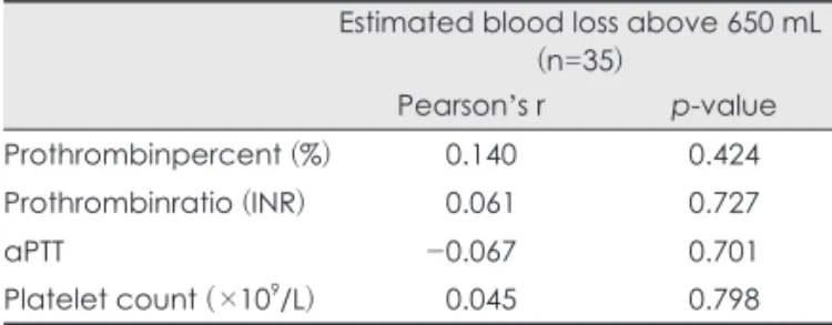
그러나 수술 중 추정 출혈량은 그 자체만으로의 수술 후 경 막외 출혈에 대한 독립된 위험요소라기보다는, 항혈소판 및 항응고제의 사용과 수술 전 응고장애가 수술 중 실혈량에 영 향을 미쳤을 것으로 생각하여, 650 mL 이상의 실혈량과 수 술 전 응고장애의 상관분석을 시행해보았다 (Table 6). 본 저 자의 예상과는 달리, 수술 중 출혈량과 수술 전 확인된 혈액 응고 파라미터(coagulation parameter)사이에는 상관관계가 없었다. 650 mL를 초과하는 수술 중 출혈은 수술 중의 불완 전한 지혈 또는 기본적인 진단검사로 식별되지 않은 응고장 애로 인해 발생했을 가능성도 생각할 수 있다.
본 연구는 몇 가지 한계점을 가지고 있다. 첫째, 후향적 연 구설계가 가지는 주관적인 치우침(selective bias)으로 인한 영향을 배제할 수 없고, 확실한 결론을 도출하기에는 모집단 인원이 적은 한계점이 있었다. 둘째, 수술 후 경막외 출혈과 연 관 될 수 있는 수술 전 출혈량에 대한 평가와 공간점유 병변 (space occupying lesion) 등의 해부학적 고려가 되지 않은 점, 정량화하기 힘든 여러 수술자의 술기의 숙련도가 결과에 영 향을 미친 부분에 대해 변수로 포함하지 못한 한계점을 가진 다. 셋째로, 대조군의 환자는 환자군의 초기 수술 3개월 이내 에 동일한 진단과 치료를 받았던 것에 기반을 두고 선정되었 다. 환자군 및 대조군의 연령은 개별적으로 연령 치우침을 최 소화하기 위해 서로 10년 이내의 유사한 연령으로 짝을 맞 추었다. 그 결과 연령, 병리 소견 등의 변수는 가능한 위험인 자에서 제외되었다. 이전 연구 중 Kalfas와 Little 등14)은 4,992 예의 두개내 수술 환자 중 40예에서 수술 후 출혈 발생을 보 고하였다 (0.8%). 병리 소견에 따른 수술 후 출혈 발생은 전
방 순환 동맥류(anterior circulation aneurysm)로 넓은 개두 술을 받은 환자에서 가장 높았고 (2.1%), 두번째는 종양 제거 를 위해 개두술을 받은 환자였다 (1.4%).
결 론
수술후 경막외 출혈은 모든 두개 내 수술의 약 1% 내에서 발생하는 심각한 합병증이다. 이 합병증의 예방을 위해서 먼 저 경막, 두개골, 근육 및 연부조직의 수술 중 완전한 지혈이 이루어져야 한다. 본 연구는 몇 가지 제한점을 가지고 있지만, 수술 중 대량의 혈액손실, 넓은 개두술 또는 두개절제술, 연 장된 PT (>11.3초) 및 수술 전 의식 상태 (GCS<8)는 두개내 수술후 경막외 출혈의 증가된 위험과 연관이 있을 수 있음을 보여준다.
■ The authors have no financial conflicts of interest.
REFERENCES
1) Altschuler E, Moosa H, Selker RG, Vertosick FT Jr. The risk and ef- ficacy of anticoagulant therapy in the treatment of thromboembolic complications in patients with primary malignant brain tumors.
Neurosurgery 27:74-76; discussion 77, 1990
2) Awad JN, Kebaish KM, Donigan J, Cohen DB, Kostuik JP. Analy- sis of the risk factors for the development of post-operative spinal epidural haematoma. J Bone Joint Surg Br 87:1248-1252, 2005 3) Ban SP, Son YJ, Yang HJ, Chung YS, Lee SH, Han DH. Analysis of
complications following decompressive craniectomy for traumatic brain injury. J Korean Neurosurg Soc 48:244-250, 2010 4) Fukamachi A, Koizumi H, Nagaseki Y, Nukui H. Postoperative ex-
tradural hematomas: computed tomographic survey of 1105 intra- cranial operations. Neurosurgery 19:589-593, 1986
5) Garber ST, Sivakumar W, Schmidt RH. Neurosurgical complica- tions of direct thrombin inhibitors--catastrophic hemorrhage after mild traumatic brain injury in a patient receiving dabigatran. J Neurosurg 116:1093-1096, 2012
6) Gentleman D, Johnston RA. Postoperative extradural hematoma as- sociated with induced hypertension. Neurosurgery 17:105-106, 1985 7) Gerlach R, Raabe A, Zimmermann M, Siegemund A, Seifert V.
Factor XIII deficiency and postoperative hemorrhage after neuro- surgical procedures. Surg Neurol 54:260-264; discussion 264-265, 8) Guangming Z, Huancong Z, Wenjing Z, Guoqiang C, Xiaosong W. 2000 Should epidural drain be recommended after supratentorial crani- otomy for epileptic patients? Surg Neurol 72:138-141; discussion 141, 9) Haft H, Liss H, Mount LA. Massive epidural hemorrhage as a com-2009
plication of ventricular drainage. J Neurosurg 17:49-54, 1960 10) Holden MP. Dangers of aspirin before cardiac surgery. BMJ 305:
365-366, 1992
11) Hussain SA, Selway R, Harding C, Polkey CE. The urgent postoperative CT scan: a critical appraisal of its impact. Br J Neurosurg 15:116-118, 2001
12) Institute SAS. The sas system for windows, version 9.2. Cary, NC:
SAS Institute, 1996
13) Jeon JS, Chang IB, Cho BM, Lee HK, Hong SK, Oh SM. Immedi- ate postoperative epidural hematomas adjacent to the craniotomy site. J Korean Neurosurg Soc 39:335-339, 2006
TABLE 6. Correlation analysis of the parameters associated with coagulopathy and intraoperative blood loss exceeding 650 mL
Estimated blood loss above 650 mL (n=35)
Pearson’s r p-value
Prothrombinpercent (%) 0.140 0.424
Prothrombinratio (INR) 0.061 0.727
aPTT -0.067 0.701
Platelet count (×109/L) 0.045 0.798 INR: international normalized ratio, aPTT: activated partial thromboplastin
14) Kalfas IH, Little JR. Postoperative hemorrhage: a survey of 4992 intracranial procedures. Neurosurgery 23:343-347, 1988 15) Le Roux AA, Nadvi SS. Acute extradural haematoma in the elder-
ly. Br J Neurosurg 21:16-20, 2007
16) Manian FA, Meyer PL, Setzer J, Senkel D. Surgical site infections associated with methicillin-resistant Staphylococcus aureus: do post- operative factors play a role? Clin Infect Dis 36:863-868, 2003 17) Palmer JD, Sparrow OC, Iannotti F. Postoperative hematoma:
a 5-year survey and identification of avoidable risk factors.
Neurosurgery 35:1061-1064; discussion 1064-1065, 1994 18) Park JE, Kim SH, Yoon SH, Cho KG, Kim SH. Risk factors predict-
ing unfavorable neurological outcome during the early period after traumatic brain injury. J Korean Neurosurg Soc 45:90-95, 2009 19) Rapanà A, Lamaida E, Pizza V. Multiple postoperative intrace-
rebral haematomas remote from the site of craniotomy. Br J Neurosurg 12:364-368, 1998
20) Sinar EJ, Lindsay KW. Distant extradural haematoma complicating removal of frontal tumours. J Neurol Neurosurg Psychiatry 49:
442-444, 1986
21) Soff GA. A new generation of oral direct anticoagulants.
Arterioscler Thromb Vasc Biol 32:569-574, 2012
22) Sokolowski MJ, Garvey TA, Perl J 2nd, Sokolowski MS, Cho W, Mehbod AA, et al. Prospective study of postoperative lumbar epi- dural hematoma: incidence and risk factors. Spine (Phila Pa 1976) 33:108-113, 2008
23) Thiagarajah S. Postoperative care of neurosurgical patients. Int Anesthesiol Clin 21:139-156, 1983
24) Troupp H. Extradural hematoma during craniotomy. Report of five cases. J Neurosurg 40:783-785, 1974
25) Yacubian EM, de Andrade MM, Jorge CL, Valério RM. Cerebellar hemorrhage after supratentorial surgery for treatment of epilepsy:
report of three cases. Neurosurgery 45:159-162, 1999
26) Yoshino Y, Aoki N, Oikawa A, Ohno K. Acute epidural hematoma developing during twist-drill craniostomy: a complication of per- cutaneous subdural tapping for the treatment of chronic subdural hematoma. Surg Neurol 53:601-604, 2000
![TABLE 1. Clinical characteristics of the control and hematoma groups Control group [n=46 (%, 66.7%)] Hematoma group [n=23 (%, 33.3%)] p-value Gender Male 30 (65.2) 13 (56.5) 0.482 Female 16 (34.8) 10 (43.5) Age Mean±SD 50.2±12.5 50.4±14.0 0.948 Diabetes me](https://thumb-ap.123doks.com/thumbv2/123dokinfo/5301862.156029/4.1020.97.872.147.869/clinical-characteristics-control-hematoma-control-hematoma-female-diabetes.webp)