Sebaceous adenomas are rare benign epithelial tumors composed of cells that exhibit sebaceous differentiation;
these tumors account for less than 0.1% of all salivary gland neoplasms (1, 2). The parotid gland is the most fre- quently involved of the salivary glands, where seba- ceous differentiation is common (3). There is little infor- mation available in English literature on the radiologic features of this rare neoplasm, especially that of cystic sebaceous adenoma. This report presents the CT find- ings of a cystic sebaceous adenoma in the parotid gland of a 60-year-old patient.
Case Report
A 60-year-old man presented with a mass on the right parotid area that had been gradually enlarging for three years without symptoms of pain or tenderness. The pa-
tient had no significant history of past illness. The differ- ential diagnosis on physical examination included be- nign parotid mass and lymphadenopathy. Laboratory tests revealed no abnormalities.
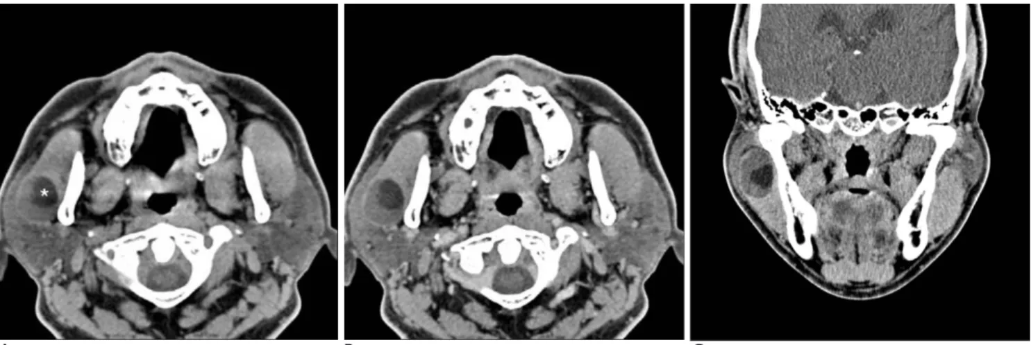
Computed tomography (CT) revealed a well-demar- cated mass measuring about 2×2.5×3 cm in the right parotid area, initially misinterpreted as arising within the right masseter muscle. On pre-contrast CT scan, the mass mainly showed low attenuation-containing fat components (-40 HU) (Fig. 1A). On contrast enhance- ment, only a thin capsule-like structure was enhanced at the periphery of the lesion (Figs. 1B and 1C). The imag- ing differential diagnosis included dermoid cyst and lipoma, and the mass was thought to originate in the right masseter muscle. No cervical lymphadenopathy was found.
Superficial parotidectomy of the right parotid gland and gross examination revealed a round mass measur- ing 3.7×3×3 cm within the right parotid gland. In sec- tions of the mass, the cut surface exhibited a unilocular cystic lesion containing old hemorrhage and grayish- white friable tissue (Fig. 2). Microscopically, the cyst wall was lined with stratified squamous epithelia with evidence of sebaceous differentiation, including the
J Korean Radiol Soc 2007;56:21-24
─ 21 ─
Cystic Sebaceous Adenoma of the Parotid Gland:
A Case Report
1Soo-Yeon Kim, M.D., Jeong-Nam Heo, M.D.2, Jinoo Kim, M.D., Moon Hyang Park, M.D.3, Chan Kum Park, M.D. 3
1Department of Diagnostic Radiology, Hanyang University Seoul Hospital,
2Department of Diagnostic Radiology, Hanyang University Kuri Hospital,
3Department of Pathology, Hanyang University Seoul Hospital Received September 13, 2006 ; Accepted October 20, 2006
Address reprint requests to : Jeong-Nam Heo, M.D., Department of Diagnostic Radiology, Hanyang University Kuri Hospital, 249-1 Kyomoon-dong, Kuri, Kyunggi-do 471-701, South Korea.
Tel. 82-31-560-2550 Fax. 82-31-560-2551 E-mail: [email protected]
Cystic sebaceous adenomas are rare neoplasms that can arise in salivary glands.
Among the salivary glands, the most commonly reported location is the parotid gland where it must be differentiated from other intraparotid masses. Unfortunately, its imaging features are not well-known as a result of its rarity. We report a case of cystic sebaceous adenoma that manifested as a gradually enlarging mass within the parotid gland of a 60-year-old man.
Index words :Neoplasms
Computed tomography (CT) Salivary glands, neoplasms
presence of foamy cytoplasm. Focal basaloid hyperpla- sia was also noted with some dysplastic change (Fig.
3A). The cystic lumen contained necrotic and papillary- growing sebaceous cells (Fig. 3B). The pathological diag- nosis of cystic sebaceous adenoma was rendered based on these findings.
Discussion
Sebaceous adenomas are rare, benign, encapsulated, epithelial tumors that comprise less than 0.1% of all sali- vary gland neoplasms; they belong to a category that in- cludes other sebaceous neoplasms, such as sebaceous lymphadenoma, sebaceous carcinoma, and sebaceous lymphadenocarcinoma, as well as sebaceous differentia- tion within other tumors (1, 2). As the name implies, these tumors originate from locations where sebaceous gland tissue exists; hence, the most common locations are the face and scalp (4, 5). However, sebaceous gland tissue has been reported in salivary glands (3), which ex- plains the discovery, although rare, of these tumors in such locations. Among the salivary glands, the parotid gland is the most common site of origin (1, 3). Other less common sites include the submandibular gland, the mi- nor salivary glands, Stenson’s duct of the buccal mu- cosa, the lower molar, and the lacrimal gland (6-8).
Patients who present with these tumors are mostly in their fifth or sixth decade with a slight male predomi- nance (2). Sebaceous adenomas involving the skin are a component of Muir-Torre syndrome (MTS), an autoso- mal dominant genodermatosis characterized by at least
a single sebaceous gland tumor and an internal malig- nancy such as colorectal or genitourinary carcinoma (5, 7). In contrast, no such relationship between these tu- mors and MTS has been documented when the tumors arise from salivary glands. In this particular case, CT evaluation of the chest and abdomen found no evidence of internal malignancy.
Grossly, the tumor size typically ranges from 0.4 to 3 cm in diameter and tumors are well-capsulated or sharply circumscribed. The tumor is usually grayish white, pinkish white or yellowish gray in color.
Histologically, in past reports, many tumors have a mi- crocystic appearance with abundant sebaceous glands, and all the neoplasms were embedded in a stromal fi-
Soo-Yeon Kim, et al: Cystic Sebaceous Adenoma of the Parotid Gland
─ 22 ─
A B C
Fig. 1. A 60-year-old man presented with a gradually enlarging mass in the right parotid area.
A. Pre-contrast axial CT scan shows a round circumscribed mass that was initially misinterpreted as located within the right mas- seter muscle; surgery and histopathology later proved it to be parotid in origin. The mass demonstrates low attenuation-containing fat components (-40 HU) (*).
B. Post-contrast axial and (C) coronal CT scans reveal only peripheral rim enhancement corresponding to the cyst wall.
Fig. 2. Gross specimen from right superficial parotidectomy.
The tumor predominantly consists of a cyst that contains old hemorrhage. The inner surface is smooth without solid por- tions.
brosis. The tumor is composed of incompletely differen- tiated sebaceous lobules containing various amounts of sebaceous cells and undifferentiated basaloid cells the cystic nature of the sebaceous adenoma resulted from necrosis of central sebaceous cells (1, 5-8).
Although sebaceous adenoma is well-known and has been reported in various literatures, it has been of little interest to radiologists because of its rarity and the fact that most sebaceous adenomas arise in superficial seba- ceous glands where imaging evaluation is rarely neces- sary. However, when it arises in the salivary gland, it must be differentiated from other salivary gland neo- plasms and understanding its imaging features becomes necessary. On CT scan, the most commonly reported appearance of sebaceous adenoma is a solid mass with various degrees of cystic change (9, 10). Differential di- agnosis of sebaceous adenomas that have a cyst-like ap- pearance includes Warthin tumor, inflammatory cyst, or first branchial cleft cyst. Some reports document hy- poattenuated areas of negative Hounsfield value within the mass, corresponding to sebaceous material, as we saw in this case. Lipoma also exhibits a negative Hounsfield value on CT due to its adipose composition, but it can be differentiated by its homogeneous fat den- sity (2, 9, 10).
In summary, a well-circumscribed mass in the parotid gland with areas of fat attenuation on CT should suggest
the possibility of a sebaceous adenoma.
References
1. Ellis GL, Auclair PL, Tumors of the salivary glands; Atlas of Tumor Pathology, Series III, Washington, DC: Armed Forces Institute of Pathology, 1996:130-133
2. Som PM, Curtin HD. Head and neck imaging. St. Louis, Mo.:
Mosby, 2003:2098-2099
3. Linhartova A. Sebaceous glands in salivary gland tissue. Arch Pathol 1974;98:320-324
4. Akhtar S, Oza KK, Roulier RG. Multiple sebaceous adenomas and extraocular sebaceous carcinoma in a patient with multiple sclero- sis: case report and review of literature. J Cutan Med Surg 2001;5:490-495
5. Abbott JJ, Hernandez-Rios P, Amirkhan RH, Hoang MP. Cystic se- baceous neoplasms in Muir-Torre syndrome. Arch Pathol Lab Med 2003;127:614-617
6. Izutsu T, Kumamoto H, Kimizuka S, Ooya K. Sebaceous adenoma in the retromolar region: report of a case with a review of the English literature. Int J Oral Maxillofac Surg 2003;32:423-426 7. Kaminagakura E, Andrade CR, Rangel AL, Coletta RD, Graner E,
Almeida OP, et al. Sebaceous adenoma of oral cavity: report of case and comparative proliferation study with sebaceous gland hy- perplasia and Fordyce’s granules. Oral Dis 2003;9:323-327 8. Liu CY, Chu PY, Li WY, Chang SY. Sebaceous adenoma in the
submandibular gland. Otolaryngol Head Neck Surg 2002;126:199- 200
9. Shen WC, Kwan PC, Ho YJ, Lee SK. CT of sebaceous adenoma of the parotid gland. AJNR Am J Neuroradiol 1994;15:1265-1266 10. Dillon WP, Mancuso AA. The oropharynx and nasopharynx. In
Newton TH, Hasso AN, Dilon WP. Computed Tomography of the Head and Neck. New York: Raven Press, 1988:56-59
J Korean Radiol Soc 2007;56:21-24
─ 23 ─
A B
Fig. 3. Photomicrographs of the histologic specimen.
A. The cystic wall is lined by stratified basaloid squamous epithelium with basal cell hyperplasia (large arrows). The cystic lumen contains nests of sebaceous cells (small arrows). (H & E, ×200)
B. The lumen of the cyst contains papillary-growing sebaceous cells. (H & E, ×100)
Soo-Yeon Kim, et al: Cystic Sebaceous Adenoma of the Parotid Gland
─ 24 ─
대한영상의학회지 2007;56:21-24
이하선의 낭성 피지선종: 증례 보고1
1한양대학교 서울병원 진단방사선과
2한양대학교 구리병원 진단방사선과
3한양대학교 서울병원 병리과
김수연・허정남2・김진우・박문향3・박찬금3
낭성 피지선종은 타액선에 발생하는 드문 종양으로 그 영상소견은 잘 알려졌지 않다. 이 종양은 타액선 중 이하 선에 가장 많이 발생하며 다른 이하선 종양과의 감별이 필요하다. 저자들은 60세 남성에서 서서히 자란 이하선 종 괴로 발현된 낭성 피지선종의 예를 영상 소견과 함께 보고하고자 한다.