Archives of Aesthetic Plastic Surgery
138
Arch Aesthet Plast Surg
비배부 히알루론산 필러주사 이후 발생한 다발성 색전증:
피부 연조직괴사, 실명, 안구운동 마비, 뇌경색의 증례보고
오 상 아
단국대학교 의과대학 성형외과학교실
Multiple Embolism after Injection of Hyaluronic acid Filler in Nasal Dorsum: A Case Report of Skin Necrosis, Blindness, Oculomotor Palsy
and Cerebral Infraction
Sangah Oh, M.D.
Department of Plastic and Reconstructive Surgery, College of Medicine, Dankook University, Cheonan, Korea As hyaluronic acid filler commonly applied in many cosmetic fields, tissue necrosis due to filler embolism is reported infrequently. Herein we report the first case of nasal hyaluronic acid filler injection resulting in nasal necrosis, multifocal brain infarction and ocular ischemia with hypotonia. A 25-year-old female, who had received an injection of hyaluronic acid filler along the nasal dorsum a month before, visited our hospital to manage the skin ulcer of nasal ala. Immediately after the injection, the patient had complained of nausea, vomiting, headache and nose pain without neurologic symptom. The skin had bluish change at her nose and forehead. And the right eyelid ptosis was observed with decreased eyeball movement and visual loss.
Multifocal punctuated infarctions were found on both frontal lobes on brain MRI. The necrotic skin of right nasal ala was healed in 3 months. But, the width of the right nasal ala was smaller than the left side, and the right eye remained blind with extropia at 8 months after injection. Although hyaluronic acid filler is safe in most cases, patients should be informed of the possibility of this rare complication.
(Archives of Aesthetic Plastic Surgery 18: 138, 2012) Key Words: Embolism, Hyaluronic acid
Ⅰ. 서 론
필러를 이용한 연조직 확대술은 쉽고 간단하여 다양한 신 체부위에 널리 시행되고 있다. 특히, 히알루론산(hyaluronic acid)은 면역반응이 적고, 교차결합으로 상당기간 지속되고, 색이 투명하다는 장점들이 있어 요즘 가장 많이 이용되는 물 질이다. 히알루론산 주사에 의한 합병증으로는 미세종괴, 혈
종, 염증반증, 감염 등이 있는데, 그 중 가장 심각한 합병증은 필러 색전증으로, 주사 물질에 의해 혈관 내부가 막히거나, 혈관외벽을 눌려 국소적인 조직 손상을 초래한다.
히알루론산 필러주사 이후 발생한 색전증의 임상증례들 이 종종 보고되고 있으며 일반적으로 미간이나 코입술주름 에서 위험성이 높다고 알려져 있다. 현재까지 보고된 히알루 론산 필러 색전증은 이마 및 미간1, 비익2, 비구순주름3에 피 부괴사가 발생한 경우가 있었고, 실명이나 안구운동장애4와 같은 기능의 손상도 보고되고 있다.
본 교실에서는 히알루론산 필러주사 시술 이후 발생한 색 전증에 의해 시력손실, 안구운동장애, 코의 괴사와 뇌경색이 동시에 발생한 증례를 경험하였기에 문한고찰과 함께 보고 하는 바이다.
Received September 10, 2012 Revised October 15, 2012 Accepted October 15, 2012
Address Correspondence: Sangah Oh, M.D., Department of Plastic and Reconstructive Surgery, College of Medicine, Dankook University, 201 Manghyang-ro, Dongnam-gu, Cheonan 330-715, Korea.
Tel: +82-41-550-6477, Fax: +82-41-554-6477, E-mail: [email protected]
*본 논문은 제 69차 대한성형외과학회 학술대회에서 포스터 발표되었음.
Vol. 18, No. 3, 138-141, 2012
Ca se R ep or t
오상아: 히알루론산 필러주사 후 다발성 색전증
139
Arch Aesthet Plast Surg
에 피부색이 검푸르게 변하여 히알루론산 분해효소를 병변 부 피하에 주사하였다. 주사 5시간 후, 우안은 상안검 하수가 발생하였고 빛감지가 불가능하였다(Fig. 1). 환자는 근처 대 학병원으로 전원되었고, 뇌 자기공명영상에서는 주요 뇌혈 관의 이상은 없었으나, 양측 전두부 다발성 점상 급성 뇌경 색(multifocal punctuate acute infarction)이 발견되어 신경과 에 입원하였다(Fig. 2). 응급실 내원 당시 우측 안저는 창백하 였고, 망막중심동맥과 정맥은 말초에서만 관찰되고 중심부 에서는 관찰되지 않았으며(Fig. 3), 우측 동공은 정상측에 비 해 확장되었고 안구하수가 있었고 안구운동은 전방향으로 제한되어 시선이 고정되어 있었다(Fig. 4). 우안의 20 prism diopter의 외사시(extropia) 및 6 prism diopter의 수직사시(hy- pertropia)가 관찰되었다. 중심망막동맥 폐색과 동안신경마 비로 진단되어 스테로이드요법을 시행하였다. 환자는 주사 1주일 후부터 형성된 이마, 비익 및 비첨, 협부에 농포성 피부 병변과 비익의 피부괴사가 발생하여 괴사조직 절제술 및 소 파술을 시행받았고, 이후 피부병변 치료를 위해 본원 외래로 전원되었다. 주사 후 3개월 경, 우안의 상안검 기능은 정상화 되었고, 피부병변은 습윤 드레싱, 항생제 치료, 스테로이드 연고 치료를 통해 치유되었으나, 환측의 비익의 폭(10 mm) 이 정상측(12 mm)에 비해 위축되어 있었다(Fig. 5). 필러주사 시술 후 8개월에 우안의 단안운동검사(duction test of Kesten- baum) 시 내측으로 -1의 제한만 남을 정도로 회복되었고 다 른 방향으로의 안구운동은 정상이었다. 20 prism diopter의 외사시는 호전이 없었으나 수직사시는 완전히 회복되었다.
주사 시술 12개월 후에도 우안은 실명된 상태로 호전되지 않 았다.
Ⅱ. 증 례
과거력상 특이사항이 없는 25세 여자 환자가 이마, 코의 염증 및 피부 부분층 괴사를 치료하기 위해 본원 성형외과 를 방문하였다. 환자는 본원 내원 1개월 전 개인의원에서 비 근에서 비첨에 이르는 부분에 히알루론산 필러(Perfectha Deep®, Obvieline, France) 1 ml로 융비술 주사시술을 받았다.
비첨부를 epinephrine이 포함된 lidocaine로 마취한 뒤 히알 루론산을 주사하였다. 주사 직후 코의 통증과 함께 오심과 구토증상을 동반하였고, 신경기능에 장애는 없었으나 극심 한 두통이 발생하였다. 주사 3시간 후, 이마, 비배부와 비측부
Fig. 1. Initial photograph of the patient. The skin was dusky and patchy. And right eyelid was ptotic, and the vision was lost.
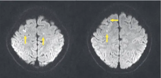
Fig. 2. Images of the brain MRI on the day of injection. Multifocal punctated acute infarctions (yellow arrow) were found in both frontal areas in diffusion images.
140
Arch Aesthet Plast Surg
Archives of Aesthetic Plastic Surgery Vol. 18, No. 3, 2012
Ⅲ. 고 찰
색전증의 사전적 의미는 외부 물질이 혈류를 통해 이동하 여 혈관을 직접 막고 분수령 구역(watershed area)에 허혈 손 상을 일으키는 것을 의미하지만, 필러 시술에서는 상기 기전 이외에도 바늘에 의한 혈관이 직접 손상되거나, 필러의 부피 에 의해 혈관이 압박되어 조직의 허혈 손상을 받을 수 있는 데 이를 임상적으로 구분하기는 어렵다.
피부손상, 뇌경색, 안동맥, 망막중심동맥에 동시에 발생한 다발성 색전증은 지방이식 후 발생한 것을 Danesh-Meyer5가 보고한 바가 있으나 본 증례와 달리 안구운동의 장애는 동반 되지 않았다. 히알루론산 필러를 주사한 경우에서 안면부의 피부조직 색전증 및 실명이 발생한 증례는 각각 보고되고 있 지만,1-4 본 교실에서 경험한 것과 같이 실명, 안구운동마비, 피부괴사, 뇌경색이 동시에 발생한 예는 현재까지 보고된 바 가 없다.
두개강과 안와, 안면부의 혈액순환 사이에 존재하는 다수 의 동맥관 문합은 다발성 색전증의 형성 기전을 설명하는 해 부학적 근거가 된다. 필러 물질이 혈관을 역행하면 문합 부 위을 통하거나 혈관의 근위부를 통해 다른 여러 혈관 분지를 막아 다발성 후향성 색전증(retrograde embolism)을 일으킬 수 있다.
코에 필러를 주사할 때에는 외비동맥(lateral nasal artery) 의 상비익분지(superior alar branch)나 비배동맥(dorsal nasal artery)을 통해 본 증례와 같은 다발성 색전증을 일으킬 수 있 다.
외비동맥의 상비익분지는 코의 하외연골(Lower lateral cartilage)의 꼬리쪽경계(caudal boder)를 따라 주행하는 혈관 으로, 외비동맥은 안각동맥(angular artery)에서 분지하거나, 드물게는 상구순동맥(superior labial artery)에서 직접 분지하 는데, 이를 통해 비익, 비순주름, 상구순 등에 필러가 이동할 수 있다.6 또한, 안각동맥은 외측으로는 하안와동맥(infraor- bital artery)과 문합하고, 위로는 비배동맥과 문합하기 때문 에 이를 통해 각각의 혈관지배 영역으로도 조직괴사를 일으 킬 수 있다.
비배부를 따라 종으로 주행하는 비배동맥은 활차상동맥 (supratrochlear artery)과 함께 안동맥(ophthalmic artery)에 서 분지되는 최종혈관으로, 상활차동맥을 통해 미간이나 이 마로 주사물질이 이동할 수 있다. 필러가 안동맥을 통해 역 류되면 망막중심동맥(central retinal artery)이나 후모양체동 맥(posterior ciliary artery)을 막아 시력을 손상시킬 수 있고, 더 근위부인 내경동맥(internal carotid artery)으로 흘러 들어 Fig. 3. Fundus of right eye on the day of injection. Optic disc was
pale, and retina was whitened with no central vessels.
Fig. 4. Eye ball movement on the day of injection. Ocular hypoto- nia developed on right eye 6 hour after the filler injection.
Fig. 5. Photograph of the patient on 6 months after injection. Skin lesion healed with alar atrophy on right side. Ocular hypotonia of the right eye was improved. Mild extropia and visual loss of right eye were permanent.
오상아: 히알루론산 필러주사 후 다발성 색전증
141
Arch Aesthet Plast Surg
립된 치료법이 없다.
필러를 이용한 성형수술은 쉽고 빠르게 자연스러운 결과 를 얻을 수 있어 환자에게나 의사에게나 보편화 되고 있다.
비록 필러 색전증이 매우 드물게 일어나는 것으로 알려져 있 으나, 그 합병증은 광범위할 수 있고 비가역적인 괴사 손상 을 줄 수 있기 때문에, 시술자들은 색전증 예방을 위하여 원 칙과 이론에 맞게 시술하고, 합병증 발생시 적절히 대응할 수 있어야 할 것이다.
REFERENCES
1. Cohen JL: Understanding, avoiding, and managing dermal filler complications. Dermatol Surg 34: s92, 2008
2. Kang MS, Park ES, Shin HS, Jung SG, Kim YB, Kim DW: Skin necrosis of the nasal ala after injection of dermal filler. Dermatol Surg 37: 375, 2011
3. Hirsch RJ, Cohen JL, Carruthers JD: Successful management of an unusual presentation of impending necrosis following a hyaluronic acid injection embolus and a proposed algorithm for management with hyaluronidase. Dermatol Surg 33: 357, 2007 4. Kim YJ, Kim SS, Song WK, Lee SY, Yoon JS: Ocular ischemia
with hypotony after injection of hyaluronic acid gel. Ophthal Plast Reconstr Surg 27: e152, 2011
5. Danesh-Meyer HV, Savino PJ, Sergott RC: Ocular and cerebral ischemia following facial injection of autologous fat. Arch Oph- thalmol 119: 777, 2001
6. Nakajima H, Imanishi N, Aiso S: Facial artery in the upper lip and nose: anatomy and a clinical application. Plast Reconstr Surg 109: 855, 2002
7. Glaich AS, Cohen JL, Goldberg LH: Injection necrosis of the gla- bella: protocol for prevention and treatment after use of dermal fillers. Dermatol Srug 32: 276, 2006
8. Schanz S, Schippert W, Ulmer A, Rassner G, Fierlbeck G: Arterial embolization caused by injection of hyaluronic acid (Restylane).
Br J Dermatol 146: 928, 2002 가면 중간내막동맥(middle meningeal artery)을 통해 뇌경색
을 일으킬 수 있다. 또한, 전교통동맥(anterior communicating artery)을 통해 반대측의 대뇌 손상도 가능하다.
필러주사에 의한 색전증을 완벽히 예방할 수 있는 방법은 없으나, (1) 국소마취제를 사용할 때 혈관수축제가 포함된 것을 사용하고, (2) 주사부위와 주사재료에 맞는 유연하고 가는 바늘을 선택하고, (3) 필러주사 전에 역류검사를 시행 하고, (4) 소량씩 주사하며, (5) 좁은 영역에 많은 양의 필러가 주사되지 않게 총량을 조절하고, (6) 해부학적으로 주요혈관 이 위치하는 곳을 피하기 위해 내측방향으로 주사하거나 얕 게 주사하는 것이 합병증 최소화하기 위한 방법일 것이다.
시술 당시 주사부위가 창백해지거나 검푸르게 변하면 일 단 피부의 색전증을 의심하고, 주사를 중단하고 부드럽게 마 사지하거나 따뜻한 거즈를 그 부위 얹어 혈관을 확장시켜야 한다. 니트로글리세린 연고를 병변부에 바르는 것도 혈관확 장에 도움이 된다.7 저분자량 헤파린을 피부의 그물모양 병 변이 사라질 때까지 2주일 동안 사용하여 색전증을 치료했 다는 보고도 있다.8 증상 초기에 혈관 주행에 맞추어 히알루 론산분해효소를 피하에 주사하면 히알루론산 젤의 분해를 촉진하여 혈관을 감압시키는 데 도움이 된다.1 일단 조직의 괴사가 진행되고 있다면, 반흔을 최소화 하기 위해 노력해야 한다. 철저히 상처를 관리하고 가피가 형성되지 않도록 하 며, 괴사조직이 감염이 되어 추가로 조직이 손실되지 않도록 해야 한다.8
필러 시술 중에 오심과 구토를 동반한 극심한 통증이나, 안 구의 기능이상을 발견하였을 경우 지체하지 말고 전문적인 치료를 받을 수 있도록 의뢰하여야 할 것이다. 뇌경색이나 외안근의 마비는 기능적인 후유증 없이 회복된 경우가 있으 나, 시력의 손상은 다양한 치료법의 시도에도 불구하고 영구 적인 시력 장애를 보이는 경우가 대부분이어서 아직까지 확