Addressing stretch myelopathy
Asian Spine Journal
Asian Spine Journal
1007Addressing Stretch Myelopathy in Multilevel Cervical Kyphosis with Posterior Surgery
Using Cervical Pedicle Screws
Bijjawara Mahesh, Bidre Upendra, Shekarappa Vijay, Kumar Arun, Reddy Srinivasa
Department of Spine Surgery, Jain Institute of Spine Care and Research (JISAR), Bhagwan Mahaveer Jain Hospital, Bangalore, India
Study Design: Technique description and retrospective data analysis.
Purpose: To describe the technique of cervical kyphosis correction with partial facetectomies and evaluate the outcome of single- stage posterior decompression and kyphosis correction in multilevel cervical myelopathy.
Overview of Literature: Kyphosis correction in multilevel cervical myelopathy involves anterior and posterior surgery. With the advent of cervical pedicle screw-rod instrumentation, single-stage posterior kyphosis correction is feasible and can address stretch myelopathy by posterior shortening.
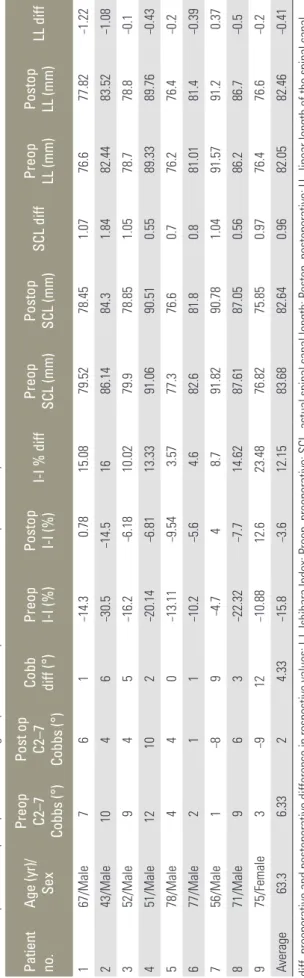
Methods: Nine patients underwent single-stage posterior decompression and kyphosis correction for multilevel cervical myelopathy using cervical pedicle screw instrumentation from March 2011 to February 2014 and were evaluated preoperatively and postopera- tively with modified Japanese Orthopaedic Association (mJOA) scoring and computed tomography scans for radiological measure- ments. Kyphosis assessment was made with Ishihara curvature index and C2–C7 Cobb’s angle. The linear length of the spinal canal and the actual spinal canal length were also evaluated. The average follow-up was 40.56 months (range, 20 to 53 months).
Results: The average preoperative C2–7 Cobb’s angle of 6.3° (1° to 12°) improved to 2° (10° to –9°). Ishihara index improved from
–15.8% (–30.5% to –4.7%) to –3.66% (–14.5% to +12.6%). The actual spinal canal length decreased from 83.64 mm (range, 76.8 to 91.82 mm) to 82.68 mm (range, 75.85 to 90.78 mm). The preoperative mJOA score of 7.8 (range, 3 to 11) improved to 15.0 (range, 13 to 17).
Conclusions: Single-stage posterior decompression and kyphosis correction using cervical pedicle screws for multilevel cervical myelopathy may address stretch myelopathy, in addition to decompression in the transverse plane. However, cervical lordosis was not achieved with this method as predictably as by the anterior approach. The present study shows evidence of mild shortening of cervical spinal canal and a positive correlation between canal shortening and clinical improvement.
Keywords: Stretch myelopathy; Multilevel cervical myelopathy; Cervical pedicle screws; Kyphosis correction; Partial facetectomies
Copyright Ⓒ 2016 by Korean Society of Spine Surgery
This is an Open Access article distributed under the terms of the Creative Commons Attribution Non-Commercial License (http://creativecommons.org/licenses/by-nc/3.0/) which permits unrestricted non-commercial use, distribution, and reproduction in any medium, provided the original work is properly cited.
Asian Spine Journal • pISSN 1976-1902 eISSN 1976-7846 • www.asianspinejournal.org
Received Feb 24, 2016; Revised Apr 9, 2016; Accepted Apr 9, 2016 Corresponding author: Bidre Upendra
Jain Institute of Spine care And Research ( JISAR), Bhagwan Mahaveer Jain Hospital, Vasanth Nagar, Bangalore 560052, India
Tel: +91-9686803495, Fax: +91-8022261153, E-mail: [email protected]
ASJ A SJ
Clinical Study Asian Spine J 2016;10(6):1007-1017 • https://doi.org/10.4184/asj.2016.10.6.1007
Introduction
The stretch component of the cervical cord seems a likely
contributing factor to cervical myelopathy [1-5] apart from non-mechanical factors like ischemia, apoptosis and inflammation [3,4]. Stretch-mediated myelopathy
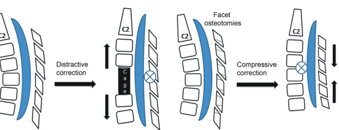
becomes more pronounced in a kyphotic cervical spine [6-8]. Furthermore, stretch-mediated diffuse axonal injury and the nonischemic apoptosis of oligodendroglial cells contributes to secondary spinal cord injury in tethered cord syndrome [5,9], whiplash injury and cervical spon- dylotic myelopathy (CSM) [10,11]. Surgical interventions planned for multilevel CSM with kyphosis need to address the stretch component on the cervical cord along with decompression in the transverse plane. Biomechanically, anterior cervical kyphosis correction with decompression over a fixed length of posterior facet joint complex may result in lengthening of the spinal canal (Fig. 1), whereas posterior compressive correction tends to decrease spinal canal length (SCL).
Kyphosis correction by the posterior approach alone requires partial facet resections and instrumented fusion under compression. However, posterior instrumenta- tion with lateral mass screws fixation is not feasible after partial facetectomies. With the advent of cervical pedicle screw-rod instrumentation, a single-stage posterior lami- nectomy and kyphosis correction with partial facetecto- mies facilitates correction of cervical kyphosis along with decompression.
The present study was done to describe the technique of cervical kyphosis correction with partial facetectomies and to evaluate the outcome of single-stage posterior de- compression and kyphosis correction in multilevel cervi- cal myelopathy.
Materials and Methods
1. Patients
Nine patients undergoing single-stage posterior decom- pression and deformity correction for multilevel cervical myelopathy with kyphosis using cervical pedicle screw (CPS) instrumentation at our institute from March 2011 to January 2014 were included in the study. Eight patients had chronic progressive spondylotic myelopathy and the remaining patient (patient 1) had sudden onset quadri- paresis with minor trauma superimposed on cervical spondylosis (Table 1). Five patients had four-level decom- pression and pedicle screw-rod instrumentation, three patients had three levels and one patient had five-level decompression and pedicle screw-rod instrumentation.
All patients were evaluated preoperatively and postopera- tively for clinical improvement using modified Japanese Orthopaedic Association (mJOA) scoring [12] and com- puted tomography (CT) scans were done postoperatively to assess pedicle screw placement.
2. Kyphosis
Radiological measurements were done to assess kypho- sis using the Ishihara curvature index (l-I [13]) and C2–
C7 Cobb’s angle preoperatively and postoperatively. Both were calculated on mid-sagittal sections of either magnetic resonance imaging (MRI) or CT scans preoperatively and postoperatively, having clear C2–C7 bony landmarks.
Fig. 1. The distractive correction of kyphosis achieved by anterior cage placement has a lengthening effect on the spinal cord as the axis of rotational correction falls on the fixed facet joint complex. The compressive correction achieved by partial facetectomies has a shortening effect on the spinal cord.
To determine the Ishihara index, the posterior inferior points of C2 and C7 were joined by a straight line denot- ing the linear length (LL) of the spinal canal. The trans- verse distance between the posterior inferior points of C3–C6 and the line LL was designated a3, a4, a5 and a6, respectively (Fig. 2). The Ishihara index was computed as (a3+a4+a5+a6)/LL×100. When postero-inferior points of C3–C6 were behind the LL, the values of a3–a6 were taken as negative.
1) Cobb’s angle, SCL, and LL
Cobb’s angle represented the angle between the inferior endplate of C2 and the superior endplate of C7 (Fig. 2).
Table 1. Preoperative and postoperative radiological parameters of patients involved in the present study Patient no.Age (yr)/ Sex
Preop C2–7 Co°)bbs (
Post op C2–7 Co°)bbs (
Cobb diff (
°) Preop I-I (%) Postop I-I (%) I-I % diffPreop SCL (mm) Postop SCL (mm)
SCL diffPreop LL (mm)Postop LL (mm)LL diff 167/Male 76 1–14.3 0.7815.0879.5278.451.0776.677.82–1.22 243/Male104 6–30.5–14.51686.1484.31.8482.4483.52–1.08 352/Male 94 5–16.2 –6.1810.0279.978.851.0578.778.8–0.1 451/Male1210 2–20.14 –6.8113.3391.0690.510.5589.3389.76–0.43 578/Male 44 0–13.11 –9.54 3.5777.376.60.776.276.4–0.2 677/Male 21 1–10.2 –5.6 4.682.681.80.881.0181.4–0.39 756/Male 1–8 9 –4.7 4 8.791.8290.781.0491.5791.2 0.37 871/Male 96 3–22.32 –7.714.6287.6187.050.5686.286.7–0.5 975/Female 3–912–10.88 12.623.4876.8275.850.9776.476.6–0.2 Average63.3 6.332 4.33–15.8 –3.612.1583.6882.640.9682.0582.46–0.41 diff, preoperative and postoperative difference in respective values; I-I, Ishihara Index; Preop, preoperative; SCL, actual spinal canal length; Postop, postoperative; LL, linear length of the spinal canal.
Fig. 2. Ishihara index (I-I) and C2–C7 Cobb’s calculation illustrated on preoperative mid-sagittal magnetic resonance imaging and postopera- tive mid-saggital computed tomography scan of patient 3. LL, linear length.
Fig. 3. Illustrates the linear length (LL) and the actual spinal canal length (SCL) being calculated in preoperative and postoperative mid- sagittal computed tomography scans of patient 2.
The SCL was determined both by linear method (LL of the spinal canal) and along the curvature of the spinal canal (actual SCL) in the mid-sagittal sections of MRI or CT scans preoperatively and postoperatively. LL was calculated by joining the posterior inferior points of C2 and C7 by a straight line (Fig. 3). The actual SCL was de- termined by lines drawn from the postero-inferior points of C2–C7 perpendicular to the posterior wall of the respective vertebrae up to the laminae (or extrapolated posterior canal limit in postoperative CT scans with lami- nectomies). The sum of the lines joining the midpoints of these lines (C3, C4, C5, C6, and C7) represented the actual SCL along the curvature of the cervical spine (Fig.
3). All measurements were made by one author (B.U.) us- ing Surgimap spine software (Surgimap Spine-1.1.2.271 Intl. 2009, Nemaris LLC) with calibrated images of MRI and CT scans. Care was taken to match the mid-sagittal preoperative and postoperative images to avoid discrep- ancies in measurement.
3. Screw placement and follow-up
Screw placement assessment was done by evaluating pedicle screw perforations graded on axial sections of CT scans: grade I perforations had <50% of the screw outside the pedicle and grade II perforations had >50% of the screw outside the pedicle walls [14]. The average follow- up was 40.56 months (range, 20 to 53 months). Clinical complications related to CPS placement and kyphosis correction were also recorded.
4. Surgical technique
A midline skin incision was made and the posterior ele- ments of the cervical vertebra to be instrumented were exposed sub-periosteally. The exposure was carried out far laterally to expose the lateral border of the lateral mass and extended cranially or caudally as required. CPS instrumentation was done at the desired levels using the medial cortical pedicle screw placement technique with partial drilling of the medial cortex, as previously described [15]. Depending on the levels of compression, laminectomies along with foraminotomies were done for central and root canal decompression. Partial facetecto- mies with removal of caudal edge of the superior facet and the cranial edge of the inferior facet were performed depending on the extent of kyphosis correction required.
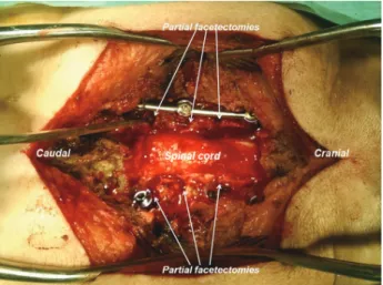
For partial facetectomies, the medial half to two-thirds of the superior articular facet and the inferior articular facet were drilled with 3-mm cutting burr taking care to stop as the posterior cortex of the superior articular facet was reached. The thinned-out portion of the superior articular facet was removed with 2 mm rongers having thin foot plates over the corresponding cervical root. Most of the facet joints became mobile once the medial half to two- thirds of the joint was excised and was compressed over pedicle screw-rods for kyphosis correction.
5. Avoiding root compression
Aggressive compression from the posterior aspect was avoided to prevent iatrogenic foraminal stenosis. After medial half-facetectomy and final compression, the space for the root was assessed by passing a blunt probe till the root exited from the foramen. Anatomically, the nerve root begins to curve ventrally after travelling the medial half of the facet (Fig. 4) and so has less chance of becom- ing compressed (Fig. 5). The authors preferred to keep a small portion of the lateral facet intact as it provided a bed for bone grafting for posterolateral fusion at the end of the procedure. The vertebral artery lodged anterior to the cervical root was safe and protected by the root dur- ing facetectomies. Correction of kyphosis was achieved by manual head elevation and finally by contoured rod inser- tion with compression as required (Fig. 5). All foramina were probed before final tightening of the screws.
6. Statistical analysis
Microsoft Word and Excel (Microsoft, Redmond, WA,
Fig. 4. Intraoperative photograph showing partial facetectomy with medial half of the facet being removed and the nerve root being visu- alized.
USA) were used to generate the tabular data. Pearson’s chi-square analysis was used to test bivariate relation- ships between outcome variables. The change in scores were computed between preoperative and postoperative measures, and bivariate relationships were determined be- tween the changed scores (I-I, SCL, LL, Cobb, and mJOA) preoperatively and postoperatively.
Results
The average age of the nine patients (8 males, 1 female)
was 63.3 years (range, 43–78 years). The average preop- erative C2–7 Cobb’s angle was 6.3° kyphosis (range, 1°
to 12°), which marginally improved to 2° (range, 10° to –9°) postoperatively. The Ishihara Index improved from –15.8% (range, –30.5% to –4.7%) to –3.66% (range, –14.5%
to +12.6%) postoperatively, accounting for an average im- provement of 76.8% (Table 1). The actual SCL decreased from an average of 83.64 mm (range, 76.8 to 91.82 mm) to 82.68 mm (range, 75.85 to 90.78 mm). LL increased postoperatively from an average of 82.0 mm (range, 76.2 to 91.5 mm) to 82.46 mm (range, 76.4 to 91.2 mm) (Table 1) indicating that the preoperative kyphosis was only cor- rected to the neutral position.
The average preoperative mJOA score of 7.88 (range, 3 to 11) improved to 15.0 (range, 13 to 17) postoperatively (Table 2). One patient (patient 2) had postoperative C5 palsy, which was improved at 8–10 weeks postoperatively.
Another patient (patient 5) expired at 30-month follow- up due to an unrelated medical event. The average blood loss was 377 mL (range, 100 to 650 mL) and the average operative time was 179 minutes (range, 90 to 270 minutes) (Table 2).
Sixty-one CPSs were inserted from C3–C7 with the me- dial cortical pedicle screw technique (Table 3) [15]. Thir- ty-eight screws were placed within the pedicle; 23 screws (37.7%) showed misplacement with 13 (21.3%) being grade I medial perforations. There was one grade II lateral perforation (Fig. 6) without any clinical consequences. No clinical complication directly related to screw placement Fig. 5. Intraoperative photograph showing decompression (laminec-
tomy with foraminotomies) with partial facetectomies and kyphosis correction using pedicle screw-rod instrumentation with compression of partial facetectomies.
Table 2. Depicts the clinical parameters with follow-up of patients involved in the study Patient
no. Age (yr)/
Sex Diagnosis Decomp.
+inst levels Blood loss
(mL) Surgical
time (min) Preop
mJOA Postop
mJOA mJOA
diff Follow-up (mo)
1 67/Male CSM 4 650 180 3 15 12 53
2 43/Male CSM 5 350 240 8 17 9 48
3 52/Male CSM 4 250 120 11 16 5 36
4 51/Male CSM 4 650 175 10 16 6 56
5 78/Male CSM 3 500 270 7 15 8 30
6 77/Male CSM 4 450 180 8 15 7 52
7 56/Male CSM 3 250 180 9 13 4 48
8 71/Male CSM 4 200 180 9 13 4 22
9 75/Female CSM 3 100 90 6 15 9 20
Average 63.3 - - 377.7 179.4 7.88 15 7.12 40.56
Decomp.+inst levels, number of decompression levels plus instrumentation levels; m-JOA, modified Japanese Orthopaedic Association score [12];
Preop, preoperative; SCL, actual spinal canal length; Postop, postoperative; CSM, cervical spondylotic myelopathy.
was observed.
There was a significant inverse correlation (p=0.031) between the LL difference with a mJOA difference follow- ing surgery (r=–0.713). There were no significant bivariate relationships between Cobb, LL, and SCI change follow- ing surgery (Table 4).
Discussion
The present results indicate that the actual SCL decreases with kyphosis correction by posterior CPS instrumenta- tion and partial facetectomies. Chiba et al. [16] reported
that, despite kyphosis, laminoplasty gave good neurologi- cal recovery and avoided a planned anterior surgery in few of their patients. They reported that a decrease in the vertical height had probably contributed to neurologi- cal recovery in their patients, despite persistent kyphosis.
Therefore, a decrease in SCL along with kyphosis correc- tion is desirable. Presently there was an average decrease of 0.96 mm in actual SCL postoperatively (Table 1). SCL decrease produces slackness in the cervical cord [16]. De- spite an increase in the LL of the spine, improvement in mJOA was seen in all patients. A decrease in the LL could have been achieved only if the authors were able to correct the kyphosis to lordosis, beyond the neutral alignment.
MRI studies have measured the cord length in cervi- cal flexion, extension, and neutral position in healthy [17] and CSM [18] patients. These studies consistently reported lengthening of the cord in flexion as compared to neutral or extension. The patho-anatomical changes of the spinal cord in CSM patients [19-21] show flattening of the cord antero-posteriorly with the demyelination and gliosis that was most pronounced in the lateral portions of the cord in a cone shaped manner with the base placed laterally. These patho-anatomical findings are not consis- tent with the theory that antero-posterior compression is the sole primary cause of CSM [22]. Levine [22] provided evidence in a biomechanical model that the findings of lateral column involvement in CSM correlated better with the dural stretch theory. In addition, the cord is tethered to the spinal canal at points of stenosis in the cervical ca- nal [23]. In a human cadaver study, Adams and Logue [24]
Table 3. Shows the number of cervical pedicle screws placed in each patient along with the misplacement grades Patient
no. Age (yr)/
Sex Diagnosis Level No. of
screws Grade-I
medial Grade-II
medial Grade-I
lateral Grade-II lateral
1 67/Male CSM 4 6 2 2 0 0
2 43/Male CSM 5 10 2 1 0 1
3 52/Male CSM 4 8 1 1 1 0
4 51/Male CSM 4 6 1 0 1 0
5 78/Male CSM 3 6 2 0 1 0
6 77/Male CSM 4 8 2 0 0 0
7 56/Male CSM 3 5 1 0 1 0
8 71/Male CSM 4 6 0 0 1 0
9 75/Female CSM 3 6 2 0 0 0
Total - - - 61 13 (21.3) 4 (6.56) 5 (8.2) 1 (1.63)
Values are presented as number (%).
CSM, cervical spondylotic myelopathy.
Fig. 6. A grade II lateral perforation seen at C3 vertebra (in patient 2). There were no intraoperative or postoperative complications due to the screw misplacement. The patient has completed 48-month follow-up.
Table 4. Statistical analysis with pearson correlation and significance of association (p-value) depicted between preoperative and postoperative differences of I-I, SCL, LL, C2–C7 Cobb angle, and mJOA
Parameters measured Correlation
I-I diff SCL diff LL diff mJOA diff
Cobb diff
Pearson correlation 0.608 0.375 0.393 –0.131
Sig. (2-tailed) p-value 0.082 0.320 0.296 0.737
I-I diff
Pearson correlation - 0.275 –0.336 0.322
Sig. (2-tailed) p-value - 0.474 0.376 0.399
SCL diff
Pearson correlation - - –0.393 0.392
Sig. (2-tailed) p-value - - 0.295 0.297
LL diff
Pearson correlation - - - –0.713
Sig. (2-tailed) p-value - - - 0.031
I-I, Ishihara Index; SCL, actual spinal canal length; LL, linear length of the spinal canal; mJOA, modified Japanese Orthopaedic Association score;
diff, preoperative and postoperative difference in respective values; Sig., significance.
Fig. 7. Examinations of patient 1 reveals the preoperative kyphotic cervical stenosis with intra-medullary signal changes on magnetic resonance imaging and postoperative X-ray and computed tomography scans. Note the effect of partial drilling of the medial cortex with increased tendency of medial perforations by the screws [15].
described the localized increase in dural stretch adjacent to the point of dural fixation by stenotic segments.
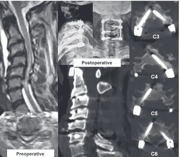
Two illustrative cases are described. Patient 1 (Fig. 7) was a 67-year-old male with sudden onset quadriparesis due to a fall 1 week before admission. He had cervical kyphosis with multilevel compressive myelopathy and was operated on using single-stage laminectomy and CPS rod instrumentation from C3–C6 with deformity cor- rection by partial facetectomies. He recovered well and has completed 53 months of follow-up, and is at present ambulatory without support. His preoperative mJOA of 3 has improved to 15. Patient 2 (Fig. 8) is 43-year-old male who presented with progressive spasticity in lower limbs for 6 months and inability to walk since 1 month before admission. His imaging showed a sigmoid cervical kyphotic deformity with myelopathic cord changes. He underwent posterior laminectomies with partial facetec- tomies for decompression and deformity correction by CPS instrumentation. He developed unilateral C5 palsy postoperatively, which gradually improved by 8–12 weeks.
He has completed 48 months of follow-up and is gainfully employed with ability to do all his indoor and outdoor activities without any support. His preoperative mJOA of 8 has improved to 17.
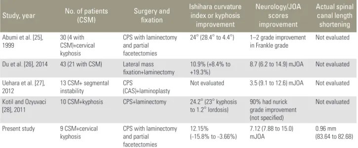
The largest study of cervical kyphosis correction with CPS instrumentation comprising of thirty patients was re- ported by Abumi et al. [25]. There were only four patients with CSM in their series (Table 5). One of the reasons for very few studies [25-28] on kyphosis correction with CPS instrumentation in CSM is the relatively high incidence of CPS misplacements in CSM patients as compared to other pathologies. The perforation rates are reported to be about 37.5% [29]. Furthermore, the numbers of lateral perfora- tions are reportedly twice that of medial perforations. In the present study, although the authors experienced a mis- placement rate of 37.7%, most were grade I medial perfo- rations (21.3%), and 9.83% (8.2% grade I; 1.63% grade II) had lateral perforations (Table 3). A multicenter study [14]
on CPS complications reported 75% of CPS perforations were lateral perforations endangering the vertebral artery,
Fig. 8. Examinations of patient 2 shows preoperative sigmoid kyphotic cervical spine on magnetic resonance imaging and com- puted tomography (CT) scans, and postoperative X-ray and CT scans along with axial sections showing placement of pedicle screws at each level.
in contrast to our results of more medial perforations.
This further substantiates the previous report [15] on the decreased lateral perforation rates seen with the medial cortical pedicle screw insertion technique.
Surgical management of multilevel CSM with kyphosis has been challenging with the literature showing no sin- gle approach to be superior. An anterior-only approach with multilevel corpectomy has been fraught with early mechanical failures with graft and implant dislodgement [30,31]. A combined anterior and posterior procedure has been found to decrease the mechanical failures with good fusion rates and neurological outcome [32]. Wen et al. [33] compared the efficacy of anterior-posterior com- bined surgery with anterior or posterior alone surgeries in a 255 patient series. Kyphosis correction was better with anterior procedures, but was complicated by implant failures in multilevel decompressions. The combined pro- cedure yielded the best results with good kyphosis correc- tion, neurological recovery and reduced mechanical fail- ures due to rigid fixation, but with increased morbidity of the combined procedure. In this context, posterior alone surgery with CPS is an additional alternative in patients with multilevel CSM with kyphosis, for better kyphosis correction with more aggressive facetectomies and rigid internal fixation, avoiding a combined approach in the elderly.
The present study is limited due to the small number of
patients and its retrospective design. However, this needs to be seen in the context of the largest series of cervical kyphosis correction (30 patients) using CPS [25], which included only four patients with CSM. The present study involves measurements made on mid-sagittal sections of preoperative and postoperative MRI or CT scans. All the measurements were made by a single author, who was also involved in the surgery, which could have led to bias in these measurements.
The present study brings to the forefront the concept of surgically addressing stretch myelopathy, which has not received much attention in surgical management of mul- tilevel CSM with kyphosis. This study provides evidence of shortening, albeit minimal, of the cervical spinal canal, with posterior deformity correction using CPS. It also shows a positive correlation between canal shortening and clinical improvement (mJOA), which might be partially due to decompression alone. Either anterior or posterior decompressive surgery provides predictable neurological recovery irrespective of the amount of kyphosis correc- tion [34]. However, long-term results of persistent kypho- sis are not encouraging with progression of deformity and deterioration of neurology [35]. Further, it is not yet clear if kyphosis correction and shortening of the SCL has any reversible effect on the stretch mediated axonal and my- elin changes in the cervical spinal cord.
Table 5. Comparison of studies evaluating results of decompression, posterior instrumentation, and kyphosis correction in multilevel cervical spondylotic myelopathy
Study, year No. of patients
(CSM) Surgery and
fixation
Ishihara curvature index or kyphosis
improvement
Neurology/JOA scores improvement
Actual spinal canal length shortening Abumi et al. [25],
1999 30 (4 with
CSM)+cervical kyphosis
CPS with laminectomy and partial
facetectomies
24° (28.4° to 4.4°) 1–2 grade improvement
in Frankle grade Not evaluated Du et al. [26], 2014 43 (21 with CSM) Lateral mass
fixation+laminectomy
10.9% (+8.4% to +19.3%)
8.7 (6.2 to 14.9) mJOA Not evaluated Uehara et al. [27],
2012 13 CSM+ segmental
instability CPS
(CAS)+laminoplasty Not evaluated 3.5 (9.1 to 12.6) mJOA Not evaluated Kotil and Ozyuvaci
[28], 2011 10 CSM+kyphosis CPS+laminectomy 24.2° (23° kyphosis
to 1.2° lordosis) 90% had nurick grade improvement (not specified)
Not evaluated
Present study 9 CSM+cervical
kyphosis CPS with laminectomy and partial
facetectomies
12.15%
(–15.8% to –3.66%) 7.12 (7.88 to 15.0)
mJOA 0.96 mm
(83.64 to 82.68) CSM, cervical spondylotic myelopathy; JOA, Japanese Orthopaedic Association score; CPS, cervical pedicle screws; mJOA, modified Japanese Orthopaedic Association score [12]; CAS, computer assisted surgery.
Conclusions
Single-stage posterior decompression and kyphosis cor- rection using CPSs for multilevel cervical myelopathy with kyphosis may address the stretch component on the spinal cord in addition to decompression in the transverse plane. However, cervical lordosis was not achieved with this method, as predictably as by the anterior approach.
Further studies are required to ascertain the reproduc- ibility of spinal canal shortening by posterior kyphosis correction using CPS and to evaluate any reversible effects on the stretch mediated axonal and myelin changes in the cervical spinal cord.
Conflict of Interest
No potential conflict of interest relevant to this article was reported.
References
1. Karadimas SK, Gatzounis G, Fehlings MG. Pathobi- ology of cervical spondylotic myelopathy. Eur Spine J 2015;24 Suppl 2:132-8.
2. Kalsi-Ryan S, Karadimas SK, Fehlings MG. Cervical spondylotic myelopathy: the clinical phenomenon and the current pathobiology of an increasingly prev- alent and devastating disorder. Neuroscientist 2013;
19:409-21.
3. Karadimas SK, Erwin WM, Ely CG, Dettori JR, Fe- hlings MG. Pathophysiology and natural history of cervical spondylotic myelopathy. Spine (Phila Pa 1976) 2013;38(22 Suppl 1):S21-36.
4. Fehlings MG, Skaf G. A review of the pathophysiol- ogy of cervical spondylotic myelopathy with insights for potential novel mechanisms drawn from trau- matic spinal cord injury. Spine (Phila Pa 1976) 1998;
23:2730-7.
5. Henderson FC, Geddes JF, Vaccaro AR, Woodard E, Berry KJ, Benzel EC. Stretch-associated injury in cervical spondylotic myelopathy: new concept and review. Neurosurgery 2005;56:1101-13.
6. Snow RB, Weiner H. Cervical laminectomy and fo- raminotomy as surgical treatment of cervical spon- dylosis: a follow-up study with analysis of failures. J Spinal Disord 1993;6:245-50.
7. Sim FH, Svien HJ, Bickel WH, Janes JM. Swan-neck
deformity following extensive cervical laminectomy.
A review of twenty-one cases. J Bone Joint Surg Am 1974;56:564-80.
8. Breig A, Turnbull I, Hassler O. Effects of mechanical stresses on the spinal cord in cervical spondylosis. A study on fresh cadaver material. J Neurosurg 1966;25:
45-56.
9. Breig A. Overstretching of and circumscribed patho- logical tension in the spinal cord: a basic cause of symptoms in cord disorders. J Biomech 1970;3:7-9.
10. Yamaura I, Yone K, Nakahara S, et al. Mechanism of destructive pathologic changes in the spinal cord un- der chronic mechanical compression. Spine (Phila Pa 1976) 2002;27:21-6.
11. Li GL, Brodin G, Farooque M, et al. Apoptosis and expression of Bcl-2 after compression trauma to rat spinal cord. J Neuropathol Exp Neurol 1996;55:280- 9.
12. Kopjar B, Tetreault L, Kalsi-Ryan S, Fehlings M. Psy- chometric properties of the modified Japanese Or- thopaedic Association scale in patients with cervical spondylotic myelopathy. Spine (Phila Pa 1976) 2015;
40:E23-8.
13. Ohara A, Miyamoto K, Naganawa T, Matsumoto K, Shimizu K. Reliabilities of and correlations among five standard methods of assessing the sagittal align- ment of the cervical spine. Spine (Phila Pa 1976) 2006;31:2585-91.
14. Nakashima H, Yukawa Y, Imagama S, et al. Compli- cations of cervical pedicle screw fixation for nontrau- matic lesions: a multicenter study of 84 patients. J Neurosurg Spine 2012;16:238-47.
15. Mahesh B, Upendra B, Mahan RS. The medial corti- cal pedicle screw: a new technique for cervical ped- icle screw placement with partial drilling of medial cortex. Spine J 2014;14:371-80.
16. Chiba K, Toyama Y, Watanabe M, Maruiwa H, Mat- sumoto M, Hirabayashi K. Impact of longitudinal distance of the cervical spine on the results of expan- sive open-door laminoplasty. Spine (Phila Pa 1976) 2000;25:2893-8.
17. Kuwazawa Y, Pope MH, Bashir W, Takahashi K, Smith FW. The length of the cervical cord: effects of postural changes in healthy volunteers using posi- tional magnetic resonance imaging. Spine (Phila Pa 1976) 2006;31:E579-83.
18. Zhang L, Zeitoun D, Rangel A, Lazennec JY, Catonne
Y, Pascal-Moussellard H. Preoperative evaluation of the cervical spondylotic myelopathy with flexion- extension magnetic resonance imaging: about a pro- spective study of fifty patients. Spine (Phila Pa 1976) 2011;36:E1134-9.
19. Brain WR, Northfield D, Wilkinson M. The neuro- logical manifestations of cervical spondylosis. Brain 1952;75:187-225.
20. Mair WG, Druckman R. The pathology of spinal cord lesions and their relation to the clinical features in protrusion of cervical intervertebral discs; a report of four cases. Brain 1953;76:70-91.
21. Ogino H, Tada K, Okada K, et al. Canal diameter, an- teroposterior compression ratio, and spondylotic my- elopathy of the cervical spine. Spine (Phila Pa 1976) 1983;8:1-15.
22. Levine DN. Pathogenesis of cervical spondylotic my- elopathy. J Neurol Neurosurg Psychiatry 1997;62:334- 40.
23. Tencer AF, Allen BL Jr, Ferguson RL. A biomechani- cal study of thoracolumbar spine fractures with bone in the canal. Part III. Mechanical properties of the dura and its tethering ligaments. Spine (Phila Pa 1976) 1985;10:741-7.
24. Adams CB, Logue V. Studies in cervical spondylotic myelopathy. I. Movement of the cervical roots, dura and cord, and their relation to the course of the ex- trathecal roots. Brain 1971;94:557-68.
25. Abumi K, Shono Y, Taneichi H, Ito M, Kaneda K.
Correction of cervical kyphosis using pedicle screw fixation systems. Spine (Phila Pa 1976) 1999;24:2389- 96.
26. Du W, Zhang P, Shen Y, Zhang YZ, Ding WY, Ren LX. Enlarged laminectomy and lateral mass screw fixation for multilevel cervical degenerative myelopa- thy associated with kyphosis. Spine J 2014;14:57-64.
27. Uehara M, Takahashi J, Ogihara N, et al. Cervical
pedicle screw fixation combined with laminoplasty for cervical spondylotic myelopathy with instability.
Asian Spine J 2012;6:241-8.
28. Kotil K, Ozyuvaci E. Multilevel decompressive lami- nectomy and transpedicular instrumented fusion for cervical spondylotic radiculopathy and myelopathy: a minimum follow-up of 3 years. J Craniovertebr Junc- tion Spine 2011;2:27-31.
29. Uehara M, Takahashi J, Hirabayashi H, et al. Perfora- tion rates of cervical pedicle screw insertion by dis- ease and vertebral level. Open Orthop J 2010;4:142-6.
30. Sasso RC, Ruggiero RA Jr, Reilly TM, Hall PV. Early reconstruction failures after multilevel cervical cor- pectomy. Spine (Phila Pa 1976) 2003;28:140-2.
31. Zdeblick TA, Hughes SS, Riew KD, Bohlman HH.
Failed anterior cervical discectomy and arthrodesis.
Analysis and treatment of thirty-five patients. J Bone Joint Surg Am 1997;79:523-32.
32. Kim PK, Alexander JT. Indications for circumfer- ential surgery for cervical spondylotic myelopathy.
Spine J 2006;6:299S-307S.
33. Wen SF, Wong IO, Long MJ, et al. Effectiveness of 3 surgical decompression strategies for treatment of multilevel cervical myelopathy in 3 spinal centers in China: a retrospective study. Spine (Phila Pa 1976) 2012;37:1463-9.
34. Kawakami M, Tamaki T, Ando M, Yamada H, Yo- shida M. Relationships between sagittal alignment of the cervical spine and morphology of the spinal cord and clinical outcomes in patients with cervical spondylotic myelopathy treated with expansive lami- noplasty. J Spinal Disord Tech 2002;15:391-7.
35. Suda K, Abumi K, Ito M, Shono Y, Kaneda K, Fujiya M. Local kyphosis reduces surgical outcomes of ex- pansive open-door laminoplasty for cervical spondy- lotic myelopathy. Spine (Phila Pa 1976) 2003;28:1258- 62.