Corresponding author: Soo A Lim, Department of Plastic and Reconstructive Surgery, Kepco Medical Center, 308 Uicheon-ro, Dobong-gu, Seoul 132-703, Korea Tel: 82-2-901-3109, Fax: 82-2-901-3104 E-mail: [email protected]
조기 박피술과 동종유래 표피세포를 이용한 깊은 2도 화상 치료의 효용성
이방석ㆍ임수아ㆍ윤용일 한전의료재단 한전병원 성형외과
The Clinical Efficacy of Early Dermabrasion and Frozen Cultured Allogenic Keratinocyte in Ma- nagement of the Deep Second Degree Burn with Thin Eschar
Bang Suk Lee, M.D., Soo A Lim, M.D. and Yong Il Yoon, M.D.
Department of Plastic and Reconstructive Surgery, Kepco Medical Center, Seoul, Korea
Purpose: As in the case of the deep second-degree burn, proper eschar elimination and early epithelization is essential for spontaneous healing without surgical intervention. Accor- dingly, we have treated with using early dermabrasion and appling frozen cultured allogenic keratinocyte patients in deep second degree burns an eschar formed.
Methods: From January 2011 to January 2012 at Kepco medical center, we selected 46 patients who were suffered from deep second degree burn formed an eschar were enrolled. Patients were divided into two parts, study and con- trol group. Study group were performed dermabrasion within first 3 days to 10 days of the injury and then applied frozen cultured allogenic keratinocyte. control group were managed moist dressing using hydrocolloid gel, form and alginate materials. We tried to prove its clinical efficacy by research- ing the period of wound healing, percentage of skin graft, and hospital days under chart review and photograph.
Results: In study group, the mean period of wound healing was 15.13±4.18 (mean±S.D.) days, and that of the hospital- ization was 16.65±5.31 (mean±S.D.) days. For the 3 patients without the epithelization, skin graft was conducted. As for the control group, the mean period of wound healing was 24.22±2.79 (mean±S.D.) days, and that of the hospital- ization was 28.30±3.33 (mean±S.D). 21 patients were con- ducted skin grafts.
Conclusion: Based on these results, we concluded that the
treatment in deep second degree burn patients eschar for- med using early dermabrasion and frozen cultured allogenic keratinocyte is effective in reducing the duration of period of wound healing, hospital day and rate of skin graft. (J Korean Burn Soc 2013;16:5-11)
Key Words: Dermabrasion, Keratinocyte, Burn
INTRODUCTION
Initial assessment of burn injury is essential to achieve optimal outcome in patient. In superficial second degree burns healing time is usually within 2 weeks without scarring. But in deep second-degree burn eschar formed, the wound is left dried or infected, it takes long times to heal and result unfavorable esthetically and functional problems. These cases skin graft is recommended. ‘How can we minimize patient’s hospitalization and operability in deep dermal burn eschar formed?’ When dermabrader is used to remove this eschar earlier, it is more time sav- ing than waiting spontaneous elimination by debriding agent and manual dressing. Eschar free and clean wound bed can promote epithelization. The use of frozen cul- tured allogenic keratinocyte (KalodermⓇ) (Tegoscience, Seoul, Korea) can accelerate healing processes also. In 1975, Green et al.1) first attempted to culture the skin keratinocytes. The cultured allogenic keratinocytes have been applied to a clinical setting and used for the treat- ment of wounds. Given the above background, we con- ducted this clinical study to examine the effectiveness of an escharectomy using a dermabrader which minimizes damages to dermis and the skin appendages in patients with a deep second-degree burn formed a thin eschar.
This treatment was followed by using dressing agents based on frozen cultured allogenic keratinocyte on the af- fected sites.
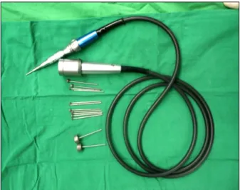
Fig. 1. Instrument. Dermabrader with a high-speed drill set and a diamond burr.
PATIENTS AND METHODS
We retrospectively reviewed the medical records and photographs of 46 patients who presented to the Kepco Medical Burn Center between January 2011 and January 2012 with a history of deep second-degree burn injury.
We separate patients two parts, the study and control group. In more accuracy for this study, we select patients who underwent deep second-degree burn formed eschar were same age, sex ,wound site, less than TBSA 5%. And no patients enrolled in this study reported a history of underlining disease as diabetes, high blood pressure, viral or bacterial infection, autoimmune disease etc. The study group of 23 patients consent to make dermabrasion and use frozen cultured allogenic keratinocyte and they in- formed about complications and benefits of procedure, the control group of 23 patients were not signed a letter of informed consent. The study group patients were com- posed of 11 men and 12 women. Their mean age was 38.30 years, ranging from 1 to 73 years. The mean range of burn size was 2.21±0.90% (mean±S.D) TBSA. The con- trol group patients were composed of 12 men and 11 women. Their mean age was 28.76 years, ranging from 1 to 70 years. The mean range of burn size was 2.47±1.23%
(mean±S.D) TBSA. Outcome measures were the period of wound healing, percentage of skin graft, and hospital days. The study group of 23 patients was performed der- mabrasion within first 3 to 10 days following the onset of injury until the healthy dermis was exposed. A der- mabrader, the experimental device used in the current study, was equipped with a high-speed drill set and a di- amond burr (AesculapⓇ), where the burr type was cylin- drical one with a maximum rotation speed of 20,000 rpm (Fig. 1). The healthy dermis was defined based on the presence of white dermis and capillary bleeding. Then we applied cultured allogenic keratinocyte, commercially available KalodermⓇ (Tegoscience, Korea). It was stored in a frozen state at a temperature of -70oC and it was subjected to a 5-min thawing time prior to laboratory use.
Dressing was covered by BactigrasⓇ (Smith & Nephew, UK) and saline gauze. By changing BactigrasⓇ and saline gauze on a daily basis, the wet environment was main- tained and the exudate was removed. The dressing with
frozen cultured allogenic keratinocyte was maintained for 5∼7 days. When the epithelization was almost achieved following the removal of dressing, the treatment was con- ducted with the use of ointment and foam agent. All these procedures were performed by identical plastic surgeons. The control group was treated by conventional dressing material with foam and hydrocolloid gel. Within 2 weeks after burn injury, if epithelization was relatively delayed or the loss of full thickness of the skin was ob- served, skin graft was done. The period of wound healing was defined as the length of time (the number of days) elapsed from the date of the burned until a 100% re-epi- thelization was achieved or until the exudate was not formed. In the patient was performed skin graft, the peri- od of wound healing was defined as the length of time (the number of days) elapsed from the date of the burned until raw surface was not observed. The confirmation of re-epithelization had made by three plastic surgeon in- cluding the surgeon who performed dermabrasion. Cate- gorical variables were compared using Fisher’s exact test or the Pearson χ2 test as appropriate, and continuous variables were compared using Student’s t-test. All tests of significance were two-tailed, and P values ≤0.05 were deemed to indicate statistical significance. Statistical anal- yses of the data were performed using the PASW sta- tistics software (version 18.0; SPSS Inc., Chicago, IL).
Patient No. Age/Sex Etiology Site TBSA (%)
Period of wound healing
(days)
Hospital day
(days) Skin graft
1 53/M Flame burn Face, neck 3 15 16 x
2 64/M Flame burn Face 3 17 18 x
3 50/F Flame burn Face 2 13 15 x
4 63/F Flame burn Face 1 22 26 o
5 34/M Flame burn Arm 2 16 16 x
6 46/F Flame burn Face 2 13 14 x
7 27/M Flame burn Hand 2 10 11 x
8 57/M Flame burn Neck 2 12 13 x
9 51/F Scalding burn Arm 2 16 17 x
10 11/M Scalding burn Chest 2 25 29 o
11 10 m/F Scalding burn Foot 1 19 20 x
12 6/F Scalding burn Chest 3 18 19 x
13 48/F Scalding burn Arm 4 12 13 x
14 12 m/F Scalding burn Thigh 2 15 16 x
15 13 m/F Scalding burn Arm 2 17 18 x
16 11 m/M Contact burn Hand 1 11 11 x
17 48/M Chemical burn Face 1 13 14 x
18 62/F Scalding burn Arm 3 23 30 o
19 72/F Flame burn Lower leg 3 9 11 x
20 56/M Flame burn Arm 2 13 14 x
21 5/M Contact burn Foot 1 12 13 x
22 73/F Scalding burn Thigh 4 11 12 x
23 53/M Scalding burn Thigh 3 16 17 x
Table 1. Period of Wound Healing, Hospital Day, and Patient Number Who Underwent Skin Graft of Study Group
Study group (number of
patient)
Control group (number of
patient)
Age (years) 1∼10 6 8
11∼20 1 2
21∼30 1 1
31∼40 1 2
41∼50 4 3
51∼60 6 4
61∼ 4 3
Sex Male 11 12
Female 12 11
Burn area Face 5 4
Upper extemities 8 10
Lower extemities 6 5
Neck, trunk 4 4
Etiology Scalding burn 10 10
Flame burn 10 10
Contact burn 2 3
Chemical burn 1 0
TBSA (%) 2.21±0.90 2.47±1.23
Table 2. Patient Profiles of Study and Control Group
Experimental group (mean±S.D)
Control group (mean±S.D)
P-value
Time to heal (days) 15.13±4.18 24.22±2.79 0.000 Hospital day (days) 16.65±5.31 28.30±3.33 0.000
Skin graft* 13% 91% 0.000
*Number of patients who were underwent skin graft Table 3. Comparison of Study & Control Groups
RESULTS
The mean period of wound healing of the study group applied by frozen cultured allogenic keratinocyte after early dermabrasion was 16.65±5.31 (mean±S.D) days. For the 3 patients out of 23, without epithelization, the skin graft was performed (Table 1). As for the control group, the mean period of wound healing was 24.22±2.79 (mean±S.D) days and that of the hospitalization was 28.30±3.33 (mean±S.D) days. Also, for the 21 patients out of 23, skin graft was performed (Table 2). Results of the comparisons between the study group and control group
Complication Number of patient
Pigmentation 9
pruritus 8
Dermatitis 5
Infection 2
Hypertrophy 5
Table 4. Complications of Study Group
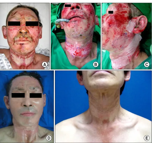
Fig. 2. Study group, case 1. A 53-year-old male patient with deep second-degree flame burn on the face and neck. (A) Preoperative view. 7 days after the burn, a 4×8 cm2 eschar was formed on the patient’s neck. (B) Intraoperative view. Eschar was eliminated using a dermabrader under general ane- sthesia. Capillary bleeding and re- ticular dermis was seen. (C) Ap- plication of the cultured allogenic skin. (D) Postoperative day 21. (E) Postoperative 18 months.
showed that the mean period of wound healing of the former was shortened by 9 days and that of the hospital- ization was also shortened by 12 days. In addition, we observed lower rate of skin graft in study group in that the number of the patients who received skin graft was 3 and 21 respectively (P=0.000) (Table 3). We also eval- uated whether the adverse effects occurred following the application of the cultured allogenic keratinocyte. This showed that 8 patients presented with pruritus but most of them had the symptoms improved within several days after the onset of them. There were 9 patients and 5 pa- tients who developed hyperpigmentation and hyper-
trophic scar, respectively (Table 4). There were 3 patients who achieved no epithelization, for whom a split-thick- ness skin graft was performed. In addition, there were no significant differences in the hematological and sero- logical parameters between prior to and following the ap- plication of the cultured allogenic keratinocyte.
DISCUSSION
Many studies demonstrate the application of frozen cul- tured human epithilial allografts not only promotes faster re-epithelization of deep and superficial partial-thickness burns but also achieve early closure of wounds and good functional outcomes2,3). Our study shows its effectiveness of timely treatment and hospitalization by application of frozen cultured allogenic keratinocyte following dermab- rasion not classical tangential excision for elimination of eschar. Early wound closure is effective in also, to reduce grafting and donor site morbidity. This is a key message of this article. Dermabrasion is a surgical procedure
Fig. 4. Control group, case. A 9 months female patient with deep second-degree scalding burn on forearm. (A) 3 days after the burn, 2×4 cm2 eschar was formed on the patient’s forearm. (B) 11 days after the burn, eschar was still observed. (C) 19 months after split thickness skin graft.
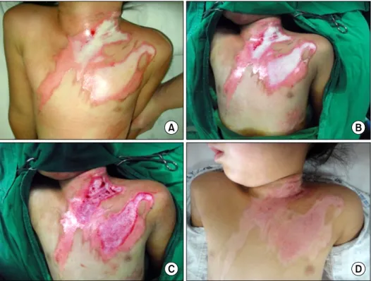
Fig. 3. Study group, case 2. A 6-year-old female patient with deep second-degree scalding burn on neck and anterior chest. (A) 3 days after the burn, a 3×7 cm2, 5×11 cm2 eschar was formed on the patient’s neck and anterior chest. (B) Preoperative view. 6 days after the burn. (C) Intraope- rative view. Afer dermabrasion, capillary bleeding was seen. (D) Postoperative day 10. Small size raw surface was left on the neck.
where the skin is carved to the desirable depth using high-speed rotary instruments. In 1953, Kurtin4) first de- veloped a motor powered wire brush. Since then, a der- mabrasion has been used to remove the scars due to acne, chicken pox or trauma as well as posttraumatic tattoo.
Especially, it would be of prime importance to recognize the current surgical depth during operation. Reticular dermis needs to be checked to decide the degree of the skin graft as its gland and hair follicles play an important role in epithelization. We can distinguish reticular dermis by checking its white color and capillary. The period of wound healing and the formation of scars following the
wound healing are dependent on the amount of skin appendages. It is therefore important to minimize the damages to the dermal layer and the skin appendages.
Esposito et al.5) noted dedicated training with an experi- enced surgeon and a careful and wise approach in not removing all of the dermis is sufficient to avoid such a complication and does not require grafting in most cases.
Therefore, it needs a careful handling of the instrument and a technical expertise of plastic surgeons. The applica- tion of frozen cultured human epithilial allografts pro- motes faster re-epithelization of Frozen cultured allogenic keratinocyte, the experimental agent used in the current
study, was taken from the culture of skin keratinocytes harvested from the epidermal tissue of newborns, and it became commercially available as a sheet form. Skin kera- tinocytes are generally cultured based on the cell culture methods of Rheinwald and Green. A piece of cellular sheet with allograft keratinocytes (7.5×7.5 cm, 56 cm2) was solely composed of approximately 1,000 million skin ker- atinocytes and it was attached to Vaseline gauze. Many studies have shown that allograft keratinocytes produce molecules increasing the proliferation and migration of the skin keratinocytes6) and thereby promoting the epithe- lization at the affected sites and these include interleukin (IL)-1α, IL-β, IL-3, IL-62, IL-63, transforming growth fac- tor α (TGF-α)7) and extracellular matrix components.
Kaloderm, which was used in this study, helps epitheliza- tion by letting both growth factor expressed by cultured allogenic keratinocyte and cytokine affect cell pro- liferation and migration. The difference between autolo- gous allogenic keratinocyte which is cultured from the patient’s own skin and cultured allogenic keratinocyte is that the former adheres directly and permanently to the injured part while the latter temporarily adheres to the wound and is replaced through the process of the move- ment and growth of self-epithelial cell located on the sur- face and marginal area. Since the late 1980’s, studies have been conducted to examine the treatment methods based on allograft tissue culture. This eventually led to the de- velopment of agents based on cultured allogenic kerati- nocyte. With the emergence of these agents, their effects have also been examined. Fratianne et al.8) reported that the period of epithelization at the affected sites was short- ened by 30∼40% with the application of cultured allo- genic keratinocyte at donor sites after a split-thickness skin graft for a superficial second-degree burn as com- pared with conventional types of dressing methods. In addition, Choi et al.9) reported that the mean period of healing was shortened by 3.4 days following the applica- tion of cultured allogenic keratinocyte. Furthermore, Braye et al.10) also reported that a prompt wound healing and stability could be achieved following the application of cultured allogenic keratinocyte in patients with a deep second-degree burn. In this study, 2 out of 3 patients in the experimental group showed contagious exudate.
Therefore, frozen cultured allogenic keratinocyte was re-
moved in 3∼4 days and pseudomonas aeruginosa was cultured in the applied wound culture. As frozen cul- tured allogenic keratinocyte is vulnerable to contagion, it is important to keep the aseptic condition when changing dressing. Also, in order to prevent hyperpigmentation and pruritus, most commonly occurred complications, keeping proper hydration and applying sun block for the few months period of epithelization are essential. In addi- tion, 3 months after discharge, hypertrophic scar was formed in 5 patients. As for 2 of them, contagious exudate was secreted in the preoperative wound and 3 patients were the ones with longest treatment period. Accordin- gly, preoperative contagion and treatment period were considered to have influence on the development of scar and complications.
CONCLUSION
Treatment of choice in burn wound is often dictated by depth and TBSA. In third degree burn involving all layers of skin, early surgical excision and grafting is indicated as soon as possible. But skin graft is not always accept- able in deep second degree burn considering sacrifice of donor skin and mismatch of color and texture around re- cipient skin. Additional, harvested donor site dressing is frustrating to patients. Furthermore, development of addi- tional scar on the donor site and long hospitalization peri- od are inevitable. So, we think about early epithelization without surgical excision and grafting. We used dermab- rader and frozen cultured allogenic keratinocyte in treat- ing patients with a deep second degree burn accompany- ing eschar. Consequently, we were able to confirm the satisfactory result which is shortening the wound healing and hospitalization period. Also, reduced rate of skin graft demonstrated its effectiveness in the reduction of donor site morbidity.
REFERENCES
1) Rheinwald JG, Green H. Serial cultivation of stains of human epidermal keratinocytes: the formation of keratinizing colo- nies from single cells. Cell. 1975;6:331-343.
2) Kurtin A. Corrective surgical planning of skin. AMA Arch Dermt Syph. 1953;68:389-397.
3) Alvarez-Diaz C, Cuenca-Pardo J, Sosa-Serrano A, Juárez-
Aguilar E, Marsch-Moreno M, Kuri-Harcuch W. Controlled clinical study of deep partial-thickness burns treated with frozen cultured human allogeneic epidermal sheets. J Burn Care Rehabil. 2000;291-299.
4) Yanaga H, Udoh Y, Yamauchi T, Yamamoto M, Kiyokawa K, Inoue Y, et al. Cryopreserved cultured epidermal allografts achieved early closure of wounds and reduced scar formation in deep partial-thickness burn wounds (DDB) and split- thickness skin donor sites of pediatric patients. Burns. 2001;
27:689-698.
5) Esposito G, Gravante G, Montone A. Use of early dermabra- sion in pediatric burn patient. Plast Reconstr Surg. 2006;
116:573-575.
6) Lavker RM, Sun TT. Heterogeneity in epidermal basal kera- tinocytes: morphological and functional correlations. Science.
1982;215:1239-1241.
7) Coffey RJ Jr, Derynck R, Wilcox JN, Bringman TS, Goustin AS, Moses HL, et al. Production and auto-induction of transforming growth factor-α in human keratinocyte. Nature.
1987;328:817.
8) Fratianne R, Papay F, Housini I, Lang C, Schafer IA.
Keratinocyte allografts accelerate healing of splitthickness do- nor sites: applications for improved treatment of burns. J Burn Care Rehabil. 1993;14:148.
9) Choi JH, Ko JH, Seo DK, Lee JW, Jeon SJ, Oh SJ, et al.
Treatment of partial thickness burn wounds with cultured epidermal homografts. J Korean Soc Plast Reconstr Surg.
2006;33:587.
10) Braye F, Pascal P, Bertin-Maghit M, Colpart JJ, Tissot E, Damour O. Advantages of using a bank of allogenic kera- tinocytes for the rapid coverage of extensive and deep second-degree burns. Med Biol Eng Comput. 2000;38:248.