When intravenous thrombolytic agents are ad- ministered to patients with aortic dissection, it may result in aortic rupture and also dilated intra- mural hematoma, leading to stenosis or distal emobolism in the aorta and branch arteries. In the case of invasion of the origin of the aorta, hemopericardium or cardiac tamponade may oc- cur, resulting in death.1,2 However, when a patient who has no previous aortic dissection is admitted to a hospital with distinct acute cerebral in- farction symptoms, and in a situation where a prompt thrombolytic therapy has to be decided, there is no time to delay thrombolytic therapy with testing for a pre-existing aortic dissection.
In several previous studies there were case reports
of discovering an aortic dissection in the process of considering or administering a thrombolytic agent in patients with acute cerebral infarction.3-5 Among these cases, there were cases of symptoms showing aortic dissection at the time of admission and in some cases, the aortic dissection symptoms newly appeared or aortic dissection was diag- nosed during the consideration or administration of thrombolytic agent. However, it is rarely re- ported that aortic dissection is discovered with a routine carotid angiography to find out the cause of cerebral infarction without newly devel- oped aortic dissection symptoms at the time of admission and before and after the administration of thrombolytic agent. The authors report the dis- Use of recombinant tissue plasminogen activator (rt-PA) for the treatment of acute cerebral infarction secondary to aortic dissection is challenging because of a narrow time window and potential life-threatening complications. An 80-year-old woman with right middle cerebral artery infarction was treated with rt-PA.
Although she had no history, symptoms, or sign of aortic dissection, carotid CT angiography revealed aortic arch dissection. Mediastinal widening, which did not show on initial chest X-ray, developed on follow-up chest X-ray. This observation indicates that physicians should monitor patient symptoms for signs of aortic dissection during thrombolysis and perform chest X-ray or carotid angiography immediately after thrombolysis even if the patient has no symptoms or signs of aortic dissection on onset of acute cerebral infarction.
Key Words: Cerebral infarction, Dissection, Thrombolytic therapy
Corresponding Author: Young Rok Do, Department of Neurology, Catholic University of Daegu School of Medicine, 33 Duryugongwon-ro 17-gil, Nam-gu, Daegu 42472, Korea
Tel: +82-53-650-3064 Fax: +82-53-654-9786 E-mail: dyr4173@cu.ac.kr
Received:
Revised:
Accepted:
Apr. 28, 2016 Jun. 30, 2016 Aug. 10, 2016
covery of aortic dissection from carotid CT an- giography routinely performed after administer- ing the intervenors thrombolytic agent in patients with acute cerebral infarction who had no symp- toms of aortic dissection before or after thrombo- lytic therapy.
CASE
An 80-year-old woman came to the emergency
room with a sudden onset of consciousness degra- dation and left hemiplegia that had occurred 32 minutes ago. The patient had no specific medical history except that she was taking an anti- hypertensive drug due to hypertension. At the time of admission, the vital signs were 110/70 mmHg blood pressure, 62 times/min pulse rate, 20 times/min respiratory rate and 36.5 degree Celsius body temperature. In the neurological ex- aminations performed on admission, the level of consciousness was a hypnoleptic state, and the
Fig. 2. Comparing with unremarkable initial chest X-ray (A), followed-up chest X-ray revealed mediastinal widening (B, black arrowheads).
Fig. 1. MR diffusion weighted images (A) and MR angiography (B) shows acute right hemispheric infarction with stenosis of the right middle cerebral artery and intracranial internal carotid artery (white arrows).
contraindication for intravenous thrombolytic therapy.
From brain CT scans performed 43 minutes after the onset of symptoms, it was confirmed that there was no cerebral hemorrhage or low shade areas.
At 66 minutes after the onset, 34.2 ㎎ (0.9 ㎎/㎏) of the recombinant tissue plasminogen activator (rt-PA, Alteplase) was administered. After admin- istration of rt-PA, consciousness deterioration and dysarthria were improved. Additionally, from the Diffusion-Weighted Brain MRI imaging performed a mild high signal intensity lesion along the cortex in the right cerebral artery region was found (Fig.
1A), and there was stenosis of the right cerebral artery and intracranial internal carotid artery stenosis in the brain magnetic resonance angiog- raphy (Fig. 1B). An intra-arterial thrombectomy was not performed because the brain lesion was large and there was no arterial occlusion. The chest X-ray performed 109 minutes after the onset was found to be normal (Fig. 2A). In the carotid CT angiography, which is routinely performed to de- termine the cause of cerebral infarction, we found that there was aortic dissection at the ascending aorta, right common carotid artery and aortic arch (Fig. 3A). Vital signs were all normal, and there was no pain in the chest, abdomen, side
DeBakey I) (Fig. 3B). A chest X-ray showed media- stinal widening that was not present in the initial X-ray (Fig. 2B). The patient underwent surgery for aortic dissection 12 hours after intravenous thrombolytic therapy and 3 months later, she is under rehabilitation with left upper and lower pa- ralysis (MRC grade I).
DISCUSSION
Intravenous thrombolytic agents may cause aort- ic rupture and dilation of dissection in patients with aortic dissection.1,2 According to the newly revised American Clinical Practice Guidelines for Stroke, intravenous thrombolytic therapy is not recom- mended for patients with aortic dissection.6 In addi- tion, routine chest X-ray is not recommended be- fore the administration of the thrombolytic agent in order to prevent the delay of intravenous throm- bolytic agent administration. However, in excep- tional circumstances when there are clinical find- ings that suggest aortic dissection, it is stated that chest X-ray or CT angiography are recommended before the administration of the thrombolytic agent. Suspected symptoms of aortic dissection include weak pulse, hypotension, pain in the
chest, abdomen, side, or back. Examination find- ings include mediastinal widening in the chest X-ray, and common carotid artery dissection (especially, on the right) in the carotid artery angiography.
Reports of newly discovered aortic dissection during the consideration or administration of in- travenous thrombolytic agent in patients with acute cerebral infarction without a history of aortic dissection have been reported frequently, including in literature reviews.3-5 In some reports, chest pain, hypotension or weak pulse at the time of admission was a sign of aortic dissection. Some other reports suggested that a newly developed chest, abdomen, side or back pain, hypotension, weak pulse, or cardiac arrest after the admission led to the diagnosis of aortic dissection. In other reports, there were no clinical findings of suspect-
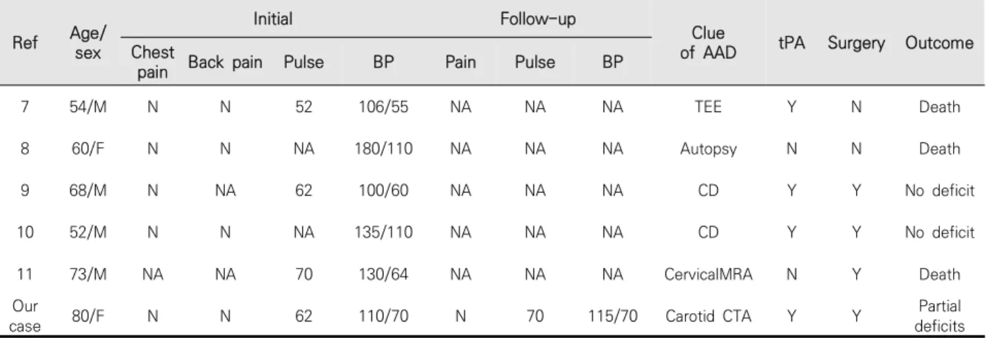
ing aortic dissection at admission, but the aortic dissection was discovered through carotid an- giography (carotid MR angiography, carotid Doppler ultrasound, or esophageal echocardiog- raphy) to determine the cause of cerebral in- farction or autopsy.7-11 However, these reports did not mention if there were any clinical findings of suspecting aortic dissection after admission (Table 1). On the other hand, in this case the pa- tient has no chest, abdomen, side, or back pain or decreased blood pressure and pulse at the time of admission and after admission. Aortic dis- section was suspected based on the findings of a carotid artery CT angiograph that is performed routinely for patients with cerebral infarction.
Finally, aortic CT angiography showed the Stanford A aortic dissection invading from the ori- gin of the aorta to ascending aorta and the Debake Fig. 3. Carotid (A) and aorta CT angiography (B) shows that aortic dissection
(Stanford A, DeBakey I) with the occlusion ofthe right common carotid artery (black arrows).
which was normal, it is thought that a slight pre- morbid asymptomatic aortic dissection caused the cerebral infarction and aortic dissection was exacerbated further due to the intravenous thrombolytic therapy or aortic dissection oc- curred as a rare complication of intravenous thrombolytic therapy in a patient with acute cere- bral infarction.
When considering intravenous thrombolytic therapy in patients with acute cerebral infarction, careful observation should be made before and after the administration of thrombolytic agents if there are any suspected aortic dissection symp- toms according to the American Clinical Practice
the aortic dissection symptoms.
REFERENCES
1. Marian AJ, Harris SL, Pickett JD, Campbell E, Fromm RE. Inadvertent administration of rtPA to a patient with type 1 aortic dissection and sub- sequent cardiac tamponade. Am J Emerg Med 1993;11:613-5.
2. Tsigkas G, Kasimis G, Theodoropoulos K, Chouchoulis K, Baikoussis NG, Aopstolakis E, et al. A successfully thrombolysed acute inferior myocardial infarction due to type A aortic dissection with lethal con-
Ref Age/
sex
Initial Follow-up
Clue
of AAD tPA Surgery Outcome Chest
pain Back pain Pulse BP Pain Pulse BP
7 54/M N N 52 106/55 NA NA NA TEE Y N Death
8 60/F N N NA 180/110 NA NA NA Autopsy N N Death
9 68/M N NA 62 100/60 NA NA NA CD Y Y No deficit
10 52/M N N NA 135/110 NA NA NA CD Y Y No deficit
11 73/M NA NA 70 130/64 NA NA NA CervicalMRA N Y Death
Our
case 80/F N N 62 110/70 N 70 115/70 Carotid CTA Y Y Partial
deficits Ref: references number, BP: blood pressure, AAD: aortic arch dissection, tPA: tissue plasminogen activator, NA:
not applicable, TEE: transesophageal echography, CD: carotid Doppler, MRA: magnetic resonance angiography, CTA: computed tomography angiography
Table 1. Summary of reported cases of aortic dissection diagnosed by routine evaluation in patients without initial clinical clue
sequences: the importance of early cardiac echocardiography. J Cardiothorac Surg 2011;6:101.
3. Grupper M, Eran A, Shifrin A. Ischemic stroke, aortic dissection, and thrombolytic therapy--the importance of basic clinical skills. J Gen Intern Med 2007;22:1370-2.
4. Hong KS, Park SY, Whang SI, Seo SY, Lee DH, Kim HJ, et al. Intravenous recombinant tissue plas- minogen activator thrombolysis in a patient with acute ischemic stroke secondary to aortic dissection. J Clin Neurol 2009;5:49-52.
5. Ramalingam VS, Sinnakirouchenan R, Sudhakar S, Brasch AV. Acute ischemic stroke in aortic dis- section: case report and review of literature.
Indian J Med Sci 2010;64:385-9.
6. Demaerschalk BM, Kleindorfer DO, Adeoye OM, Demchuk AM, Fugate JE, Grotta JC, et al. Scientific Rationale for the Inclusion and Exclusion Criteria for Intravenous Alteplase in Acute Ischemic Stroke: A Statement for Healthcare Professionals From the American Heart Association/American
Stroke Association. Stroke 2016;47:581-641.
7. Fessler AJ, Alberts MJ. Stroke treatment with tissue plasminogen activator in the setting of aortic dissection. Neurology 2000;54:1010.
8. Villa A, Molgora M, Licari S, Omboni E. Acute ischemic stroke, aortic dissection, and thrombo- lytic therapy. Am J Emerg Med 2003;21:159-60.
9. Yamashiro S, Arakaki R, Kise Y, Kuniyoshi Y.
Emergency operation for aortic dissection with ischemic stroke. Asian Cardiovasc Thorac Ann 2014;22:208-11.
10. Hama Y, Koga M, Tokunaga K, Takizawa H, Miyashita K, Iba Y, et al. Carotid Ultrasonography Can Identify Stroke Patients Ineligible for Intravenous Thrombolysis Therapy due to Acute Aortic Dissection. J Neuroimaging 2015;25:671-3.
11. Matsumoto H, Yoshida Y, Hirata Y. Usefulness of cervical magnetic resonance imaging for de- tecting type A acute aortic dissection with acute stroke symptoms. Magn Reson Imaging 2016;34:
902-7.