• 검색 결과가 없습니다.

The Effect of Real-time Ultrasound Imaging Feedback during Abdominal Hollowing in Four Point Kneeling to Healthy Men

N/A
N/A
Protected

Academic year: 2021

Share "The Effect of Real-time Ultrasound Imaging Feedback during Abdominal Hollowing in Four Point Kneeling to Healthy Men"

Copied!
6
0
0

로드 중.... (전체 텍스트 보기)

전체 글

(1)

Du-jin Park, MS

Department of Physical Therapy, College of Health Sciences, Catholic University of Pusan

Purpose: This study investigated the effects of visual feedback during abdominal hollowing (AH) in four point kneeling  position, using real‐time ultrasound imaging through measurement of the changes in the thickness of transversus abdominis  (TrA), internal abdominal oblique (IO), and external abdominal oblique (EO).

Methods: The subjects of this study were 32 healthy males who were divided intothe experimental group of 16 subjects and  the control group of 16 subjects. The real‐time ultrasound feedback was applied to the experimental group while they were  educated on the AH exercise in four point kneeling whereas only general education and training were given to the control  group.  After  the  training,  the  changes  in  the  thickness  of  abdominal  muscles  during  AH  in  four  point  kneeling  were  compared between the experimental group and the control group.

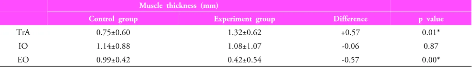
Results:  The  differences  of  the  changes  in  the  thickness  of  TrA  and  EO  between  the  two  groups  were  statistically  significant.

Conclusion: The experimental group experienced a higher increase in the thickness of TrA than the control group while the  thickness of IO and EO of the experimental group decreased. Therefore, real‐time ultrasound feedback was effective for the  selective contraction of TrA while reducing the activities of IO and EO during the AH exercise.

Keywords: Four‐point kneeling, Abdominal hollowing, Real‐time ultrasound imaging, Feedback Received: September 15, 2010

Revised: November 25, 2010 Accepted: December 6, 2010

Corresponding author: Du-jin Park, [email protected]

Abdominal Hollowing in Four Point Kneeling to Healthy Men

The Journal Korean Society of Physical Therapy

I. Introduction

In recent years, spinal stabilization exercises are gradually gaining popularity as a treatment of low back pain. Two systematic reviews

1,2

have provided evidence for the effects of spinal stabilization exercises, thus continuing their popularity.

The first step in the treatment process of patients with low back pain is to stimulate the selective activity of deep abdominal muscles such as TrA. To achieve this first step, clinicians have tried abdominal hollowing (AH) exercise targeted on deep abdominal muscles. Richardson and Jull

3

recommended four point kneeling as the easiest position for activating TrA.

The four point kneeling positionis recommended together with the prone position since it can achieve the separate contraction of the lower fibers of the internal oblique abdominal muscle more consistently compared to the prone position

4

and it

minimizes external load and pain during the rehabilitation of the local system.

3

Furthermore, it is a non-anti gravity position that can reduce the load on the spine and easily maintain balance in a spinal neutral position compared to other positions.

5

Due to these advantages, AH is often performed clinically in four point kneeling. Chanthapetch et al

6

reported, however, that it is difficult to induce the independent contraction of TrA by removing the activity of EO during AH. Park and Lee

7

recommended inducing the selective contraction of TrA using real-time ultrasound imaging because it is difficult to remove the activity of the EO.

Real-time ultrasound imaging is a non-invasive tool that

provides visual images and enables the measurement of deep

structures in the trunk.

8

It is cost-effective and very practical

among imaging modalities. Ultrasound imaging visualizes

muscles in real time and can measure the activity, size and

(2)

Figure 1. Real-time ultrasound imaging of abdominal muscles during the abdominal hollowing.

TrA: Transversus abdominis, IO: Internal abdominal oblique, EO: External abdominal oblique

thickness of deep trunk muscles

.9-11

The reliability of ultrasound imaging as a tool for measuring muscle thickness has been already demonstrated.

8

Furthermore, it has been proven that ultrasound imaging improves performance of AH

12

by providing individuals with visual feedback to the movement and changing thickness of deep abdominal muscles through the real-time ultrasound screen, and that it is very effective in the improvement of multifidus (MF) muscle.

13

As indicated above, real-time ultrasound imaging is widely used for the treatment and management of low back pain and for evaluation of the effects of rehabilitation treatment, but studies using ultrasound imaging with regard to stabilization exercises are still insufficient in the country. Accordingly, this study is intended to investigate the effects of visual feedback through real-time ultrasound imaging during AH in four point kneeling position.

II. Methods

1. Subjects

The subjects of this study were 32 healthy male adults who voluntarily agreed to participate in the experiments after listening to the purpose and method of study. They were divided into the experimental group of

16

subjects who received training with visual feedback and the control group of 16 subjects who received training without visual feedback. Those who had pain or dysfunction in upper limbs or lower limbs during exercise, who had not experienced AH exercise, who had experienced LBP during the last 6 months, who had underwent surgical treatment, or who had a disease in their nervous system were excluded from the experiment.

2. Experiment method 1) Measuring instruments

Sonoace X4 from Medison, Korean was used to measure the thickness of TrA, IO and EO muscles during AH. The thicknesses of muscles were measured using 7.5 MHz linear transducers.

2) Exercise method

According to the protocol by Richardson and Jull,

3

before the experiment the subjects were trained for AH to slowly pull the navel inward and upward while not moving the spine, ribs, and

pelvis. The experimental group received training for AH with visual feedback through real-time ultrasound imaging whereas the control group received the same education without visual feedback. Depending on the level of understanding of the subjects, the education time ranged from 15 to 20 min. Once their navel moved closer to their spine, they had to maintain abdominal contraction for 10 seconds while breathing normally.

Three measurements were made for each posture while they repeated this procedure three times.

6

In order to prevent muscle fatigue, they had a rest of 2 minutes after abdominal hollowing.

The exercise procedure was as follows: In the four point kneeling position, their eyes were directed to the floor, and while their shoulder and eyes were in line, their wrists were put under their shoulders and their knees under their hips. The thicknesses of TrA, IO, and EO were measured first during resting in this position, and then the thicknesses of the same muscles were measured again during AH in the same position. The averages of three measurements were calculated and compared.

3) Ultrasound recordings

The transducer was transversly placed halfway between the 11th

costal cartilage and the iliac crest.

14

Furthermore, to standardize

the location of the catheter, it was set in such a way that the space

where TrA and thoracolumbar fascia (TLF) meet would appear

at the right end of the ultrasound image (Figure 1).

15

To control

the effects of respiration,

16

the subjects were educated to perform

AH during exhalation. Ultrasound images were captured at the

end of the exhalation. The examiner measured the thicknesses of

(3)

Table 1. Change in muscle thicknesses between experimental group and control group during AH in four point kneeling Muscle thickness (mm)

Control group Experiment group Difference p value

TrA 0.75±0.60 1.32±0.62 +0.57 0.01*

IO 1.14±0.88 1.08±1.07 -0.06 0.87

EO 0.99±0.42 0.42±0.54 -0.57 0.00*

*Significant difference (p<0.05) AH: Abdominal hollowing

TrA: Transversus abdominis, IO: Internal abdominal oblique, EO: External abdominal oblique

muscles at the right-hand side of the subject while the subject was doing AH. The education and measurement of the experimental group and the control group were carried out by the same researchers.

3. Reliability testing

Inter-rater reliability was examined using interclass correlation coefficients (ICC). ICC was demonstrated for 3 repeat measurements of each muscle, whilst resting and AH state, and in both two groups in 9 sets of data. ICC of TrA, IO and EO were shown 0.98, 0.98 and 0.97 in the experimental group. ICC of TrA, IO and EO were shown 0.97, 0.98 and 0.97 in control group.

4. Data analysis

An independent t-test was conducted to compare the change in the thicknesses of abdominal muscles during AH between the experimental group and the control group. For statistical significance, the level of significance was set to =0.05 and the collected data were analyzed using the SPSS 12.0 for Windows.

III. Results

1. Characteristics of the subjects

The mean age, mean height, mean weight, and BMI of the experimental group were 22.5±1.79 years, 174.88±4.95 cm, 68.06±7.42 kg, and 22.31±2.68 kg/m

2

, respectively. The mean age, mean height, mean weight, and BMI of the control group were 22.13±3.81 years, 176.31±6.05 cm, 69.19±10.49 kg, and 22.16±2.32 kg/m

2

, respectively. There were no significant differences between the two groups.

2. Changes in the thicknesses between two groups The thicknesses of TrA, IO, and EO of the control group during resting were 5.10±0.75, 8.67±1.89, and 5.78±1.29 mm, respectively. The thicknesses of TrA, IO, and EO of the control group during AH exercise were 5.84±0.84, 9.81±1.98, and 6.76±1.35 mm, respectively. The differences in the thicknesses of TrA, IO, and EO of the control group between resting and AH exercise were 0.75±0.60, 1.14±0.88, and 0.99±0.42 mm, respectively. The thicknesses of TrA, IO, and EO of the experimental group during resting were 4.31±0.78, 8.03±2.16, and 5.60±1.09 mm, respectively. The thicknesses of TrA, IO, and EO of the experimental group during AH exercise were 5.63±1.10, 9.10±2.89, and 6.01±1.28 mm, respectively. The differences in the thicknesses of TrA, IO, and EO of the experimental group between resting and AH exercise were 1.32±0.62, 1.08±1.07, and 0.42±0.5 4 mm, respectively. The differences in the thicknesses of the muscles between the two groups during AH exercise are shown in Table 1. The differences of the changes in the thicknesses of TrA and EO between the two groups were statistically significant.

IV. Discussion

Even though many domestic studies are being conducted using ultrasonography

17

or ultrasound,

18,19

there are few studies that use ultrasound imaging during spinal stabilization exercises.

Hence, thisstudy is intended to investigate the effects of visual

feedback through real-time ultrasound imaging during AH

which is a spinal stabilization exercise. For the experimental

group of this study, the thicknesses of TrA, IO, and EO

increased by approximately 30.6%, 13.3%, and 7.3%,

respectively during AH exercise compared to resting. For the

control group, the thicknesses of the same muscles increased by

(4)

approximately 14.5%, 13.1%, and 17%, respectively. Although the AH exercise stimulates the selective contraction of TrA which is a deep abdominal muscle,

15

the subjects of the control group showed similar changes in thickness of TrA to those of other muscles, indicating that no separate contraction of TrA could be achieved. Furthermore, in the study examined thicknesses of abdominal muscles between resting and abdominal hollowing in four point kneeling by Critchley and Coutts

20

using ultrasound, the thicknesses of TrA, IO, and EO in control subjects during AH exercise increased by approximately 51%, 18%, and 17%, respectively compared to resting. Even though the change in the thickness of TrA increased considerably, the changes in the thicknesses of IO and EO were also high. This supported the findings of prior studies

6,7

that the separate contraction of TrA isdifficult during AH because the thicknesses of IO and EO also change considerably. For the experimental group of this study, however, the change in the thickness of TrA was higher and the change in the thickness of EO, in particular, was lower compared to the control group. The reason for this appears to be that the feedback through real-time ultrasound imaging improved the motor relearning of specific muscles by providing knowledge of performance.

21

Knowledge of performance is information concerning the movement itself by real-time ultrasound imaging. This type of feedback is an effective method of motor relearning.

To compare the differences of the changes in the thicknesses of TrA between resting and AH exercise in two groups, the thicknesses of the experimental group increased by approxi- mately 76% more than those of the control group, and this difference was statistically significant. The change in the thickness of IO of the experimental group was lower than the control group by approximately 5.4%, but this difference was not statistically significant. However, the change in the thickness of EO of the experimental group was lower than the control group by approximately 58%, and this difference was statistically significant. The result of IO is due to the activity of IO in maintaining a neutral pelvis and spine positions in four point kneeling. It could be seen that, IO maintained more constant activity than EO in four point kneeling.

The study examined the activities of the abdominal muscles during abdominal hollowing Park and Lee

7

using EMG reported that only 8.3% of the subjects showed the selective contraction

of muscles from EO to TrA in four pointskeeling, indicating the difficulty of the selective contraction of TrA. For the experimental group of this study, however, 75% of the subjects (12/16) induced the separate contraction of TrA while reducing the thickness of EO. The real-time ultrasound feedback applied to the experimental group appears to have given great assistance to the accuracy of the AH exercise by increasing the activity of TrA while reducing the activity of EO. The study examined differences among the 3 feedback groups by Henry and Westervelt

12

using ultrasound also found that the number of trainings for AH exercise decreased in the group to which real-time ultrasound feedback was applied compared to a different feedback group. Another prior study

13

also reported that the motor learning of the group to which real-time ultrasound feedback was given improved compared to another group to which only the knowledge of result was applied. These results show the advantage of real-time ultrasound feedback which can simultaneously provide visual feedback

22

and precise feedback

23

which can improve skill acquisition. Furthermore, these results also support the findings of prior studies

12,21

which recommended real-time ultrasound feedback during AH training.

Another study

24

on the effects of real-time ultrasound feedback for patients with low back pain reported positive results. This study investigatedthe recurrence of low back pain between the experimental group who performed the contraction exercise of MFmuscle and the control group who received general medical care using real-time ultrasound feedback for 4 weeks. The recurrence rate of low back pain after 1 year was approximately 30% for the experimental group, but it was approximately 84% for the control group. After 2-3 years, it was 35% for the experimental group and 75% for the control group.

While many such studies found positive results after applying real-time ultrasound feedback, Teyhen et al

25

reported that no improvement in motor learning was observed. As shown by this example, different opinions exist because research on the effects of real-time ultrasound feedback is still insufficient. Thus, we hope that more active studies on real-time ultrasound feedback will be conducted.

This study demonstrated that use of real-time ultrasound

imaging feedback while teaching AH to subjects without LBP

was a beneficial teaching tool. To aid innate difficulties in

learning and teaching, we suggest the use of real-time ultrasound

(5)

imaging feedback to achieve correct AH exercise in clinical field.

However, this study didn’t demonstrate that real-time ultrasound imaging feedback was effective for LBP subjects during AH. Thus, further studies to examine the effects of real-time ultrasound imaging feedback in subjects with LBP during AH will be necessary.

V. Conclusion

This study compared the changes in the thicknesses of abdominal muscles during AH exercise in the four point kneeling positionbetween the experimental group, to which real-time ultrasound feedback was applied, and the control group who only received general training. The experiment group experienced a higher increase in the thickness of TrA thanthe control group while the thickness of IO and EO of the experiment group decreased. Therefore, real-time ultrasound feedback was effective for the selective contraction of TrA while reducing the activities of IO and EO during the AH exercise.

More in-depth research on the effects of real-time ultrasound feedback on various postures and exercises is needed.

Author Contributions Research design: Park DJ Acquisition of data: Park DJ

Analysis and interpretation of data: Park DJ Drafting of the manuscript: Park DJ Researchsupervision: Park DJ

References

1. Ferreira PH, Ferreira ML, Maher CG et al. Specific stabilisation exercise for spinal and pelvic pain: a systematic review. Aust J Physiother. 2006;52(2):79-88.

2. Rackwitz B, de Bie R, Limm H et al. Segmental stabilizing exercises and low back pain. What is the evidence? A systematic review of randomized controlled trials. Clin Rehabil.

2006;20(7):553-67.

3. Richardson CA, Jull GA. Muscle control-pain control. What exercises would you prescribe? Man Ther. 1995;1(1):2-10.

4. Beith ID, Synnott RE, Newman SA. Abdominal muscle activity during the abdominal hollowing manoeuvre in the four point

kneeling and prone positions. Man Ther. 2001;6(2):82-7.

5. McGill SM. Low back exercises: evidence for improving exercise regimens. Phys Ther. 1998;78(7):754-65.

6. Chanthapetch P, Kanlayanaphotporn R, Gaogasigam C et al.

Abdominal muscle activity during abdominal hollowing in four starting positions. Man Ther. 2009;14(6):642-6.

7. Park DJ, Lee HO. Activation of abdominal muscles during abdominal hollowing in four different positions. J Phys Ther Sci. 2010;22(2):203-7.

8. Norasteh A, Ebrahimi E, Salavati M et al. Reliability of B-mode ultrasonography for abdominal muscles in asymptomatic and patients with acute low back pain. J Bodyw Mov Ther.

2007;11(1):17-20.

9. Mew R. Comparison of changes in abdominal muscle thickness between standing and crook lying during active abdominal hollowing using ultrasound imaging. Man Ther. 2009;

14(6):690-5.

10. Vasseljen O, Fladmark AM. Abdominal muscle contraction thickness and function after specific and general exercises: a randomized controlled trial in chronic low back pain patients.

Man Ther. 2010;15(5):482-9.

11. Mannion AF, Pulkovski N, Gubler D et al. Muscle thickness changes duringabdominal hollowing: an assessment of between-day measurement error in controls and patients with chronic low back pain. Eur Spine J. 2008;17(4):494-501.

12. Henry SM, Westervelt KC. The use of real-time ultrasound feedback in teaching abdominal hollowing exercises to healthy subjects. J Orthop Sports Phys Ther. 2005;35(6):338-45.

13. Van K, Hides JA, Richardson CA. The use of real-time ultrasound imaging for biofeedback of lumbar multifidus muscle contraction in healthy subjects. J Orthop Sports Phys Ther. 2006;36(12):920-5.

14. Teyhen DS, Gill NW, Whittaker JL et al. Rehabilitative ultrasound imaging of the abdominal muscles. J Orthop Sports Phys Ther. 2007;37(8):450-66.

15. Springer BA, Mielcarek BJ, Nesfield TK et al. Relationships among lateral abdominal muscles, gender, body mass index, and hand dominance. J Orthop Sports Phys Ther. 2006;

36(5):289-97.

16. Hodges PW, Gandevia SC. Activation of the human diaphragm during a repetitive postural task. J Physiol. 20005;22 Pt 1:165-75.

17. Seo SK. The analysis of ultrasonography on gastrocnemius

medialis of patient with stroke. J Kor Soc Phys Ther.

(6)

2007;19(2):11-9.

18. Min SG, Chun JS, Yong JH et al. The effects of pulsed ultrasound on the bone healing process in fracture model of diabetes mellitus rat. J Kor Soc Phys Ther. 2006;18(2):47-58.

19. Kim HA, Han JM. The effect of ultrasound irradiation on the neural cell adhesion molecules (NCAM) expression in rat spinal cord after the sciatic nerve crush injury. J Kor Soc Phys Ther.

2007;19(2):41-55.

20. Critchley DJ, CouttsFJ. Abdominal muscle function in chronic low back pain patients: measurement with real-time ultrasound scanning. Physiotherapy. 2002;88(6):322-32.

21. Hides JA, Richardson CA, Jull GA. Use of real-time ultrasound imaging for feedback in rehabilitation. Man Ther. 1998;

3(3):125-31.

22. Kim HJ, Kramer JF. Effectiveness of visual feedback during isokinetic exercise. J Orthop Sports Phys Ther. 1997;26(6):

318-23.

23. Wright DL, Smith-Munyon VL, Sidaway B. How close is too close for precise knowledge of results? Res Q Exerc Sport.

1997;68(2):172-6.

24. Hides JA, Jull GA, Richardson CA. Long-term effects of specific stabilizing exercises for first-episode low back pain. Spine (Phila Pa 1976). 2001;26(11):E243-8.

25. Teyhen DS, Miltenberger CE, Deiters HM et al. The use of ultrasound imaging of the abdominal drawing-in maneuver in subjects with low back pain. J Orthop Sports Phys Ther.

2005;35(6):346-55.

수치

Figure 1. Real-time ultrasound imaging of abdominal  muscles during the abdominal hollowing.
Table 1. Change in muscle thicknesses between experimental group and control group during AH in four point kneeling Muscle thickness (mm)

참조

관련 문서

The index is calculated with the latest 5-year auction data of 400 selected Classic, Modern, and Contemporary Chinese painting artists from major auction houses..

The “Asset Allocation” portfolio assumes the following weights: 25% in the S&amp;P 500, 10% in the Russell 2000, 15% in the MSCI EAFE, 5% in the MSCI EME, 25% in the

1 John Owen, Justification by Faith Alone, in The Works of John Owen, ed. John Bolt, trans. Scott Clark, &#34;Do This and Live: Christ's Active Obedience as the

Regarding SI or BI EMG measured at upright posture on hard platform, bio-feedback group under the condition with opening both eye and aqua group under the condition with

Understanding the visual language demands the ‘Visual clue’ which helps the information audiences comprehend the structure of the visual language and grasp

The government strives to create venture ecosystem for realizing creative economy, at the same time, the Mistry of Education is spending huge resources

ƒ Give feedback to each other in non-threatening supportive ways (a good way to do this is to focus on the group goals and how a particular issue assists group objectives

This research suggests a need for an instructor education program for effective feedback practice and the enhancement of feedback quality in higher education..