Introduction
Dislocation of the temporomandibular joint (TMJ) occurs when one or both mandibular condyles are displaced in front of the articular eminence. It may be reducible when it returns spontaneously to the glenoid cavity, or irreducible when one or two condyles remain dislocated. In this posi- tion, the mouth remains open due to the action of the ele- vator muscles with or without lateral deviation, depending on whether the dislocation is unilateral or bilateral.1,2Acute mandibular dislocation is usually managed by manually pressing the mandible downwards and then pulling it back upwards in an attempt to try relocating the condyle in the
glenoid fossa. If the condyle continues to dislocate several times, it is described as chronic recurrent TMJ dislocation.
Dislocation of TMJ is generally of unknown origin, while several theories put forward to explain its onset. It is com- monly associated with poor development of the articular fossa, laxity of the temporomandibular ligament or joint capsule, and excessive activity of the lateral pterygoid and infrahyoid muscles due to drug use or disease. Additionally some disorders of collagen metabolism such as ligamentous hyperlaxity and Ehler-Danlos syndrome might be related.1-3 There were well documented surgical and nonsurgical treatment protocols of chronic recurrent TMJ dislocation in the literatures.1,4Recently autologous blood injection (ABI) to TMJ has been applied for the treatment of chro- nic recurrent TMJ dislocation. The procedure is easy to perform and it causes no foreign body reaction. There have been some successful clinical studies in the literatures. On the other hand, there were some concerns about the pro-
Autologous blood injection to the temporomandibular joint: magnetic resonance imaging findings
Celal Candirli, Serdar Yüce*, Umut Yücel Cavus**, Kayihan Akin***, Banu Cakir***
Department of Oral and Maxillofacial Surgery, Faculty of Dentistry, Karadeniz Technical University, Trabzon, Turkey
*Department of Plastic, Reconstructive Surgery, Fatih University Hospital, Ankara, Turkey
**Department of Emergency Medicine, Fatih University Hospital, Ankara, Turkey
***Department of Radiology, Fatih University Hospital, Ankara, Turkey ABSTRACT
Purpose : The aim of this study was to investigate the effect of the autologous blood injection (ABI) for chronic recurrent temporomandibular joint (TMJ) dislocation using magnetic resonance imaging (MRI).
Materials and Methods : ABI was applied to 14 patients who had chronic recurrent TMJ dislocation. MRIs of the patients were taken and compared before and one month after the injection.
Results : All of the patients had no dislocations of their TMJs on clinical examination one month after the injection.
In the pre-injection, unilateral or bilateral TMJ dislocations were observed on MRIs in all patients. One month after the injection, TMJ dislocations were not observed in MRI evaluation of any patients. A significant structural change that caused by ABI was not observed.
Conclusion : The procedure was easy to perform and it caused no foreign body reaction. However, it was unclear how the procedure prevented the dislocation. (Imaging Sci Dent 2012; 42 : 13-8)
KEY WORDS : Temporomandibular Joint; Dislocations; Magnetic Resonance Imaging
Received November 29, 2011; Accepted December 28, 2011 Correspondence to : Dr. Celal Candirli
Department of Oral and Maxillofacial Surgery, Faculty of Dentistry, Karadeniz Technical University, Kalkinma Street, Kanuni Campus 61080, Trabzon, Turkey Tel) 90-312-2035352, Fax) 90-312-2213670, E-mail) [email protected]
Copyright ⓒ 2012 by Korean Academy of Oral and Maxillofacial Radiology
This is an Open Access article distributed under the terms of the Creative Commons Attribution Non-Commercial License (http://creativecommons.org/licenses/by-nc/3.0) which permits unrestricted non-commercial use, distribution, and reproduction in any medium, provided the original work is properly cited.
Imaging Science in Dentistry∙pISSN 2233-7822 eISSN 2233-7830
cedure such as fibrous or bony ankylosis and articular car- tilage degeneration. The aim of this study was to evaluate the pathophysiology of the ABI and observe the mechanism of the procedure using magnetic resonance imaging (MRI).
Materials and Methods
Fourteen patients (5 males) between the ages of 17 and 74 who had chronic recurrent TMJ dislocation visited Fatih University Hospital from the year of 2009 to 2010 (Table 1). They were diagnosed with chronic recurrent TMJ dis- location based on the clinical and radiographic criteria. Six of the fourteen patients had bilateral chronic recurrent TMJ dislocation. The patients’ maximal mouth opening (just before dislocation) measured between maxillary and man-
dibular incisal edges, ranged from 36 to 48 mm, with an average of 41 mm. All of these patients complained of dis- location of their joints at least twice a week. No patients had other TMJ disorders such as disc displacement or osteo- arthritis. The patients underwent AB˙I (6 bilateral injections).
Local anesthesia was given to the auriculatemporal nerve.
The articular fossa was assumed as located at a point 10 mm anterior to the tragus of the ear and 2 mm inferior to the tragal-canthal line. Five mL blood was withdrawn from the patients’ anticubital fossa. Four mL blood with a 21- gauge needle was injected in the articular cavity and 1 mL was injected in the pericapsular tissue (Fig. 1). After the completion of the injection, an elastic bandage was applied and left for 24 hours to constrain the joint movements. All of the patients were scanned before and 1 month after the therapy with a 1.5 T MRI scanner (Achieva; Philips Medi- cal Systems, Best, The Netherlands) using a multichannel head coil. Both of the joints were imaged in each patient.
First, the axial scout section was used to localize the mandi- bular condyle. Based on the axial scout view, oblique sagittal T1-weighted spin echo (SE) sequences were acqu- ired (repetition time [TR]: 450 msec, echo time [TE]: 15 msec, matrix: 180×320, slice thickness: 3 mm, field of view [FOV]: 130 mm, number of excitation [NEX]: 3) in closed mouth position. Eight sagittal images for each joint were obtained. Subsequently, oblique sagittal T1 3-dimen- sional water only WATS images (TR: 30 msec, TE: 3.9 msec, matrix: 180×256, slice thickness: 1 mm, FOV: 130 mm, NEX: 3, FA: 20�) were acquired in closed mouth position.
Twenty-five images were acquired for each TMJ. Also, oblique sagittal T2-weighted fast spin echo (FSE) images
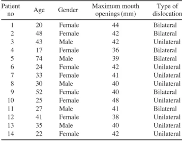
Table 1.Patients treated with autologous blood injection Patient Age Gender Maximum mouth Type of
no openings (mm) dislocation
1 20 Female 44 Bilateral
2 48 Female 42 Bilateral
3 43 Male 42 Unilateral
4 17 Female 36 Bilateral
5 74 Male 39 Bilateral
6 24 Female 42 Unilateral
7 33 Female 41 Unilateral
8 30 Male 40 Unilateral
9 52 Female 40 Bilateral
10 25 Female 48 Unilateral
11 27 Male 41 Bilateral
12 41 Female 38 Unilateral
13 35 Male 40 Unilateral
14 22 Female 42 Unilateral
Fig. 1. A. The reference line for locating the articular fossa. B. Auto- logous blood injection.
A B
(MOVIE) (TR: 131 msec, TE: 14 msec, matrix: 240×192, slice thickness: 3 mm, FOV: 160 mm, NEX: 1, FA: 30�) were acquired in six different mouth opening positions.
The patients have been instructed to open their mouth gradually.
Results
All the patients tolerated the procedure well. There were no infections or any other complications in any of the pat- ients. The post operative pain was tolerable in all the pat- ients and lasted for only few days after the procedure.
During four weeks after the operation, all the patients had no dislocations of their TMJs, while one patient showed a recurrent dislocation in the second month after the injection.
Two patients with unilateral TMJ dislocation mentioned that the incidence of dislocation had been reduced, but it had not been completely disappeared. After the study, ABI was repeated by the request of the patients who had failed treatment and achieved good results. In pre-injection MRI evaluation, there was no articular cartilage degeneration,
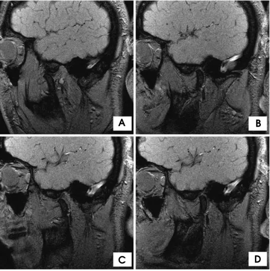
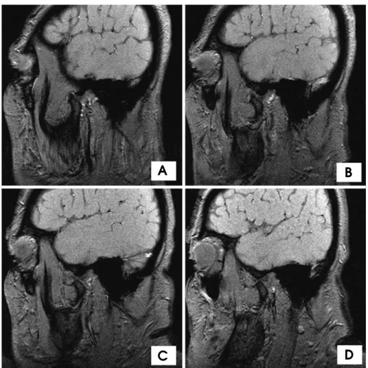
disc displacement, or osteoarthritis in any patients. Before administration of the injection, it was identified that the patients had unilateral or bilateral condyles anterior to the eminence with their mouths in the open position. In the first month after the injection, MRIs of all patients taken with the open-mouth position revealed that the condyle was either at the apex of the eminence or posterior to it (Figs. 2 and 3). However, there was no structural variation that described the prevention of the dislocations such as fibrosis in MRI findings. Hematoma was disappeared with- out any alteration of the joint tissues.
Discussion
Chronic recurrent TMJ dislocation is a painful and alarm- ing illness that patients usually cannot reduce dislocated joints by themselves and need a doctor or an infirmarian for the treatment. Non surgical methods are usually first applied to the patients before decision of surgery. There were some successful nonsurgical treatment models des- cribed in the literatures such as injection of a sclerosing
Fig. 2.Case 1: MRI images of the left TMJ. A. Open-mouth position before injection. B. Close-mouth position before injection. C. Open- mouth position after injection. D.
Close-mouth position after injection.
A B
C D
solution into the joint cavity (tincture of iodine, alcohol and sodium psylliate). However, many side effects and the possible risk of facial paralysis or traumatic arthritis have hindered its widespread usage.1,5Another procedure with less side effects is the use of botox.4The toxin tem- porarily causes denervation of the muscles that draw the chin down. The muscle of choice for injection is the lateral pterygoid muscle. In this way, the displacement of the condyle is prevented even when the mouth is opened exces- sively.6,7Recently ABI has been used for the treatment of chronic recurrent TMJ dislocation. It can be thought that ABI is more advantageous because of the less complication probability regarding to the other nonsurgical methods.
ABI can be readily carried out under local anesthesia in outpatient clinic and the treatment can be performed repea- tedly with minimal complications.5There were some studies that obtained good results regarding this method. However, it has not gained much popularity as the mechanism of action has not been explicitly defined. The pathophysilogy of the procedure has not been introduced with a comment.
Additionally there are some concerns with this procedure
such as fibrous or bony ankylosis and articular cartilage degeneration.1,5,8In our knowledge, this was the first study that evaluated the effect of the ABI procedure by MRI.
Machon et al1mentioned that ABI for chronic TMJ dis- location was first reported in the German literature in 1964 by Brachmann. He had treated 60 patients using this tech- nique and declared good results. Schulz treated 16 patients in 1973 by ABI to the affected TMJ twice a week for 3 weeks followed by immobilization via intermaxillary fixa- tion for 4 weeks. The symptoms were disappeared in 10 patients at 1-year follow-up. In 1981, Jacobbi-Hermanns published the experience with 19 patients who received only 1 ABI and had intermaxillary fixation for 14 days.
At 18 months follow-up, 17 patients were symptom free.1 Hasson reported the successfully treated 3 patients with ABI (4 cc superior joint space and 1 cc pericapsular tissues).
After the treatment, elastic bandage was applied and left for the first 24 hours.2Kato et al presented the treatment of chronic recurrent TMJ dislocation by ABI (3 cc superior joint space and 1 cc pericapsular tissues) in an 84-year-old female under local anesthesia. The mandible had been
Fig. 3.Case 2: MRI images of the open-mouth position. A. The left TMJ before injection. B. The right TMJ before injection. C. The left TMJ after injection. D. The right TMJ after injection.
A B
C D
fixed with a bandage and the use of bandage had been continued for 1 month as a precaution.5Pinto et al success- fully treated an 83-year-old female patient by injecting 10 cc of ABI into the superior joint space and the periarticular tissue.9They used a face lift bandage for one month after the procedure. Machon et al reported that 20 among 25 patients were treated successfully with 1 or 2 injections (2 cc in the superior joint space and 1 cc in the pericapsular tissue). Based on their number of patients, there was no benefit after 2 injections of blood to the TMJ, and surgical intervention should then be pursued.1 In Daif’s study, 30 patients having chronic recurrent TMJ dislocation were randomly divided into 2 equal groups (15 patients in each).
Group A was treated only by 2 cc autologous blood injec- tion into the superior joint space (SJS), whereas group B received 2 cc ABI to the SJS and 1 cc in the pericapsular tissues (PT). After the completion of the injection proce- dures, an elastic bandage was applied and left for the first 24 hours. At the end of the follow-up period of 1 year, the results of the current study showed that the ABI to the SJS and PT gave a higher success rate (80%) than its injection only into the SJS (60%). Moreover, the decrease of maximal mouth opening of group B was larger than that of group A.
Also, the digital radiographic imaging of the joints in group B only showed the condylar head posterior to the articular eminence in open position, instead of being anterior to it before the injection. In both groups, no destructive changes to the bony components of the joint have been observed.8
The principal aim of the ABI is the limitation of move- ment of the mandible. It is thought that the mechanism occurs by the formation of a local recess of fibrous tissue in the pericapsular space during the wound healing process.
The injection given into the cavity over the joint leads to its formation of adhesions in the cavity. However, this anticipated mechanism has not been studied experimentally or radiologically. The studies mentioned above have just approved the success of the procedure by clinical obser- vations. Some researchers claimed that the contact of car- tilage with blood caused the changes in chondrocyte meta- bolism and thus cartilage destruction.10,11 Other studies showed that these changes were temporary, not permanent.
It is thought that ABI procedure could cause articular degeneration and subsequent arthritis.5
The pathophysiology of blood injection resembles that of knee or elbow joint bleeding. Initially, the capsule swells and stretches. In the next few hours or days, an inflamma- tory reaction starts via mediators released from neighbor- ing platelets, wounded and dead cells. As a result, neighbor- ing tissues swell and the joint has difficulty in motion.
Thereafter, organized blood clots and fibrous tissue struc- tures lead to joint stiffness. These tissues cause permanent limitation in joint motion. The contact of cartilage with blood leads to impairment of the cartilage matrix cycle.
The injected blood artificially triggers an inflammatory reaction, which in turn leads to fibrosis, adhesions and scars in the neighboring tissues. Immobilization prevents the early tension produced by new fibrous tissue.1In the experimental studies on the joints concerning injection of blood into TMJs, there were no late period pathologies even in cases of frequent injections. The impairment in the cartilage matrix which could be seen in the early period recovered to complete normal state later.12Pathologies in late period have only been identified in patients who fre- quently bore their weight on the extremity joints.13,14Con- sidering their studies, we performed the MRI investiga- tion to determine the early effect of the procedure after a month. Our previous experimental study using rabbits also showed that there was no pathology in their TMJs on the histopathological evaluation 1 month after ABI.15
The relatively simple procedures of this method, the very low amount of blood involved (the amount is the same as that in open surgery), the restriction of movement after the procedure, and the prevention of weight bearing on the mandible might be the possible reasons for the occurrence of no complication. Some studies applied controlled-exer- cise programs to patients to potentially limit mandibular movement after the procedure.1
In this study AB˙I was applied to 14 patients according to the methods. At the end of the 1 year follow-up period, their symptoms were improved in 13 patients successfully.
The results of the treatment were evaluated by physical examination and together with MRI evaluation. These results of treatment were in agreement with the previous studies as mentioned above. However, contrary to the studies mentioned above, a structural change such as fibro- sis or other mechanisms were not observed on MRI.
In conclusion, the ABI in the treatment of chronic recur- rent TMJ dislocation had advantages such as no require- ment of dissection, few or no post operative complications such as facial nerve injury, loss of sensation, swelling, infection and pain, no necessity to stay in hospital, and easiness of administration under local anesthesia. To sum up, ABI for patients with chronic recurrent TMJ disloca- tion was an effective, safe, and simple technique that could be used before surgery. However, more clinical and experi- mental studies should be performed to evaluate the efficacy of the treatment modality and to describe the pathophysio- logy of the treatment.
References
1. Machon V, Abramowicz S, Paska J, Dolwick MF. Autologous blood injection for the treatment of chronic recurrent tempo- romandibular joint dislocation. J Oral Maxillofac Surg 2009;
67 : 114-9.
2. Hasson O, Nahlieli O. Autologous blood injection for treat- ment of recurrent temporomandibular joint dislocation. Oral Surg Oral Med Oral Pathol Oral Radiol Endod 2001; 92 : 390- 3.
3. Oztan HY, Ulusal BG, Turegun M, Deveci M. Titanium screw implantation to the articular eminence for the treatment of chronic recurrent dislocation of the temporomandibular joint.
Int J Oral Maxillofac Surg 2005; 34 : 921-3.
4. Ziegler CM, Haag C, Mühling J. Treatment of recurrent tempo- romandibular joint dislocation with intramuscular botulinum toxin injection. Clin Oral Investig 2003; 7 : 52-5.
5. Kato T, Shimoyama T, Nasu D, Kaneko T, Horie N, Kudo I.
Autologous blood injection into the articular cavity for the treatment of recurrent temporomandibular joint dislocation: a case report. J Oral Sci 2007; 49 : 237-9.
6. Fu KY, Chen HM, Sun ZP, Zhang ZK, Ma XC. Long-term efficacy of botulinum toxin type A for the treatment of habi- tual dislocation of the temporomandibular joint. Br J Oral Maxillofac Surg 2010; 48 : 281-4.
7. Vázquez Bouso O, Forteza González G, Mommsen J, Grau VG, Rodríguez Fernández J, Mateos Micas M. Neurogenic temporomandibular joint dislocation treated with botulinum
toxin: report of 4 cases. Oral Surg Oral Med Oral Pathol Oral Radiol Endod 2010; 109 : e33-7.
8. Daif ET. Autologous blood injection as a new treatment moda- lity for chronic recurrent temporomandibular joint dislocation.
Oral Surg Oral Med Oral Pathol Oral Radiol Endod 2010; 109 : 31-6.
9. Pinto AS, McVeigh KP, Bainton R. The use of autologous blood and adjunctive ‘face lift’ bandage in the management of recurrent TMJ dislocation. Br J Oral Maxillofac Surg 2009;
47 : 323-4.
10. Lafeber FP, Miossec P, Valentino LA. Physiopathology of haemophilic arthropathy. Haemophilia 2008; 14 Suppl 4 : 3-9.
11. Roosendaal G, Lafeber FP. Blood-induced joint damage in hemophilia. Semin Thromb Hemost 2003; 29 : 37-42.
12. Roosendaal G, TeKoppele JM, Vianen ME, van den Berg HM, Lafeber FP, Bijlsma JW. Blood-induced joint damage: a canine in vivo study. Arthritis Rheum 1999; 42 : 1033-9.
13. Hooiveld MJ, Roosendaal G, Jacobs KM, Vianen ME, van den Berg HM, Bijlsma JW, et al. Initiation of degenerative joint damage by experimental bleeding combined with loading of the joint: a possible mechanism of hemophilic arthropathy.
Arthritis Rheum 2004; 50 : 2024-31.
14. Hooiveld M, Roosendaal G, Vianen M, van den Berg M, Bijlsma J, Lafeber F. Blood-induced joint damage: longterm effects in vitro and in vivo. J Rheumatol 2003; 30 : 339-44.
15. Candrl C, Yüce S, Yldrm S, Sert H. Histopathologic evalua- tion of autologous blood injection to the temporomandibular joint. J Craniofac Surg 2011; 22 : 2202-4.