Korean Type Distal Radius Anatomical Volar Plate System: A Preliminary Report
Jeong Hwan Kim, MD, Hyuk Jin Lee, MD*, Jihyeung Kim, MD
†, Min Bom Kim, MD*, Seung Hwan Rhee, MD
†, Hyun Sik Gong, MD, Young Ho Lee, MD*, Goo Hyun Baek, MD*
Department of Orthopedic Surgery, Seoul National University Bundang Hospital, Seongnam,
*Department of Orthopedic Surgery, Seoul National University Hospital, Seoul National University College of Medicine, Seoul,
†Department of Orthopedic Surgery, SMG-SNU Boramae Hospital, Seoul National University College of Hospital, Seoul, Korea
Received July 6, 2013; Accepted October 22, 2013 Correspondence to: Goo Hyun Baek, MD
Department of Orthopedic Surgery, Seoul National University Hospital, Seoul National University College of Medicine, 101 Daehak-ro, Jongno-gu, Seoul 110-744, Korea
Tel: +82-2-2072-3787, Fax: +82-2-764-2718, E-mail: ghbaek@snu.ac.kr Co-Correspondence to: Hyuk Jin Lee, MD
Department of Orthopedic Surgery, Seoul National University Hospital, Seoul National University College of Medicine, 101 Daehak-ro, Jongno-gu, Seoul 110-744, Korea
Tel: +82-2-2072-3787, Fax: +82-2-764-2718, E-mail: hjleemd@gmail.com
Background: Distal radius fracture is the most common fracture of the upper extremity, and approximately 60,000 distal radius fractures occur annually in Korea. Internal fixation with an anatomical volar locking plate is widely used in the treatment of un- stable distal radius fractures. However, most of the currently used distal radius anatomical plate systems were designed based on the anatomical characteristics of Western populations. Recently, the Korean-type distal radius anatomical volar plate (K-DRAVP) system was designed and developed based on the anatomical characteristics of the distal radius of Koreans. The purpose of this study was to evaluate the preliminary results of the new K-DRAVP system, and to compare its radiologic and functional results with those of the other systems.
Methods: From March 2012 to October 2012, 46 patients with acute distal radius fractures who were treated with the K-DRAVP system at three hospitals were enrolled in this study. Standard posteroanterior and lateral radiographs were obtained to assess fracture healing, and three radiographic parameters (volar tilt, radial inclination, and radial length) were assessed to evaluate radiographic outcomes. The range of motion and grip strength, the Gartland and Werley scoring system, and the disabilities of the arm, shoulder and hand (DASH) questionnaire were used to assess clinical and functional outcomes.
Results: All radiologic parameters were restored to normal values, and maintained without any loosening or collapse until the time of final follow-up. Grip strength was restored to 84% of the value for the unaffected side. The mean range of motion of the wrist at final follow-up was restored to 77%−95% of the value for the unaffected side. According to the Gartland and Werley scor- ing system, there were 16 excellent, 26 good, and 4 fair results. The mean DASH score was 8.4 points. There were no complica- tions after surgery.
Conclusions: The newly developed K-DRAVP system could be used to restore and maintain good anatomical parameters, and provide good clinical outcomes with low complication rates. This system is a promising surgical option for the treatment of distal radius fractures in the Korean population.
Keywords: Radius, Distal radius fracture, Volar locking plate, Anatomical plate
Copyright © 2014 by The Korean Orthopaedic Association
This is an Open Access article distributed under the terms of the Creative Commons Attribution Non-Commercial License (http://creativecommons.org/licenses/by-nc/3.0) which permits unrestricted non-commercial use, distribution, and reproduction in any medium, provided the original work is properly cited.
Clinics in Orthopedic Surgery • pISSN 2005-291X eISSN 2005-4408
Distal radius fracture is the most common fracture of the upper extremity, with an annual incidence of 2 to 4 per 1,000 persons.1) The annual incidence of distal radius frac- ture is increasing in elderly population, due to an increase in life expectancy, as well as in young population, due to sports activities. Approximately 60,000 distal radius frac- tures occur annually in Korea, and the residual lifetime risk is about 21.7% for women aged 50 years.2)
Various treatment options are available for distal ra- dius fractures. Stable distal radius fractures can be success- fully treated by conservative methods, such as splinting or casting. However, some cases of unstable distal radius frac- tures and displaced intra-articular fractures require surgi- cal treatment. Surgical options for the treatment of distal radius fractures include percutaneous pinning, internal fixation, and external fixation. Among these methods, the use of internal fixation using a volar locking plating sys- tem is the most common.3,4) At present, volar locking plate is generally used in the treatment of unstable distal radius fracture because of its advantages and the advancements presented with plate fixation systems.
Various types of distal radius anatomical plates have been developed and are in widespread use. However, these anatomical plates were designed in Western countries, based on the anatomical characteristics of Western popu- lations. Koreans have different anatomical features from those of the Western populations, such as a relatively small and short radius, especially in elderly women. And the angle of volar cortex is comparatively more acute than that of Western populations.5) Therefore, the conventional ana- tomical plate systems specific for the Western populations do not always anatomically fit in Korean patients. In some patients, although the smallest conventional anatomical plate system was chosen, the plate size was too large for small radius of elderly Korean women. And in some pa- tients, the precontoured plates do not appropriately con- tact the volar cortex. These mismatches of the anatomi- cally precontoured plate system may cause complications, such as failure to achieve anatomic reduction, failure to achieve firm contact of the plate on the cortex, and ten- don or nerve irritation.6) Thus, Korean type of anatomical volar plate system was required to solve these problems.
The Korean-type distal radius anatomical volar plate (K- DRAVP) system was designed and developed based on anatomical characteristics of distal radius of Koreans.5)
The purpose of this study was to evaluate the pre- liminary results of the new K-DRAVP system, and to compare its radiologic and clinical results with those of the other systems.
METHODS
Our Institutional Review Board approved this study, and all of the patients provided informed consent. From March 2012 to October 2012, 46 patients with acute distal radius fractures, who were treated with the K-DRAVP system at three hospitals, were enrolled in this study. Indications for surgical treatment of the distal radius fracture were as fol- lows: (1) failure of initial closed reduction or maintenance in a cast; (2) an intra-articular step-off greater than 2 mm;
and (3) need of surgical treatment to allow for early motion.
Exclusion criteria were as follows: (1) inability to attend follow-up examinations for more than 6 months; (2) treat- ment with other surgical options, such as pinning and other plate systems; (3) presence of additional injuries in the af- fected upper limb; and (4) refusal to enroll in this study.
The participants included 16 men and 30 women with a mean age of 62 years (range, 28 to 90 years) at the time of injury. The mean period from injury to surgery was 4.6 days (range, 1 to 12 days). The mean follow-up time was 10.1 months (range, 6 to 14 months). The left wrist was affected in 25 patients, and the right wrist was affected in 21 patients. The fractures affected the dominant hand in 27 patients (59%). The most common cause of fracture was a simple slip down or fall onto an outstretched hand (39 cases, 85%). Other causes of fractures were a fall down from more than 2-m height (3 patients, 7%), a traffic accident (3 patients, 7%), and an injury caused by twisting (1 patient, 2%). All fractures were categorized and classified according to the Association for Osteosynthesis/
Association for the Study of Internal Fixation (AO/ASIF) classification. All fractures were closed fractures, and there were 11 cases of A2, 9 of A3, 7 of C1, 4 of C2, and 15 of C3 classification. Three patients had other injuries in addition to the distal radius fracture: one patient had a traumatic intracranial hemorrhage with facial bone fractures; one patient had a femoral intertrochanteric fracture; and one patient had a liver laceration with rib and facial bone frac- tures.
Surgical Technique
Under general or regional anesthesia, a volar approach with an 8-cm zigzag skin incision along the radial side of the flexor carpi radialis (FCR) tendon was used. After splitting the forearm fascia, the FCR tendon, flexor tendons, and median nerve were retracted to the ulnar side, and the pro- nator quadratus muscle was detached from the radius. Re- duction was performed under direct vision and confirmed with an image intensifier, and the bone was fixed tempo- rarily with Kirschner wires. Then, the plate was inserted
and fixed using a 2.7-mm cortical screw in the gliding hole.
Correct positioning was verified using an image intensifier.
The distal portion of the plate was fixed to the radius using 2.4-mm locking screws, and the proximal portion was fixed to the radius using 2.7-mm locking screws. After place- ment of the plate, the detached pronator quadratus muscle was re-attached with absorbable sutures. After wound closure, a compressive dressing and a splint were applied.
Postoperatively, the wrist was immobilized for 6 weeks with application of a long-arm cast for 3 weeks and application of a short-arm cast for 3 weeks. Range of motion exercises for the wrist were started at 6 weeks after surgery.
Plate System
The K-DRAVP system (BK Meditech, Hwaseong, Korea) was developed by the Department of Orthopedic Surgery of Seoul National University Hospital, and is patented in Korea (Patent No. 10-0784362). This system is fabricated from titanium alloy (Ti-6Al-4V ELI) that conforms to the standards of the American Society for Testing and Materi- als (ASTM) F136. This system has some advantages over other plate systems. First, this low-profile plate system is precontoured to provide the best position on the radius based on the anatomic characteristics of Koreans.5) Second, the distal edge of the plate is specially contoured to follow the watershed line. Third, the plate has specially contoured thin and narrow end at the distal margin to minimize ten- don or nerve irritations. The plate is of a single size and is available for the right and left wrists. The distal portion of the plate has threaded locking holes that can accept both locking and non-locking 2.4-mm screws. In addition, the plate system has a dynamic compression hole for 2.7-mm cortical screws at its shaft, and 3 threaded locking holes for both locking and non-locking 2.7-mm screws (Fig. 1).
Radiologic Examination and Clinical Evaluation
All patients were assessed every other week until union was achieved. After fracture union, the patients were as- sessed at 3 months, 6 months, and 1 year after surgery.
Standard posteroanterior and lateral radiographs of the wrist were obtained to assess fracture healing. Three ra- diographic parameters (volar tilt, radial inclination, and radial length) were measured: before surgery, immediately after surgery, and at the final follow-up evaluation. Two of the authors (JHK, HJL) evaluated radiographs of the wrist.
Both these authors were orthopedic surgeons and blinded to other information regarding this study. We evaluated the intraobserver reliability by repeating all radiographic assessments after 2 weeks, and we evaluated the interob- server reliability by having the two examiners assess all radiographic parameters independently. The intra- and interobserver reliabilities of radiographic assessments were tested using interclass correlation coefficients (ICCs).
ICC value of the intraobserver reliability of radiographic assessment was 0.922 (95% confidence interval), and that of the interobserver reliability was 0.889 (95% confidence interval). Thus we used the radiographic parameters mea- sured by one of the authors in the analysis.
The range of motion of the affected wrist and the contralateral side was measured using a goniometer. Grip strength was measured using the Jamar dynamometer (Asimow Engineering, Los Angeles, CA, USA) with the elbow flexed at 90° and the forearm in neutral position.
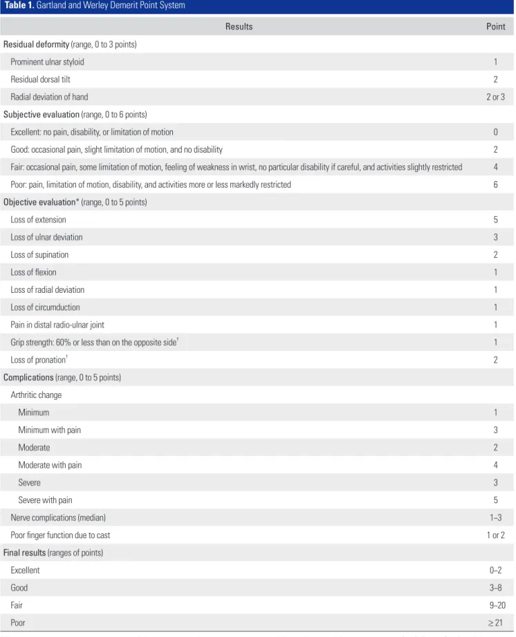
The disabilities of the arm, shoulder and hand (DASH) questionnaire was used to assess functional outcomes in activities of daily living. Final clinical and functional out- comes were assessed and graded using the scoring system of Gartland and Werley7) modified by Sarmiento et al.8) (Table 1).
Statistical Analysis
The paired t-test was used to compare each radiographic parameter. A p < 0.05 was considered statistically signifi- cant. The intra- and interobserver reliabilities of radio- graphic assessments were tested using ICCs. ICC values of > 0.8 were considered as excellent reliability. Statistical analyses were performed using IBM SPSS ver. 20.0 (IBM Co., Armonk, NY, USA).
RESULTS
Radiologic Outcomes
Bony union was achieved in all patients, and the mean time for union was 42 days (range, 37 to 50 days) (Fig. 2).
None of the patients required an autologous or artificial Fig. 1. The Korean-type distal radius anatomical volar plate system.
Table 1. Gartland and Werley Demerit Point System
Results Point
Residual deformity (range, 0 to 3 points)
Prominent ulnar styloid 1
Residual dorsal tilt 2
Radial deviation of hand 2 or 3
Subjective evaluation (range, 0 to 6 points)
Excellent: no pain, disability, or limitation of motion 0
Good: occasional pain, slight limitation of motion, and no disability 2
Fair: occasional pain, some limitation of motion, feeling of weakness in wrist, no particular disability if careful, and activities slightly restricted 4 Poor: pain, limitation of motion, disability, and activities more or less markedly restricted 6 Objective evaluation* (range, 0 to 5 points)
Loss of extension 5
Loss of ulnar deviation 3
Loss of supination 2
Loss of flexion 1
Loss of radial deviation 1
Loss of circumduction 1
Pain in distal radio-ulnar joint 1
Grip strength: 60% or less than on the opposite side† 1
Loss of pronation† 2
Complications (range, 0 to 5 points) Arthritic change
Minimum 1
Minimum with pain 3
Moderate 2
Moderate with pain 4
Severe 3
Severe with pain 5
Nerve complications (median) 1−3
Poor finger function due to cast 1 or 2
Final results (ranges of points)
Excellent 0−2
Good 3−8
Fair 9−20
Poor ≥ 21
*The objective evaluation is based on the following ranges of motions as being the minimum for normal function: extension (45°), flexion (30°), radial deviation (15°), ulnar deviation (15°), pronation (50°), and supination (50°). †Criteria added by Sarmiento et al.8)
bone graft. The preoperative volar tilt was –8.0° ± 16.3°
(range, –47.4° to 34.2°), the immediate postoperative vo- lar tilt was 8.5° ± 2.8° (range, 1.2° to 13.7°), and the final follow-up volar tilt was 8.4° ± 2.7° (range, 1.2° to 13.4°).
The preoperative radial inclination was 16.3° ± 6.2° (range, 2.5° to 27.9°), the immediate postoperative radial inclina- tion was 22.4° ± 2.3° (range, 17.7° to 26.4°), and the final follow-up radial inclination was 22.2° ± 2.3° (range, 17.7°
to 26.3°). Preoperative radial length was 8.3 ± 3.2 mm
(range, 1.6 to 14.8 mm), the immediate postoperative radi- al length was 11.3 ± 1.6 mm (range, 8.7 to 14.2 mm), and the final follow-up radial length was 11.3 ± 1.6 mm (range, 8.7 to 14.2 mm). There was a significant difference in each parameter between the preoperative and the immediate postoperative values (p < 0.001). However, there was no statistically significant difference between each parameter immediately postoperative and the final follow-up evalua- tion (Fig. 3).
Fig. 2. (A) Initial radiograph of C1 fracture in a 73-year-old woman. (B) Radiograph taken at postoperative 6 months. Anato- mical reduction was restored and well maintained.
Fig. 3. Changes of radiologic parameters. (A) Volar tilt. (B) Radial inclination. (C) Radial length.
Clinical Outcomes
Grip strength was restored to 84% (range, 69% to 100%) of the value of the unaffected side at the final follow-up evaluation. The mean range of motion of the wrist at the final follow-up was 56.2° ± 13.2° of extension (range, 25°
to 80°; 86% compared with the unaffected side), 51.3° ± 11.2° of flexion (range, 25° to 75°; 77% compared with the unaffected side), 15.6° ± 4.3° of radial deviation (range, 10° to 20°; 85% compared with the unaffected side), 25.9°
± 7.8° of ulnar deviation (range, 15° to 35°; 87% compared with the unaffected side), 81.0° ± 10.9° of pronation (range, 65° to 85°; 95% compared with the unaffected side), and 79.6° ± 11.7° of supination (range, 60° to 90°; 87% com- pared with the unaffected side). According to the Gartland and Werley scoring system, there were 16 excellent, 26 good, and 4 fair results. The mean follow-up time was the same for patients who showed excellent or good results (10.4 months; range, 7 to 14 months). The mean follow- up period for patients with fair results (7 months; range, 6 to 8 months) was relatively shorter compared with other patients with excellent or good results. The mean DASH score was 8.4 points (range, 0 to 20 points). All clinical outcomes are shown in Table 2.
Complications
One patient requested removal of the plate at 10 months after surgery. There were no cases of infection, complex regional pain syndrome, tendon rupture, nerve irritation, or implant failure.
DISCUSSION
In this study, the K-DRAVP system appropriately fit the anatomy of the distal radius of Korean patients. The sys- tem provided good maintenance of radiologic alignment after reduction of the fracture, and most patients (91%) achieved excellent or good results according to the Gart- land and Werley scoring system. There was no statistically significant deterioration in any of the radiologic parame- ters, such as volar tilt, radial inclination, and radial length.
Grip strength and range of motion at the final follow-up were restored to approximately 85% of the values of the unaffected side. The mean DASH score (8.4 points) indi- cated little discomfort in the activities of daily living.
Volar plating with a locking screw system has the advantages of an easy surgical procedure, relatively low risk of complications, and early functional mobilization.
Several authors have reported good outcomes for various types of volar plates used for treatment of distal radius fractures. Drobetz and Kutscha-Lissberg9) reported the results from 50 patients treated with a locking plate system (Mathys, Salzburg, Austria). According to the Gartland and Werley scoring system, 26 patients showed excellent results, 20 patients showed good results, 3 patients showed fair results, and 1 patient showed a poor result after a mean follow-up of 26 months. Kamano et al.10) reported the re- sults from 40 patients with distal radius fractures treated with palmar plates (Biotechini Co., Ciotat, France). They reported that 12 patients showed excellent results and 28 patients showed good results after a mean follow-up of 12 months, according to the Gartland and Werley scoring system. Wong et al.11) reported the results from 35 patients with dorsally displaced distal radius fractures treated using the Stryker plating system with SmartLock locking screws after a mean follow-up of 10 months. The mean Mayo Clinic wrist score was 90 points and 20 patients achieved an excellent result. Figl et al.12) reported the results from 80 patients with unstable distal radius fractures treated using the APTUS plate (Medartis AG, Basel, Switzerland) after a mean follow-up of 7 months. The mean DASH score was 25 points, and according to the Castaing score, 30 patients showed perfect results, 49 patients showed good results, and 1 patient showed an adequate result. Minegishi et al.13) reported the results from 15 patients with unstable distal radius fractures treated using the Acu-Loc distal radius plate (Acumed, Hillsboro, OR, USA) after a mean follow-up of 15.5 months. In their study, according to the Cooney’s clinical scoring chart, 5 patients showed excel- lent results, 7 patients showed good results, and 3 patients showed fair results. Lattmann et al.14) reported a relatively Table 2. Clinical Outcomes
Variable Mean ± SD Range Restoration (%)*
Range of motion (°)
Extension 56.2 ± 13.2 25–80 86
Flexion 51.3 ± 11.2 25–75 77
Radial deviation 15.6 ± 4.3 10–20 85
Ulnar deviation 25.9 ± 7.8 15–35 87
Pronation 81.0 ± 10.9 65–85 95
Supination 79.6 ± 11.7 60–90 87
Grip power (%) 69–100 84
DASH score (point) 8.4 0–20
Gartland and Werley scoring system: excellent (16), good (26), fair (4).
DASH: disabilities of the arm, shoulder and hand.
*Compared with the unaffected side.
large series of 228 patients with distal radius fractures treated with LC-T plates (Synthes, Bettlach, Switzerland).
Grip strength was 91% of that on the contralateral side, and the assessed Patient-Rated Wrist Evaluation score was 8 points. Few studies have evaluated the results of Asian- type distal radius volar plates. Osada et al.15) reported the results of 49 patients with distal radius fractures treated with a distal radius volar locking plate (DRV-LP, Mizuho Ikakogyo Co., Tokyo, Japan). After 1 year of follow-up, the mean DASH score was 6.1 points (range, 0 to 30 points) and all patients showed excellent or good results according to the Gartland and Werley scoring system. Yasuda and Ando16) also reported good outcomes with a new variable angle distal screw locking volar plate system (Nakashima Propeller Co., Okayama, Japan).
Our radiologic and clinical results are similar to those of other studies using different types of distal radius volar plates (Table 3).9-15,17) Furthermore, in our study, there were no mechanical complications such as irritations of flexor tendons or the median nerve. Some studies have reported on mechanical irritations after volar plate fixa- tion.18-20) Kim et al.18) reported 2 cases of multiple flexor tendon ruptures after volar plate (LC-T, plate; Synthes) fixation for distal radius fractures. They reported that the prominent distal portion of the volar plate could cause damage to the flexor tendons. Lee et al.19) reported 2 com- plications of mechanical irritations after volar plate (Acu- Loc System; Acumed) fixation for distal radius fractures.
Lee et al.20) reported mechanical irritation of the median nerve after volar plate fixation for distal radius fractures. In this case, there were no mechanical complications because the K-DRAVP system was initially designed as a low-pro- file system with a specially contoured thin and narrow end of the distal margin in order to minimize these mechanical complications, such as tendon or nerve irritations.
This study has some limitations. First, this was not a randomized controlled study, and we did not compare our results with patients who were treated with other types of distal radius plates. Therefore, we compared our data with those of previous reports. Further prospective randomized controlled studies are needed. Second, we had a relatively short follow-up period because this was a preliminary report. Patients who showed fair results according to the Gartland and Werley scoring system had relatively shorter follow-up periods (mean, 7 months; range, 6 to 8 months) than those of patients who showed excellent or good results (mean, 10.4 months; range 7 to 14 months).
This finding suggests that more favorable outcomes can be achieved after a longer follow-up period. Third, the K-DRAVP plate system had some problems. On the basis Table 3. Results of Recent Studies on Volar Locking Plate Systems StudyCase no.Plate systemMean follow-up period (mo)Result Drobetz and Kutscha-Lissberg (2003)9) 50Mathys plate system (Salzburg, Austria)26Excellent (26), good (20), fair (3), poor (1)* Kamano et al. (2005)10) 40Palmar plate (Biotechini Co., Ciotat, France)12Excellent (12), good (28)* Wong et al. (2009)11) 35SmartLock (Stryker, Kalamazoo, MI, USA)10Excellent (20), good (12), fair (2), poor (1)† Figl et al. (2009)12) 80APTUS (Medartis AG, Basel, Switzerland)7DASH 25 points: perfect (30), good (49), adequate (1)‡ Minegishi et al. (2011)13) 15Acu-Loc (Acumed, Hillsboro, OR, USA) 15.5Excellent (5), good (7), fair (3)§ Matschke et al. (2011)17) 1173.5 mm LCP-DR (Synthes, Bettlach, Switzerland)24DASH 11.2 points: excellent (59), good (37), fair/poor (12)* Lattmann et al. (2011)14) 228LC-T plates (Synthes)12Grip strength 91%: PRWE 8 points Osada et al. (2008)15) 49DRV Locking Plate (Mizuho Ikakogyo Co., Tokyo, Japan)12DASH 6.1 points: excellent (47), good (2)* Current study46Korean type distal radius anatomical volar plate (BK Meditech, Hwaseong, Korea)10.1DASH 8.4 points: excellent (16), good (26), fair (4)* DASH: disabilities of the arm, shoulder and hand, PRWE: Patient-Rated Wrist Evaluation. *By Gartland and Werley score. † By Mayo wrist score. ‡ By Castaing score.§ By Cooney’s clinical scoring chart.
of our experience with the K-DRAVP system, we recom- mended some improvements in the plate design. First, the drilling guides for the screws were small and separated, and management of these guides was difficult and time- consuming. Second, plates of variable sizes and lengths need to be designed. One female patient could not be treated with the K-DRAVP system because the diameter of her radius was smaller than that of this plate. Third, another plate design for juxta-articular type distal radius fractures is needed for fixation of small distal juxta-artic- ular fragments. After we reported our suggestions to the manufacturer, a revised version of the K-DRAVP system has been developed and is currently being used. We will report the results of the revised version of the plate in due course.
Although this study was only a preliminary report of the newly developed K-DRAVP system, we found that this plate system could be used to restore and maintain the anatomical parameters, and it provided good clinical outcomes with low complication rates because it is the first anatomical distal radius volar plate designed based on the anatomical characteristics of Koreans. The K-DRAVP sys- tem is a promising surgical option for treatment of distal radius fractures in the Korean population.
CONFLICT OF INTEREST
No potential conflict of interest relevant to this article was reported.
REFERENCES
plating system for Colles' fractures: a preliminary report. J Hand Surg Am. 2005;30(4):750-5.
11. Wong TC, Yeung CC, Chiu Y, Yeung SH, Ip FK. Palmar fixa- tion of dorsally displaced distal radius fractures using lock- ing plates with Smartlock locking screws. J Hand Surg Eur Vol. 2009;34(2):173-8.
12. Figl M, Weninger P, Liska M, Hofbauer M, Leixnering M.
Volar fixed-angle plate osteosynthesis of unstable distal radius fractures: 12 months results. Arch Orthop Trauma Surg. 2009;129(5):661-9.
13. Minegishi H, Dohi O, An S, Sato H. Treatment of unstable distal radius fractures with the volar locking plate. Ups J Med Sci. 2011;116(4):280-4.
14. Lattmann T, Meier C, Dietrich M, Forberger J, Platz A. Re- sults of volar locking plate osteosynthesis for distal radial fractures. J Trauma. 2011;70(6):1510-8.
15. Osada D, Kamei S, Masuzaki K, Takai M, Kameda M, Tamai K. Prospective study of distal radius fractures treated with a volar locking plate system. J Hand Surg Am. 2008;33(5):691- 700.
16. Yasuda M, Ando Y. A new variable angled locking volar plate system for Colles' fracture: outcome study and time- course improvement of objective clinical variables. Hand Surg. 2009;14(2-3):93-8.
17. Matschke S, Marent-Huber M, Audige L, Wentzensen A;
LCP Study Group. The surgical treatment of unstable distal radius fractures by angle stable implants: a multicenter pro- spective study. J Orthop Trauma. 2011;25(5):312-7.
18. Kim JY, Kang HJ, Yi Y. Multiple flexor tendon injuries after 1. Thompson PW, Taylor J, Dawson A. The annual incidence
and seasonal variation of fractures of the distal radius in men and women over 25 years in Dorset, UK. Injury. 2004;
35(5):462-6.
2. Park C, Ha YC, Jang S, Jang S, Yoon HK, Lee YK. The inci- dence and residual lifetime risk of osteoporosis-related frac- tures in Korea. J Bone Miner Metab. 2011;29(6):744-51.
3. Orbay JL. The treatment of unstable distal radius fractures with volar fixation. Hand Surg. 2000;5(2):103-12.
4. Chung KC, Shauver MJ, Birkmeyer JD. Trends in the United States in the treatment of distal radial fractures in the el- derly. J Bone Joint Surg Am. 2009;91(8):1868-73.
5. Lim ST, Yeom JS, Lee CH, Lee YH, Chang CB, Baek GH.
Development of anatomical plating system for treatment of distal radius fractures. J Korean Soc Surg Hand. 2007;
12(3):95-104.
6. Arora R, Lutz M, Hennerbichler A, Krappinger D, Espen D, Gabl M. Complications following internal fixation of unstable distal radius fracture with a palmar locking-plate. J Orthop Trauma. 2007;21(5):316-22.
7. Gartland JJ Jr, Werley CW. Evaluation of healed Colles' frac- tures. J Bone Joint Surg Am. 1951;33(4):895-907.
8. Sarmiento A, Pratt GW, Berry NC, Sinclair WF. Colles' frac- tures: functional bracing in supination. J Bone Joint Surg Am. 1975;57(3):311-7.
9. Drobetz H, Kutscha-Lissberg E. Osteosynthesis of distal radial fractures with a volar locking screw plate system. Int Orthop. 2003;27(1):1-6.
10. Kamano M, Koshimune M, Toyama M, Kazuki K. Palmar
volar plate fixation for distal radius fracture: two cases re- port. J Korean Soc Surg Hand. 2012;17(1):47-51.
19. Lee SJ, Bae JY, Cho HJ, Suh KT. Short term results of AO type C fractures of the distal radius treated with volar lock- ing plating system. J Korean Soc Surg Hand. 2011;16(4):
191-7.
20. Lee SU, Park IJ, Kim HM, Lee JY, Yoo HH, Jeong C. K-wire fixation supplemented with external fixator versus volar locked plating for unstable fractures of the distal radius. J Korean Soc Surg Hand. 2010;15(4):157-63.