37 37
THE EWHA MEDICAL JOURNAL THE EWHA MEDICAL JOURNAL
Amphetamine-Like Weight Reduction Drug Induced Acute Cardiomyopathy with Left Ventricular Thrombosis
Jeong Min Kim
1, Sung Kee Ryu
1, Jae Woong Choi
1, Dong Geum Shin
2, Yung Hee Lee
1, Hye Ran Kang
1, Won Young Chae
1, Ji Sang Park
11Department of Internal Medicine, Eulji University College of Medicine,
2Yonsei Cardiovascular Hospital, Yonsei University College of Medicine, Seoul, Korea
Introduction
Obesity has become one of the main health issues, and vari- ous attempts to reduce weight have been tried. Many people take drugs that suppress appetite and the absorption of food and increase the metabolic rate. However, some of these drugs have unpredictable complications, including cardiovascular side ef- fects. Some legally marketed amphetamine-like drugs such as fenproporex and diethylpropione are also prescribed for weight reduction for their appetite reduction effect, but these drugs share the side effects of amphetamines [1]. There are multiple reported cases of acute myocardial infarction and stroke after the use of amphetamines [2-10]. However, to our knowledge there are no published case reports of intracardiac thrombosis with an
amphetamine-like drug without myocardial infarction. We report the case of a 37-year-old female presenting with a left ventricle (LV) thrombus after amphetamine intake.
Case
A 37-year-old female patient with dyspnea on exertion and peripheral edema was admitted to our hospital. For one and a half years, the patient had been taking various drugs and supple- ments to reduce weight such as phentermine hydrochloride, theae folium powder, orthosiphon powder, saposhnikoviae radix pow- der, and other herbal medicines. Two weeks before admission, a progressive cough, febrile sense, and myalgia had developed.
The patient complained of dyspnea (New York Heart Association
Case Report
Ewha Med J 2014;37(Suppl):S37-S40 http://dx.doi.org/10.12771/emj.2014.37.S.S37 pISSN 2234-3180 • eISSN 2234-2591
A 37-year-old female patient admitted due to dyspnea on exertion and peripheral edema. For one and a half years, the patient had been taking various drugs and supple- ments to reduce weight, including amphetamine-like drugs. The patient had no major cardiovascular risk factors except three pack-years of smoking. A chest computed tomography showed a 1.7 cm diameter, capsulated space-occupying lesion in the left ventricle (LV) and 2-dimensional echocardiography showed LV systolic dysfunction (Left ventricular ejection fraction [LVEF], 30%) with a mobile cystic mass (1.1×1.8 cm) that was attached to the LV apex, which was increased in size and number the next day, even with low dose low-molecular-weight heparin. With an increased dose of antico- agulation medication and heart failure management with diuretics and angiotensin receptor II blocker, LV dysfunction was recovered and the LV thrombus disappeared.
(Ewha Med J 2014;37(Suppl):S37-S40)
Received June 27, 2014 Accepted August 21, 2014 Corresponding author Sung Kee Ryu
Department of Internal Medicine, Eulji University School of Medicine and Division of Cardiology, Eulji General Hospital, 68 Hangeulbiseong-ro, Nowon-gu, Seoul 139- 711, Korea
Tel: 82-2-970-8671, Fax: 82-2-970-8621 E-mail: [email protected]
Key Words
Amphetamine; Cardiomyopathies; Thrombosis
Copyright ⓒ 2014, The Ewha Medical Journal
cc This is an Open Access article distributed under the terms of the Creative Commons Attribution Non Commercial License (http://creativecommons.org/licenses/by-nc/3.0/) which permits unrestricted non commercial use, distribution, and reproduction in any medium, provided the original work is properly cited.
38 THE EWHA MEDICAL JOURNAL Kim JM, et al
[NYHA] III~IV) and peripheral edema a week before admission.
The patient had no history of hypertension, hyperlipidemia, dia- betes mellitus or other cardiovascular or cerebrovascular diseases, and the patient had no family history of cardiovascular disease.
She did not take oral contraceptives, but she was a current smok- er with a three pack-year smoking history. At admission, her blood pressure was 100/60 mmHg, heart rate was 80 beats per minute, respiratory rate was 20 per minute and body temperature
36.9oC. The patient appeared acutely ill with an alert mental state.
A crackling sound was auscultated on both lower lung fields, and her heartbeat was regular, without significant murmur. The ex- amination of her abdomen was unremarkable; a grade IV pitting edema was observed. Her body weight had increased from 64 kg to 79.2 kg over one week. On simple chest radiography, a pulmo- nary edema was seen in both lung fields with cardiomegaly (Fig.
1), and an electrocardiography (ECG) showed sinus tachycardia with poor R progression on the chest lead and T wave inversion in the limb leads. Under the impression of congestive heart failure
Fig. 1. Initial chest X-ray showed cardiomegaly with pulmonary con-
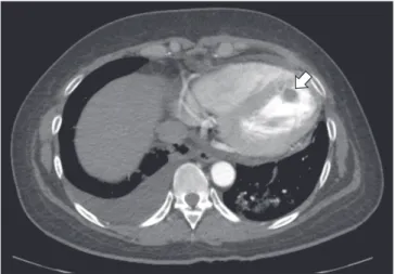
gestion. Fig. 2. Chest computed tomography shows capsulated space occupy-
ing lesion with 1.7 cm in diameter in left ventricle (arrow).
A B
C
Fig. 3. Two-dimensional echocardiography follow-up. (A) Initially mobile cystic mass (1.1×1.8 cm) is attached to septal area of apex of left ventricle and small mass like lesion was attached to papillary muscle (arrows). (B) Number and size of masses increased in one day (arrows). (C) Masses disappeared on discharge.
39
THE EWHA MEDICAL JOURNAL Amphetamine-Like Drug Induced LV Dysfunction and Thrombosis
(CHF), diuretics (furosemide 20 mg/day, spironolactone 50 mg/
day) and angiotensin receptor II blocker were given, and to pre- vent deep vein thrombosis, a half dose of low molecular weight heparin (LMWH) was injected. A chest computed tomography (CT) study showed a capsulated space-occupying lesion 1.7 cm in diameter in the LV (Fig. 2). Two-dimensional echocardiography showed reduced LV systolic function (LVEF, 30%), with moder- ate-to-severe global LV hypokinesis. There was no significant valvular lesion or significant LV enlargement (LV end-diastolic diameter, 54 mm). Neither right ventricular enlargement nor significant pulmonary hypertension was found (right ventricular systolic pressure, 27 mmHg).
A mobile cystic mass (1.1×1.8 cm) was attached to the septal area of the apex of the LV (Fig. 3A). Follow-up echocardiogra- phy, which was performed the next day, revealed newly devel- oped cystic masses that were attached to the papillary muscle (Fig. 3B). With this finding, we concluded that the cystic mass was a thrombus and increased the dose of LMWH. After that, warfarin was also given. Follow-up echocardiography was done two days afterwards, and it showed improved LV dysfunction (LVEF 60%), and remarkably reduced and collapsed previous cystic mass-like lesions. On the eighth day in hospital, LV func- tion was completely normalized, and the mass-like lesion had almost disappeared (Fig. 3C). The patient was discharged with warfarin and ARB, and followed up in out patient department without any adverse events over six months.
Discussion
Amphetamine-like medicines are popular as weight reduction drugs for their appetite suppressing effects. Amphetamines have sympathetic activators with the stimulation of α- and β-adrenergic receptors and various effects on the cardiovascular system such as hypertension and tachyarrhythmia [10]. Acute cardiomyopa- thy associated with amphetamines or similar drug use was also reported [11,12]. In this case, the patient presented with acute car- diomyopathy with multiple thrombi. Echocardiography revealed significantly reduced LV systolic function without aneurysmal change; therefore, besides the LV dysfunction, hypercoagulable status may have a role in the formation of multiple thrombi. Left ventricular thrombi can be developed by the stasis of blood flow associated with myocardial infarction or cardiomyopathy ac- companying severe LV dysfunction, especially apical aneurysm,
valvular heart disease, and endomyocardial disease. Hyperco- agulable status can be associated with this complication. The incidence of LV thrombus complicating an anterior acute myo- cardial infarction is estimated at 5~15%, and the incidence of left ventricular thrombus complicating with dilated cardiomyopathy and congestive heart failure varies from 10% to 30% [13]. With the progress in treatment of acute myocardial infarction, this in- cidence may be reduced; LV dysfunction with hypercoagulable status is still one of the primary causes of LV thromboembolism.
The mechanism of thrombus formation in amphetamine use is associated with catecholamine. Catecholamine is known to induce platelet activation and aggregation. Also, amphetamine stimulates release of norepinephrine and blockade of reuptake at the sympathetic synaptic receptors, resulting acute ventricular dysfunction. Therefore, high catecholamine levels after exposure to amphetamine may trigger thrombosis by acute ventricular dysfunction and hypercoagulable status [9,14]. Echocardiography is useful in the diagnosis of LV thrombus, and it appears as an echo dense mass with definite margin during the whole course of systole and diastole. Echogenicity may be homogenous, but sometimes appears with central lucency, which mimics a cystic mass [15]. We report a case of multiple thrombi with acute car- diomyopathy after long-term treatment with a weight reduction drug that was successfully treated with conventional heart failure medication and anticoagulation. Amphetamine use is illegal in most countries, but amphetamine-like drugs are widely used legally that can share its cardiovascular complications. When we use these drugs, more attention should be paid to the cardiovas- cular complications.
References
1. Mariotti KC, Rossato LG, Froehlich PE, Limberger RP. Amphet- amine-type medicines: a review of pharmacokinetics, pharma- codynamics, and toxicological aspects. Curr Clin Pharmacol 2013;8:350-357.
2. Bashour TT. Acute myocardial infarction resulting from am- phetamine abuse: a spasm-thrombus interplay? Am Heart J 1994;128:1237-1239.
3. Carson P, Oldroyd K, Phadke K. Myocardial infarction due to amphetamine. Br Med J 1987;294:1525-1526.
4. De Silva DA, Wong MC, Lee MP, Chen CL, Chang HM. Amphet- amine-associated ischemic stroke: clinical presentation and proposed pathogenesis. J Stroke Cerebrovasc Dis 2007;16:185- 186.
5. Huang CN, Wu DJ, Chen KS. Acute myocardial infarction
40 THE EWHA MEDICAL JOURNAL Kim JM, et al
caused by transnasal inhalation of amphetamine. Jpn Heart J 1993;34:815-818.
6. Hung MJ, Kuo LT, Cherng WJ. Amphetamine-related acute myo- cardial infarction due to coronary artery spasm. Int J Clin Pract 2003;57:62-64.
7. Orzel JA. Acute myocardial infarction complicated by chronic amphetamine use. Arch Intern Med 1982;142:644.
8. Packe GE, Garton MJ, Jennings K. Acute myocardial infarc- tion caused by intravenous amphetamine abuse. Br Heart J 1990;64:23-24.
9. Ragland AS, Ismail Y, Arsura EL. Myocardial infarction after am- phetamine use. Am Heart J 1993;125:247-249.
10. Waksman J, Taylor RN Jr, Bodor GS, Daly FF, Jolliff HA, Dart RC.
Acute myocardial infarction associated with amphetamine use.
Mayo Clin Proc 2001;76:323-326.
11. Call TD, Hartneck J, Dickinson WA, Hartman CW, Bartel AG.
Acute cardiomyopathy secondary to intravenous amphetamine
abuse. Ann Intern Med 1982;97:559-560.
12. Croft CH, Firth BG, Hillis LD. Propylhexedrine-induced left ven- tricular dysfunction. Ann Intern Med 1982;97:560-561.
13. Waller BF, Rohr TM, McLaughlin T, Grider L, Taliercio CP, Fet- ters J. Intracardiac thrombi: frequency, location, etiology, and complications: a morphologic review: part II. Clin Cardiol 1995;18:530-534.
14. Haft JI, Kranz PD, Albert FJ, Fani K. Intravascular platelet ag- gregation in the heart induced by norepinephrine. Microscopic studies. Circulation 1972;46:698-708.
15. Pepi M, Evangelista A, Nihoyannopoulos P, Flachskampf FA, Athanassopoulos G, Colonna P, et al. Recommendations for echocardiography use in the diagnosis and management of cardiac sources of embolism: European Association of Echocar- diography (EAE) (a registered branch of the ESC). Eur J Echo- cardiogr 2010;11:461-476.