| Abstract |
1)PURPOSE: This study examined the effects of active release technique on pain, Oswestry Disability Index, and pelvic asymmetry in chronic low back pain patients.
METHODS: Thirty five outpatients diagnosed with chronic low back pain were enrolled in this study. The patients were divided randomly into an active release technique therapy group(experimental group; n=18) and myofascial release technique therapy group(control group; n=17). These groups performed their respective therapy for a 40-minute session occurring twice a week over six weeks. The Visual Analogue Scale(VAS) was used to measure the subjects’
pain, and the Korean Oswestry Disability Index(KODI) was used to measure the subjects’ dysfunction. To assess the patients’ pelvic asymmetry, their pelvic tilt and pelvic
†Corresponding Author : Seung-Min Nam
[email protected], https://orcid.org/0000-0002-9215-0545 This is an Open Access article distributed under the terms of the Creative Commons Attribution Non-Commercial License (http://creativecommons.org/licenses/by-nc/3.0) which permits unrestricted non-commercial use, distribution, and reproduction in any medium, provided the original work is properly cited.
rotation were measured using X-ray imaging.
RESULTS: Both the experimental group and control group exhibited significant decreases in their VAS and KODI scores after the therapy(p<.05). The experimental group exhibited a significant decrease in their pelvic tilt and pelvic rotation after therapy(p<.05). A significant difference was observed between the experimental group and the control group (p<.05).
CONCLUSION: These results suggest that active release technique is effective in decreasing the level of pain and dysfunction in chronic low back pain patients. In addition, the active release technique is considered to be more effective in improving the pelvic tilt and pelvic rotation than myofascial release technique. This can be an effective method for the non-pharmacological and non-surgical treatment of chronic low back pain.
Key Words: Active release technique, Chronic low back pain, Pelvic asymmetry, KODI
Ⅰ. 서 론
허리통증은 전 세계적으로 70%의 사람들이 가장 흔히 경험하는 증상으로써, 현대 사회의 산업 발전과 신체활
Research Article Open Access
능동이완기법이 만성 허리통증 환자의 통증, 요통장애지수 및 골반비대칭에 미치는 영향
이승후⋅남승민
†대구대학교 물리치료학과
Effects of Active Release Technique on Pain, Oswestry Disability Index and Pelvic Asymmetry in Chronic Low Back Pain Patients
Seung-Hoo Lee, PT, MS, Seung-Min Nam, PT, PhD
†Department of Physical Therapy, Daegu University Received: November 11, 2019 / Revised: November 25, 2019 / Accepted: December 19, 2019
ⓒ 2020 J Korean Soc Phys Med
동의 감소로 허리통증을 호소하는 사람들이 증가하고 있다[1]. 허리통증은 일반적으로 허리와 엉치부를 중심 으로 나타나고, 신경근이 자극을 받아 무릎 밑으로 진행 되는 방사통이 나타날 수 있다. 또한 허리통증은 근력, 지구력 및 유연성의 감소, 감각이상, 몸통형태의 변화 등으로 이어져 신체적 활동이 제한된다[2]. 이러한 허리 통증의 대부분은 2주 이내 호전되지만, 20%는 통증이 지속되고 만성 허리통증으로 발전한다고 보고되었다[3].
이러한 만성 허리통증은 일상생활 및 사회활동을 수행하 는데 있어 불편함을 느끼게 하며, 일상생활의 동작과 관 련된 요통장애지수의 증가로 이어진다고 보고되었다[4].
허리통증의 주요원인은 다양하며, 대표적으로 직접 적인 척추의 병변에 의하여 발생하는 척추성 허리통증, 장기의 질환으로 발생하는 허리통증, 척추 및 골반의 구조적인 문제 및 기능 저하에 의하여 발생하는 생체역 학적 요인 등이 있다[5]. 특히 만성 허리통증 환자의 50-70%에서 골반의 비대칭이 흔히 관찰되며, 골반의 비대칭은 엉치엉덩관절을 중심으로 이루어진다[4]. 나 쁜 자세와 습관으로 인하여 비정상적인 척추의 만곡은 엉치엉덩관절 주변 근육의 기능부전 및 엉치엉덩관절 의 구조적인 불균형을 초래하고 허리와 엉치 주변 연부 조직이 불필요하게 사용되어 만성 허리통증의 원인이 된다[6]. 또한 골반의 비대칭은 척추의 측만이 유발되 고, 엉치엉덩관절면에 비대칭적인 부하가 가해져 엉치 엉덩관절의 퇴행성 변화 및 통증이 촉진된다[6].
엉치엉덩관절의 비대칭 및 허리와 엉치 주변근육의 기능부전은 만성 허리통증의 주된 원인이며, 다양한 임상적 증상을 나타낸다[6,7]. 따라서 임상에서는 만성 허리통증을 치료하기 위해서 수술치료, 약물치료, 물리 치료, 도수치료 등의 방법이 사용된다. 특히 도수교정, 근막이완기법 등과 같은 도수치료를 받는 환자들은 일 반적인 물리치료를 받은 경우에 비해 3배 정도의 만족 감을 보였다고 보고되었다[8].
도수치료 접근법 중에서 근막이완기법(myofascial release technique)은 근막에 초점을 두는 치료법으로써 인체에서 통증을 유발시키는 긴장된 조직을 이완시키 는 방법이다[9]. 근막은 신체의 인접 조직들이 서로 원 활하게 움직일 수 있도록 일종의 윤활유 역할을 한다 [9]. 이렇듯 근막이완기법은 근막의 수직배열을 개선시
켜 짧아진 연부조직을 늘려주고, 근육 및 신경 등이 적절하게 기능할 수 있도록 넓은 공간을 제공함으로써, 인체의 불균형 개선 및 통증을 완화시키는 치료법이다 [10]. 하지만, 최근 연구에 의하면 수동적인 운동보다 능동적인 운동이 더 효과적이라고 보고되었다[11]. 선 행연구에 의하면 능동적 스트레칭이 수동적 스트레칭 보다 뒤넙다리근의 유연성에 대해 더 좋은 효과가 있다 고 보고되었으며, 도수치료 접근법 중 능동이완기법 (active release technique)은 기존의 다른 도수치료 접근법 과는 다르게 능동적인 움직임을 이용하여 통증, 단축, 약화 등 기능부전의 원인이 되는 근육 및 연부조직이 늘어나는 자세로 압박 및 능동적 스트레칭을 병행하는 치료방법이다[11,12]. 이러한 능동이완기법은 반흔 조직 을 기계적으로 늘려주어 의도한대로 움직일 수 있도록 특별한 구조로 변화시켜 주며, 단축된 조직의 섬유 결 방향을 따라 종적으로 접촉 후 조직이 짧아지는 자세에 서 늘어나는 자세로 압박 및 능동적 스트레칭을 병행하 여 연부조직의 유착을 해소시킨다[13]. 즉, 조직에서 발 생할 수 있는 섬유화와 유착의 제거를 통해 조직의 장력 완화 및 힘줄, 신경 및 근섬유 등의 연부조직을 치료하여 통증 감소 및 기능회복을 시키는데 목적이 있다[14].
능동이완기법의 효과를 증명한 연구에 의하면 만성 목통증 환자를 대상으로 능동이완기법을 실시한 결과 일반적인 물리치료 및 수동적인 근막이완기법에 비해 통증 및 기능장애지수 완화에 유의한 효과가 있다고 보고되었다[15]. 여러 선행연구들에서 능동이완기법의 효과가 보고되었지만, 만성 허리통증 환자에게 능동이 완기법을 실시한 연구는 부족한 상황이다. 이에 만성 허리통증 환자를 대상으로 능동이완기법과 수동적인 근막이완기법을 허리통증 및 골반의 비대칭을 유발하 는 근육 및 연부조직에 적용하여, 능동이완기법이 만성 허리통증 환자의 통증, 요통장애지수 및 골반 비대칭에 미치는 영향을 알아보고 만성 허리통증 환자에게 효율 적인 도수치료방법을 제시하고자 한다.
Ⅱ. 연구방법
1. 연구대상
본 연구는 2019년 1월부터 2019년 4월까지 6주간
대구광역시 소재 H재활의원에 외래로 내원한 환자 중 임상적 소견과 X-ray 등의 의료장비를 통해 전문의로부 터 만성 허리통증으로 진단 받은 환자 40명을 대상으로 연구를 진행하였다. 구체적인 대상자 선정기준은 3개 월 이상 허리통증을 호소하는 자, 기능적 다리길이 검 사에서 양쪽의 차이가 5mm 이상인 자로 선정하였다 [16]. 또한 선행연구에 의하면 허리통증에 의해 치료를 받을 경우, 오스웨스트리 기능장애 지수 5점 미만인 자는 치료의 효과를 거의 보지 못한다고 보고되어, 본 연구에서는 한국판 오스웨스트리 기능장애 지수 5점 이 상인 자로 선정하였다[17,18]. 연구 대상자 전원에게 사 전에 연구의 목적 및 실험 내용을 설명한 후 자발적 참가 동의를 얻었으며, 본 연구는 대구대학교 생명윤리위원 회의 승인(1040621-201811-HR-016-08)을 받아 그 절차 에 따라 진행하였다. 선정된 40명을 대상으로 능동이완 기법 치료를 실시한 20명의 실험군(EG, n=20), 근막이완
기법 치료를 실시한 20명의 대조군(CG, n=20)으로 무작 위 배치하였으며, 실험 도중 중도포기 자 5명을 제외한 실험군 18명, 대조군 17명이 최종 실험을 완료하였다.
2. 치료방법
1) 능동이완기법
본 연구에서는 허리통증 및 골반의 비대칭을 유발하 는 근육 중 넙다리근막긴장근(tensor fasciae latae), 엉덩 허리근(iliopsoas), 궁둥이상근(piriformis), 중간볼기근 (gluteus medius), 작은볼기근(gluteus minimus)에 능동이 완기법을 적용하였다[19]. 능동이완기법은 6주 동안 주 2회 실시하였으며, 1회당 40분의 치료시간을 가졌다.
각각 근육에 적용된 능동이완기법은 7분의 치료시간을 가지고, 각 치료 사이에는 1분간의 휴식시간을 가졌다.
구체적인 치료방법은 다음과 같다(Table 1).
Muscle Start Position Manual Therapy Method
Tensor Fasciae Latae
Flexion hip and knee in sidelying position to
minimize tension
(1) Recognize the maxillary hip extension, knee flexion by passive movement for muscle stretching
(2) After applying pressure to the pain trigger point, move to the position where the muscle is shortened again
(3) Instructs you to remain active and stay active while stretching your muscles
Iliopsoas
Flexion hip and knee in sidelying position to
minimize tension
(1) Recognize the maxillary hip extension, knee extension by passive movement for muscle stretching
(2) After applying pressure to the pain trigger point, move to the position where the muscle is shortened again
(3) Instructs you to remain active and stay active while stretching your muscles
Piriformis
Extension hip and knee in sidelying position to
minimize tension
(1) Recognize the maxillary hip extension, external rotation and knee extension by passive movement for muscle stretching
(2) After applying pressure to the pain trigger point, move to the position where the muscle is shortened again
(3) Instructs you to remain active and stay active while stretching your muscles
Gluteus Medius
Extension hip and knee in sidelying position to
minimize tension
(1) Recognize the maxillary hip flexion, external rotation and knee flexion by passive movement for muscle stretching
(2) After applying pressure to the pain trigger point, move to the position where the muscle is shortened again
(3) Instructs you to remain active and stay active while stretching your muscles
Gluteus Minimus
Extension hip and knee in sidelying position to
minimize tension
(1) Recognize the maxillary hip flexion, internal rotation and knee flexion by passive movement for muscle stretching
(2) After applying pressure to the pain trigger point, move to the position where the muscle is shortened again
(3) Instructs you to remain active and stay active while stretching your muscles
Table 1. Active Release Technique
2) 근막이완기법
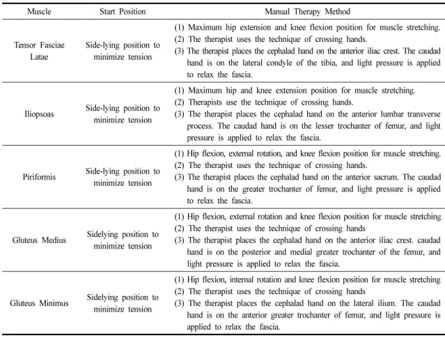
본 연구에서는 허리통증 및 골반의 비대칭을 유발하 는 근육 중 넙다리근막긴장근, 엉덩허리근, 궁둥이상 근, 중간볼기근, 작은볼기근에 근막이완기법을 적용하 였다[19]. 근막이완기법은 6주 동안 주 2회 실시하였으 며, 1회당 40분의 치료시간을 가졌다. 각각 근육에 적용 된 근막이완기법은 7분의 치료시간을 가지고, 각 치료 사이에는 1분간의 휴식시간을 가졌다. 구체적인 치료 방법은 다음과 같다(Table 2).
3. 측정방법
1) 시각적 상사 척도
허리통증의 통증 정도를 알아보기 위해 시각적 상사 척도(Visual Analogue Scale; VAS)를 이용하였다. 100
mm의 수평자에 왼쪽 끝은 통증이 없는 아주 편안한 상태, 오른쪽 끝은 극심한 통증으로 정의하도록 하여 피험자가 느끼는 주관적인 허리통증 정도를 표시하도 록 하는 방법이다. 시각적 사상 척도는 임상에서 통증 의 정도를 측정하는데 가장 널리 사용되는 방법 중 하나 이며, 치료효과를 판정하는데 유용한 평가도구이다 [20]. 평가도구의 신뢰도는 Cronbach’s α=.76-.84로 나타 났다[21].
2) 한국판 오스웨스트리 기능장애 지수
허리통증으로 인한 기능장애 정도를 평가하기 위해 서 한국판 오스웨스트리 기능장애 지수(Korean Oswestry Disability Index) 를 이용하였다. 한국판 오스웨스트리 기능장애 지수는 기능장애 변화에 민감한 자가인지 도 구이며, 임상에서 척추질환 관리에 널리 사용되는 방법
Muscle Start Position Manual Therapy Method
Tensor Fasciae Latae
Side-lying position to minimize tension
(1) Maximum hip extension and knee flexion position for muscle stretching.
(2) The therapist uses the technique of crossing hands.
(3) The therapist places the cephalad hand on the anterior iliac crest. The caudad hand is on the lateral condyle of the tibia, and light pressure is applied to relax the fascia.
Iliopsoas Side-lying position to minimize tension
(1) Maximum hip and knee extension position for muscle stretching.
(2) Therapists use the technique of crossing hands.
(3) The therapist places the cephalad hand on the anterior lumbar transverse process. The caudad hand is on the lesser trochanter of femur, and light pressure is applied to relax the fascia.
Piriformis Side-lying position to minimize tension
(1) Hip flexion, external rotation, and knee flexion position for muscle stretching.
(2) The therapist uses the technique of crossing hands.
(3) The therapist places the cephalad hand on the anterior sacrum. The caudad hand is on the greater trochanter of femur, and light pressure is applied to relax the fascia.
Gluteus Medius Sidelying position to minimize tension
(1) Hip flexion, external rotation and knee flexion position for muscle stretching (2) The therapist uses the technique of crossing hands
(3) The therapist places the cephalad hand on the anterior iliac crest. caudad hand is on the posterior and medial greater trochanter of the femur, and light pressure is applied to relax the fascia.
Gluteus Minimus Sidelying position to minimize tension
(1) Hip flexion, internal rotation and knee flexion position for muscle stretching (2) The therapist uses the technique of crossing hands
(3) The therapist places the cephalad hand on the lateral ilium. The caudad hand is on the anterior greater trochanter of femur, and light pressure is applied to relax the fascia.
Table 2. Myofascial Release Technique
이다. 총점은 45점 이며, 점수가 높을수록 허리통증으 로 인한 기능장애가 심한 것을 의미한다. 평가도구의 신뢰도는 Cronbach’s α=.89로 나타났다[21].
3) X-ray 촬영
골반의 비대칭을 평가하기 위하여 본 연구에서는 방사선 촬영장치(BL-50, DK medical system co, korea)을 사용하였으며, X-ray 분석은 Gonstead technique으로 분 석하였다[22]. 골반 경사(pelvic tilt)와 골반 회전(pelvic rotation)을 측정하기 위해서 제2엉치뼈(S2)를 기준으로 AP view 를 촬영하였다. 모든 피험자는 바로 선 자세에 서 촬영을 실시하였다.
(1) 골반경사(pelvic tilt)
골반경사는 좌․우 넙다리뼈머리(femur head)의 꼭대 기(apex)를 연결한 넙다리뼈머리선(femoral head line)을 긋고, 그와 평행인 선을 각각 엉덩뼈(ilium) 최상단과 궁둥뼈(ischium) 최하단에 수평선을 그은 후, 기준선과 수직이 되는 선을 그어 엉덩뼈 최상단과 궁둥뼈 최하단 의 길이를 측정하여, 골반경사를 간접적으로 평가할 수 있다. 좌․우 길이의 차이는 엉덩뼈의 앞돌림 변위 (anteversion) 및 뒤돌림 변위(retroversion)를 의미하며, 길이가 짧은 쪽을 위앞쪽(anterior superior) 변위, 긴 쪽
을 아래뒤쪽(posterior inferior) 변위로 평가할 수 있다 (Fig. 1).
(2) 골반회전(pelvic rotation)
골반회전은 제2엉치뼈(S2)의 중앙에 점을 찍은 후, 그 점에서 좌, 우 엉치뼈의 바깥쪽까지 연결한 선의 길이를 측정하여, 골반회전을 간접적으로 평가할 수 있다. 좌․우 길이의 차이는 엉치뼈의 앞돌림 변위 (anteversion) 및 뒤돌림 변위(retroversion)를 의미하며, 길이가 짧은 쪽을 위앞쪽(anterior superior) 변위, 긴 쪽 을 아래뒤쪽(posterior inferior) 변위로 평가할 수 있다 (Fig. 1).
4. 통계분석
본 연구에서 얻어진 실험의 결과는 평균±표준편차 (Mean±SD)로 기술하였다. 각 그룹 내 운동 전․후 차이를 검증하기 위하여 대응 표본 t검정(Paired T-test)을 실시 하였고, 그룹 간의 비교를 위하여 독립 표본 t검정 (Independent Sample T-test)을 실시하였다. 통계 처리는 통계처리 프로그램 SPSS 22.0 for Windows를 이용하여 분석하였다. 통계적 유의수준은 p<.05로 정의하였다.
Ⅲ. 연구결과
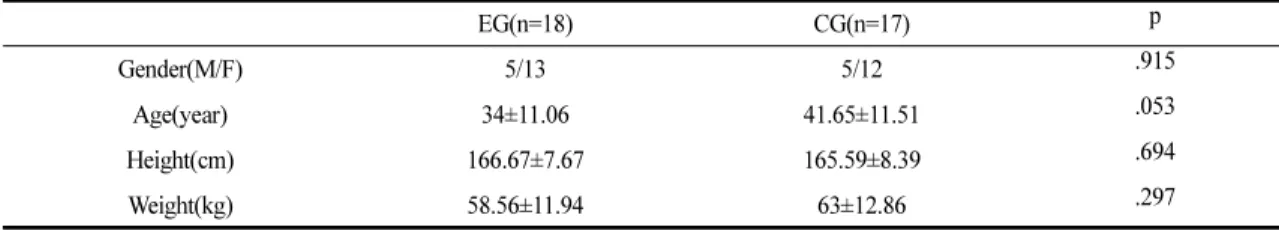
연구대상자의 동질성 검정을 실시한 결과 그룹 간에 통계학적으로 유의한 차이가 없었다(p>.05)(Table 3).
연구결과 통증 및 요통장애 지수는 실험군과 대조군 모두 치료 전․후 유의한 감소가 있었다(p<.05). 집단 간 의 차이 검정 결과 유의한 차이가 없었다(p>.05). 골반 비대칭 변화 비교에서 골반경사 및 골반회전은 실험군 에서 치료 전․후 유의한 감소가 있었으나(p<.05), 대조군 에서는 유의한 감소가 없었다(p>.05). 집단 간의 차이 검정 결과 유의한 차이가 있었다(p<.05)(Table 4).
Ⅳ. 고 찰
본 연구는 만성 허리통증의 효과적인 치료방법을
제시하기 위하여 만성 허리통증 환자를 대상으로 능동
Fig. 1. pelvic tilt and pelvic rotation.
이완기법 치료가 어떠한 영향을 미치는지 알아보았다.
허리통증 및 골반의 비대칭을 유발하는 근육 및 연부조 직에 능동이완기법을 실험군에 적용하고, 대조군에는 근막이완기법을 각각 6주 동안 주 2회 적용하였으며, 치료의 효과를 알아보기 위해 각각의 치료 전과 치료 후에 통증의 평가, 기능장애의 평가, X-ray 영상을 이용 하여 골반 비대칭의 변화를 평가하였다.
연구결과 시각적 상사척도 점수 및 한국판 오스웨스 트리 기능장애 지수는 실험군과 대조군 모두 치료 전
․ 후 통계적으로 유의하게 감소되었고, 치료 후 집단 간 비교에서는 통계적으로 유의한 차이가 없었다. 이러 한 연구결과는 만성 목통증 환자 24명을 대상으로 능동 이완기법을 실시한 결과 통증이 완화되었다고 보고된 연구결과와 일치하였으며, 만성 허리통증 환자를 대상
EG(n=18) CG(n=17) p
Gender(M/F) 5/13 5/12 .915
Age(year) 34±11.06 41.65±11.51 .053
Height(cm) 166.67±7.67 165.59±8.39 .694
Weight(kg) 58.56±11.94 63±12.86 .297
*p<.05
Mean±SD : mean±standard deviation EG: Active Release Technique group CG: Myofascial Release Technique group Table 3. General Characteristics of the Subjects
EG(n=18) CG(n=17) p
VAS
Pre 4.56±1.76 4.71±1.45 .785
Post 1.89±1.13 2.47±.80 .090
P .000
*.000
*KODI
Pre 16.78±6.39 16.94±5.88 .938
Post 9.11±3.80 10.59±4.58 .306
P .000* .000
*Pelvic Tilt
Pre .32±.22 .29±.15 .605
Post .17±.13 .33±.23 .016*
P .000
*.314
Pelvic Rotation
Pre .31±.21 .25±.21 .446
Post .12±.09 .34±.17 .000*
P .004
*.198
*