Reduction Aortoplasty with Suture Plication Technique for Dilatation of the Ascending Aorta associated with Aortic Valve Disease
Man-Jong Baek, M.D., Ph.D.*, Chan-Young Na, M.D., Ph.D.*,Sam-Sae Oh, M.D.*
Chang-Ha Lee, M.D., Ph.D.*, Seong Wook Whang, M.D.*, Cheol Lee, M.D.*
Hong Gook Lim, M.D.*, Jae Hyun Kim, M.D.*, Hong Ju Seo, M.D.*, Gun Gyk Kim, M.D.*
Background: Reduction aortoplasty has been advocated for dilatation of the ascending aorta associated with aortic valve disease in older, high-risk patients. We report our results with modification of reduction aortoplasty and aortic valve replacement. Material and Method: Between July 2001 and December 2002, 14 consecutive patients who underwent modification of reduction aortoplasty, suture plication technique without excision of the dilated aortic wall, were reviewed. The mean age was 63.7±6.7 (50 to 75) years. Ten patients had congenital bicuspid aortic valve.
Twelve patients had severe aortic valve stenosis and 6 had regurgitation of grade III∼IV. The diameter of the ascending aorta was measured before and immediately after surgery and 6 and 12 months postoperatively using echocardiography or computed tomography. Follow-up was complete in an average of 14.7±5.4 (7 to 24) months.
Result: There were no early postoperative deaths and no bleeding complications. Reduction aortoplasty with suture plication technique decreased the diameter of ascending aorta from 49.4±3.5 mm preoperatively to 33.2±3.4 mm postoperatively (p<0.001). During follow-up, there were no late deaths and no aneurysm recurrence on the ascending aorta. Conclusion: Suture plication technique of reduction aortoplasty without excision of the dilated aortic wall offers good early and short-term results in older, high-risk patients with dilatation of the ascending aorta associated with aortic valve disease. Surgical long-term results of our technique should be evaluated in further studies.
(Korean J Thorac Cardiovasc Surg 2005;38:221-228) ꠏꠏꠏꠏꠏꠏꠏꠏꠏꠏꠏꠏꠏꠏꠏꠏꠏꠏꠏꠏꠏꠏꠏꠏꠏꠏꠏꠏꠏꠏꠏꠏꠏꠏꠏꠏꠏꠏꠏꠏꠏꠏꠏꠏꠏꠏꠏꠏꠏꠏꠏꠏꠏꠏꠏꠏꠏꠏꠏꠏꠏꠏꠏꠏꠏꠏꠏꠏꠏꠏꠏꠏꠏꠏꠏꠏꠏꠏꠏꠏꠏꠏꠏꠏꠏꠏꠏꠏꠏꠏꠏꠏ Key words: 1. Aorta
2. Aneurysm 3. Aortic valve 4. Aortoplasty
*부천세종병원 흉부외과
Department of Thoracic and Cardiovascular Surgery, Sejong General Hospital, Sejong Heart Institute 논문접수일:2004년 11월 8일, 심사통과일:2005년 1월 29일
책임저자:백만종 (422-232) 경기도 부천시 소사구 소사본2동 91-121, 부천세종병원 흉부외과 (Tel) 032-340-1151, (Fax) 032-340-1236, E-mail: [email protected]
본 논문의 저작권 및 전자매체의 지적소유권은 대한흉부외과학회에 있다.
대동맥판막 질환과 동반된 상행대동맥 확장에 대한 봉합봉축법에 의한 대동맥 축소성형술
백만종*․나찬영*․오삼세*․이창하*․황성욱*
이 철*․임홍국*․김재현*․서홍주*․김근직*
INTRODUCTION
Many surgeons frequently encounter dilatation of the ascending aorta associated with aortic valve disease. The dilated or aneurysmal ascending aorta is at risk for spon- taneous rupture or dissection. Graft replacement is the most commonly performed procedure for the ascending aortic aneurysm[1]. However, in older or high-risk patients, a short aortic cross-clamp time might be advantageous during aortic valve replacement. A less radical operation than graft replacement, such as reduction aortoplasty with excision of the aneurysmal wall and external wrapping of the ascending aorta, was reported as one of treatment for dilatation of the ascending aorta[2]. Many authors have reported on the surgical results using this technique for dilatation of the ascending aorta[3-7].
However, this technique involving the excision of the ascending aortic wall may need more time and careful suture technique to securely close the long incision of the dilated thin aortic wall. On July 2001, we modified the excisional technique of reduction aortoplasty to a simple suture plication technique, without excision of the dilated aortic wall, to decrease the diameter of the aneurysm and remodel the ascending aorta in older or high-risk patients. We have used this technique, as one of the reduction aortoplasties in our institution. The purpose of this study was to describe our surgical technique and to report the results of clinical experience for reduction aorotoplasty by the suture plication technique.
MATERIAL AND METHOD
1) Patients
From July 2001 to December 2002, 14 consecutive patients with dilatation of the ascending aorta associated with aortic valve disease were operated on using reduction aorotoplasty with a suture plication technique and aortic valve replacement at Sejong Heart Institute. We used this technique on older or high-risk patients with severe ventricular dysfunction. There were 6 males and 8 females. The mean age at operation was 63.7±6.7 (50 to 75) years, and the mean body weight was 57.6±11.5 (40 to 81) kg. None had undergone previous car-
diac or aortic operations. Symptoms included congestive heart failure in 13 patients, angina in 5, and syncope in 1. The median functional class of New York Heart Association (NYHA) was 3, and its class was I in 1 patient, II in 3, III in 8, and IV in 2. Three patients were known to have hypertension. The mean ejection fraction of the left ventricle was 46.8±14.0% (20 to 65)%, and 8 patients had ejection fraction less than 45%. The mean diastolic diameter of the left ventricle was 60.6±11.9 mm. Ten patients were found to have congenital bicuspid valves. The remaining 4 patients with tricuspid aortic valve had rheumatic changes. Aortic valve stenosis was severe in 12 patients whose leaflet and annulus of the aortic valve were heavily calcified, and moderate in 2. The mean aortic valve area was 0.8±0.2 cm
2. Aortic valve regurgitation was combined in 11 patients, and its grade was I in 2 patients, II in 3, III in 4 and IV in 2.
The peak and the mean pressure gradient through the aortic valve measured by Doppler echocardiography were 97.7±
33.9 (68 to 175) mmHg and 58.5±22.1 (37 to 120) mmHg,
respectively. Coronary angiography revealed no stenosis or
abnormalities in all patients. The diameter of the ascending
aorta was measured by echocardiography, aortography, and by
computed tomographic scan of the chest. Most of the patients
had aneurysm of the fusiform type dilated along the greater
curvature of the ascending aorta. The mean diameter of the
ascending aorta was 49.4±3.5 (45 to 56) mm in all patients,
and 49.8±4.1 mm in 10 patients with bicuspid aortic valve
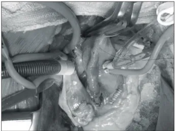
Fig. 1. The fusiform aneurysm of the ascending aorta asso-
ciated with aortic valve disease.
(Fig. 1). Although this diameter was not the definite indi- cation for graft replacement or aortoplasty, we applied our strategy to decrease the dilated segment of the aortic wall to remodel the ascending aorta. We performed reduction aorto- plasty with suture plication technique without resection of the dilated aortic wall to decrease the diameter of the aneurysm in older or high-risk patients. Follow-up echocardiography was performed at an average of 9 postoperative days.
2) Surgical Technique
The heart and the ascending aorta were exposed through the usual median sternotomy. The ascending aorta and the proximal aortic arch were evaluated. The ascending aorta was separated from the main pulmonary artery. Aortic cannula was placed in the proximal transverse aortic arch in all patients, and either right atrial or bicaval venous cannulation was utilized. Routine techniques of cardiopulmonary bypass was established with moderate hypothermia. The left ventricle was vented through the right upper pulmonary vein. The aorta was then cross-clamped just proximal to the innominate artery, and a conventional oblique aortotomy was done and carried into the noncoronary sinus of Valsalva. Myocardial protection was achieved by direct infusion of cold crystalloid or blood cardioplegic solution to the exposed coronary
orifices and local ice slush. The aortic valve was examined, excised, and replaced with a St. Jude mechanical prosthesis in 8 patients and a Hancock II bioprosthesis in 6. Concomitant cardiac operations were performed including mitral valve replacement in 3 patients, Maze operation in 1, and tricuspid valve repair in 1. During the systemic rewarming after the completeness of all cardiac procedures, the conventional aortotomy was closed in the usual manners.
Then reduction aortoplasty by suture plication technique was performed without excision of the dilated aortic wall in the oblique longitudinal fashion. Preoperatively we calculated the partial circumferential length of the ascending aorta which should be intraoperatively excluded by suture plication to bring the widest aortic diameter down to about 35 mm in diameter (Fig. 2A). If the patient's maximal diameter of the ascending aorta is measured 50 mm, the whole circumference of the dilated aorta equals 157 mm by 2πr. To decrease this patient's aortic diameter to about 35 mm, the entire cir- cumference should be decreased to 109 mm by reduction.
The partial circumference length of 48 mm should be excluded, and its half, 24 mm, should be plicated by first row of two rows of 4-0 polypropylene suture. The first row was advanced in continuous mattress suture from the sinotubular junction of the noncoronary sinus, through the right lateral border of the dilated aorta, to the distal Fig. 2. Schematic diagrams of suture plication technique of
reduction aortoplasty. The dilated wall of ascending aorta that should be excluded by suture plication is calculated and outlined (A) (dotted area). The dilated wall of ascending aorta is plicated by continuous mattress suture using a first row of two rows of 4-0 polypropylene (B). The plicated aortic wall is reinforced by a continuous over-and-over suture of the second row in a beating heart state (C).
A B C
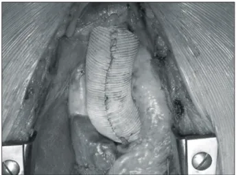
Fig. 3. Suture plication of the dilated ascending aorta by
continuous mattress suture using a first row of two rows of 4-0
polypropylene at an arrested heart.
ascending aorta just proximal to the aortic clamp (Fig. 2B, Fig. 3). After the aortic wall was plicated by the first row of 4-0 polypropylene suture, aortic cross-clamp was released.
Since this procedure was possible within two to three minutes, we were able to decrease the aortic cross- clamp time. The plicated aortic wall was then reinforced by continuous over-and-over suture of the second row in a beating heart state (Fig. 2C, Fig. 4). After the cardio- pulmonary bypass was weaned, external wrapping of the ascending aorta using a butterfly shaped Hemashield vascular graft and the proximal and distal anchoring sutures of 4-0 pledgetted polypropylene was performed in all patients (Fig.
5). The mean total cardiopulmonary bypass and aortic cross- clamp time were 146.9±38.4 minutes and 103.4±36.5 minutes, respectively.
3) Follow-up
Patients were contacted directly at the beginning of out- patient clinic and were analyzed for late mortality and event of aneurysm recurrence. Follow-up was complete in all patients, with a mean time of 14.7±5.4 (7 to 24) months.
We evaluated the diameter of the ascending aorta by echo- cardiography at the early postoperative course. At the out- patient clinic, chest radiography and electrocardiography were performed routinely. Echocardiography or computed tomo- graphic scan of the chest was done at postoperative 6 months
and 12 months for evaluation of the ascending aorta.
4) Statistical Analysis
All continuous data and results were expressed as the mean
±standard deviation and the significance of differences between the preoperative and postoperative data was assessed by one-way analysis of variance. A value of p less than 0.05 was considered statistically significant.
RESULTS 1) Early Mortality and Morbidity
There were no perioperative or early postoperative deaths and no bleeding complications requiring exploration. Severe ventricular dysfunction developed, immediate postoperatively after weaning of cardiopulmonary bypass, in three patients whose ventricular function was severely impaired preopera- tively. They were weaned from the cardiopulmonary bypass with temporary assistance. Among these patients, one patient suffered prolonged chest drainage and pericardial effusion requiring pericardiostomy, and the other revealed poor general condition. However, they were alive at the last follow-up with New York Heart Association functional class II. Another patient had a progressed septal hyperterophy despite of aortic valve replacement with a bioprosthesis for severe aortic stenosis and is now medically controlled. Transient left Fig. 4. Reinforcement of the plicated aortic wall by a conti-
nuous over-and-over suture of the second row in a beating heart state.
Fig. 5. External reinforcement of the ascending aorta using a
butterfly shaped vascular graft.
phrenic nerve palsy developed in one patient.
At the early postoperative echocardiographic study, the mean ejection fraction of the left ventricle was 50.9±12.2%, and there was no difference to the preoperative value (p=
0.193). But the mean end-diastolic diameter of the left ventricle decreased significantly to 50.6±8.3 mm compared to the preoperative value (p<0.001).
2) Follow-up Results
There was no late death, and there were no reoperations on the ascending aorta or aortic valve during the follow-up period. All patients were alive in NYHA functional class I (11 patients) or II (3 patients) at the time of the last follow-up. One patient had complete atrioventricular block at the postoperative 6 months, requiring implantation of the per- manent pacemaker. There were no endocarditis, cerebro- vascular and thromboembolic complications.
3) Changes of Diameter of the Ascending Aorta The reduction aortoplasty with suture plication technique for dilatation of the ascending aorta resulted in significant reduction of the ascending aortic diameter in all patients. The mean diameter of the ascending aorta decreased from 49.4±
3.5 mm preoperatively to 33.2±3.4 (28 to 38) mm imme- diate postoperatively (p<0.001)(Fig. 6). During follow-up with a mean time of 14.7±5.4 months, there was no evi- dence of enlargement of the ascending aorta in all patients on
the chest CT scan or echocardiography. The mean diameter of the ascending aorta measured at postoperative 6 months and 12 months were 33.3±2.7 mm and 33.8±2.2 mm, respectively. There were no significant differences in the changes of diameter of the ascending aorta during the follow- up period after reduction aortoplasty using suture plication technique (Fig. 6).
4) Comment
The aneurysmal dilatation of the ascending aorta is frequently associated with aortic valve disease. Currently, there are various operative techniques available for surgical treatment of the dilated ascending aorta combined with aortic valve disease. These include reduction aortoplasty with external wrapping of the ascending aorta[2], separate re- placement of the ascending aorta and aortic valve[8], root replacement with composite graft[8], valve-sparing root replacement[10,11], and root replacement with pulmonary autograft[12]. The resection and graft replacement is the most commonly applied procedure for the treatment of the ascending aortic aneurysm[1].
However, in older or high-risk patients, a short aortic cross-clamp time might be advantageous during aortic valve replacement. The least radical method is reduction aortoplasty that was first described by Robicsek[2]. This technique consists of decreasing the aortic diameter by excising the oval segment of the ascending aorta after longitudinal aortotomy followed by placing a well-tailored Dacron vascular graft around the ascending aorta. It has the advantage of sim- plicitiy, and more importantly, preserves the endothelial lining of the ascending aorta. Thereafter many surgical results of reduction aortoplasty with or without external wrapping in older or high-risk patients with ascending aortic aneurysm associated with aortic valve disease have been reported[3,5-7].
Also recently, modification to reduce the diameter of the moderately dilated ascending aorta has been reported by Baumgartner and colleagues as a modified Z-plasty with S-shaped incision and excision of the ascending aorta[4].
The dilated or aneurysmal ascending aorta associated with aortic valve disease have the risk of spontaneous rupture or dissection, as emphasized by Robicsek[13]: Most aneurysms are caused by abnormally high wall stress, and most aneu- Fig. 6. Changes of diameter of the ascending aorta after
reduction aortoplasty. NS=not significant; Preop=preoperatively;
mon=month
rysms rupture because of abnormally high wall stress-a force that acts on the aortic wall and is directly proportional with the aortic diameter and inversely with wall thickness and strength. Fusiform aneurysms that develop in the ascending aorta tend to expand anteriorly and toward the right, away from the pulmonary artery, and the ascending aorta frequently elongates vertically, resulting in an abnormally lengthy aorta with thin wall. In these conditions, longitudinal incision and excision of the dilated segment of a vertically elongated aorta during reduction aortoplasty may need more time and secure suture technique to close the long aortic incision. For these reasons, to decrease the diameter of the ascending aorta in older or high-risk patients, we modified the technique of reduction aortoplasty with long incision and resection of the thin aortic wall to a simple suture plication technique without incision and resection of the aortic wall.
Since the suture plication technique for the dilatation of the ascending aorta does not need secure suture to close the long aortic incision and can be done within 2 to 3 minutes during the aortic cross-clamp, it may be useful for patients with severe cardiac disease in whom prolonged cardiopulmonary bypass and aortic cross-clamping time could not be tolerated.
Since July 2001, we have used this technique as one of the reduction aortoplasty combined with aortic valve replacement.
In our experience with this technique, we had no early death and bleeding complication. This result is comparable to that of reduction aortoplasty using the longitudinal aortotomy and resection technique for dilatation of the ascending aorta performed by other groups. Carrel and co-workers[3] found lower operative mortality of 1.8% and lower incidence of postoperative bleeding and significantly shorter aortic cross- clamp and total bypass times in 164 patients undergoing tailoring aortoplasty with Dacron wrap. Also Bauer and colleagues[7] reported excellent results without perioperative mortality and major complications. However, hospital mor- tality in the study of Barnett and colleagues[5] and Mueller and co-workers[6] were 17.6% in 17 patients and 12% in 17 patients, respectively.
Late survival of reduction aortoplasty using the excision technique has shown good results. Mueller and co-workers[6]
reported 86.7% of survival at 7 years and Bauer and colleagues[7] reported 5-year survival rate of 94%. In a study
comparing tailoring aortoplasty and Dacron wrap with both composite valved graft replacement and supracoronary graft replacement of the ascending aorta, Carrel and colleagues[3]
found the best 8-year survival of 89.6% in those who had tailoring aortoplasty and wrap. However, as our study had the short-term follow-up period with a mean time of 14.7 months, we could not predict the late survival of reduction aortoplasty using our technique. We had no death and complications related to aortoplasty only in the short-term results.
Reduction aortolplasty using an ovoid or S-shaped excision of the ascending aorta have decreased the diameter of the ascending aorta effectively. Our suture plication technique without resection of the aortic wall could also significantly decrease the diameter of the ascending aorta. The mean diameter of the ascending aorta was significantly decreased from 49.4±3.5 mm preoperatively to 33.2±3.4 mm imme- diate postoperatively. During follow-up, we evaluated the diameter of the ascending aorta using the routine control echocardiographic or computed tomographic study at regular intervals. After follow-up with a mean time of 14.7 months, we had no recurrence of dilatation of the ascending aorta.
The diameter of the ascending aorta measured at post- operative 6 months and 12 months were not significantly different to that of the early postoperative study. Considering that the average interval from operation to recurrence was 63 months in the study by Mueller and co-workers[6] and 65 months in the group of McCready and Pluth[14], the follow- up time in our study was too short to detect recurrent aneurysm. However, our result indicates that reduction aor- toplasty using a suture plication technique can significantly decrease the diameter of the ascending aorta, and may be effective for prevention of dilatation of the ascending aorta in the short-term control study performed at regular intervals.
Long-term follow-up is a prerequisite for evaluation of aneu- rysmal recurrence after reduction aortoplasty using our suture plication technique.
It is not known whether the development of ascending
aortic aneurysm is caused by the intrinsic aortic wall defect
or by altered hemodynamic wall stress associated with aortic
valvular pathology. In patients with stenotic aortic valve
pathology, hemodynamic stress has been suggested as the
primary factor in the development of poststenotic dilatation [15,16]. Also, recent studies have suggested that intrinsic aortic wall abnormalities in patients with bicuspid aortic valve may also play a role in the development of ascending aortic dilatation[17-19]. In our study, 10 patients were found to have congenital bicuspid aortic valve. Aortic valve stenosis was severe in 12 patients, and moderate in 2. Aortic valve regurgitation was combined in 11 patients, and among them, 6 had severe regurgitation. We believe that the development of ascending aortic aneurysm is caused by the intrinsic aortic wall defect or by altered hemodynamic wall stress associated with aortic valve stenosis or regurgitation. No recurrence of aneurysm after reduction aortoplasty were found in our patients without respect to the type of aortic valve pathology.
However, because of the small number of patients and short term follow-up, we could not conclude definitely the etio- logical factors for recurrence of the ascending aortic aneu- rysm. Mueller and co-workers[6] mentioned that patients with aortic valve regurgitation reveal high recurrence rate of the ascending aortic aneurysm. Egloff and colleagues[20] already concluded that a tissue factor must be important and at least partly responsible for recurrent dilatation and dissection. Also, Carrel and co-workers[3] confirmed that dilatation and dissection pathology can develop in further segments of the ascending arch or descending or abdominal aorta after operations on the ascending aorta. These studies strongly suggest that careful follow-up is very important to detect aneurysmal recurrence and prevent late event of rupture or dissection.
In my opinion, external wrapping using a vascular graft can play an important role in preventing aneurysm recurrence after reduction aortoplasty using a suture plication technique.
We performed reduction aortoplasty with external wrapping in all patients. We had no aneurysm recurrence. Previous surgical studies using excision of the ascending aortic wall have also reported that, as the unsupported aortoplasty had high aneurysm recurrence rates, the ascending aorta should be supported from the outside with a Dacron graft to avoid later aneurysmal recurrence[2,3,6,7,14,20]. For these reasons, we agree with the comment by Robicsek that “aortoplasty elimi- nates the aneurysm, but it does not prevent recurrence” and
“the normal aortic geometry, thus restored, should be further
secured by external application of a well fitted Dacron vascular graft”[13].
Reduction aortoplasty using a suture plication technique is relatively safe and offers good short-term results in patients with dilatation of the ascending aorta combined with aortic valve disease. This technique can also decrease the diameter of the ascending aorta significantly and may be effective in preventing dilatation of the ascending aorta at short-term follow-up.
5) Limitations
Although our patients undergoing reduction aortoplasty with suture plication technique had no recurrence, this study has had small number of patients and short follow-up periods that limits us from detecting the recurrence of the ascending aortic aneurysm and drawing definite conclusions. Now, we have used this technique in older or high-risk patients with dilatation of the ascending aorta associated with aortic valve disease. Long-term results should be evaluated in further studies to investigate the effectiveness in preventing aneu- rysmal recurrence.
REFERENCES
1. Gillum RF. Epidemiology of aortic aneurysm in the United
States. J Clin Epidemiol 1995;48:1289-98.
2. Robicsek F. A new method to treat fusiform aneurysms of
the ascending aorta associated with aortic valve disease: an alternative to radical resection. Ann Thorac Surg 1982;34:
92-4.
3. Carrel T, von Segesser L, Jenni R, et al. Dealing with
dilated ascending aorta during aortic valve replacement:
advantages of conservative surgical approach. Eur J Car-
diothorac Surg 1991;5:137-43.4. Baumgartner F, Omari B, Pak S, Ginzton L, Shapiro S, Milliken J. Reduction aortoplasty for moderately sized as-
cending aortic aneurysms. J Card Surg 1998;13:129-32.
5. Barnett MG, Fiore AC, Vaca KJ, Milligan TW, Barner HB.
Tailoring aortoplasty for repair of fusiform ascending aortic aneurysms. Ann Thorac Surg 1995;59:497-501.
6. Mueller XM, Tevaearai HT, Genton CY, et al. Drawback of
aortoplasty for aneurysm of the ascending aorta associated with aortic valve disease. Ann Thorac Surg 1997;63:762-7.
7. Bauer M, Pasic M, Schaffarzyk R, et al. Reduction aorto-
plasty for dilatation of the ascending aorta in patients with
bicuspid aortic valve. Ann Thorac Surg 2002;73:720-4.
8. Yun KL, Miller DC, Fann JI, et al. Composite valve graft
versus separate aortic valve and ascending aortic replace- ment: is there still a role for the separate procedure? Cir-
culation 1997;96(Suppl II):368-75.9. Bentall H, DeBono A. A technique for complete replacement
of the ascending aorta. Thorax 1968;23:338-9.
10. Sarsam MA, Yacoub M. Remodelling of the aortic valve
annulus. J Thorac Cardiovasc Surg 1993;105:435-8.
11. David TE, Amstrong S, Ivanov J, Webb GD. Aortic valve
sparing operations: an update. Ann Thorac Surg 1999;67:
1840-2.
12. Ross D. Replacement of the aortic valve with a pulmonary
autograft: the “switch” operation. Ann Thorac Surg 1991;
52:1346-50.
13. Robicsek F. Invited commentary on: Barnett MG, Fiore AC, Vaca KJ, Milligan TW, Barner HB. Tailoring aortoplasty for
repair of fusiform ascending aneurysms. Ann Thorac Surg
1995;59:501.14. McCready RA, Pluth JR. Surgical treatment of ascending
aortic aneurysms associated with aortic valve insufficiency.
Ann Thorac Surg 1979;28:307-16.
15. Jarchow BH, Kincaid OW. Poststenotic dilatation of the
ascending aorta: its occurrence and significance as a roentgenologic sign of aortic stenosis. Proc Staff Meet Mayo
Clin 1961;36:23-33.16. Robicsek F. Post-stenotic dilatation of the great vessels.
Acta Med Scand 1955;151:481-5.
17. Hahn RT, Roman MJ, Mogtader AH, Devereux RB. Asso-
ciation of aortic dilation with regurgitant, stenotic and functionally normal bicuspid aortic valves. J Am Coll Car-
diol 1992;19:283-8.18. Pachulski RT, Weinberg AL, Chan KL. Aortic aneurysm in
patients with functionally normal or minimally stenotic bicuspid aortic valve. Am J Cardiol 1991;67:781-2.
19. Lindsay J. Coarctation of the aorta, bicuspid aortic valve
and abnormal ascending aortic wall. Am J Cardiol 1988;61:
182-4.
20. Egloff L, Rothlin M, Kugelmeier J, Senning A, Turina M.