Original Article
Role of high risk-human papilloma virus test in the follow-up of patients who underwent conization of the
cervix for cervical intraepithelial neoplasia
Jeong-Yeol Park1, Jaeman Bae2, Myong Cheol Lim2, So Yi Lim2, Dong-Ock Lee2, Sokbom Kang2, Sang-Yoon Park2, Byung-Ho Nam3, Sang-Soo Seo2
1Department of Obstetrics and Gynecology, University of Ulsan College of Medicine, Asan Medical Center, Seoul,
2Center for Uterine Cancer, 3Cancer Biostatistics Branch, Research Institute and Hospital, National Cancer Center, Goyang, Korea
Objective: To examine whether the presence of high risk-human papilloma virus (HR-HPV) after conization of the cervix was a risk factor for persistence or recurrence of cervical intraepithelial neoplasia (CIN) and whether HR-HPV test could be a guideline for post-therapy surveillance.
Methods: The study retrospectively analyzed data from 243 patients who underwent LLETZ or CKC of the cervix due to CIN.
Results: A positive HR-HPV test result which was performed between 3 and 6 months after procedure was a risk factor for persistent or recurrent cytological (p<0.001, odds ratio [OR]=22.51, 95% confidence interval [CI]=
9.74-52.02) and pathological (p<0.001, OR=18.28, 95% CI=5.55-60.20) abnormalities.
Conclusion: HR-HPV positive patients between 3 and 6 months after procedure should undergo frequent and meticulous post-therapy surveillance, while HR-HPV negative patients do not require such high-level surveillance and could undergo routine surveillance.
Key Words: HR-HPV, Conization, CIN, Recurrence
Received June 14, 2009, Revised June 19, 2009, Accepted June 21, 2009
Address reprint requests to Sang-Soo Seo
Center for Uterine Cancer, Research Institute and Hospital, National Cancer Center, 809, Madu 1-dong, Ilsan-gu, Goyang 411-351, Korea Tel: 82-31-920-1646, Fax: 82-31-920-1238
E-mail: ssseomd@ncc.re.kr
This work was supported by grants from the National Cancer Center, Korea (0410080-2).
INTRODUCTION
Conization of the uterine cervix such as large loop excision of the transformation zone (LLETZ) and cold knife conization (CKC) is not only a diagnostic procedure but also an appro- priate treatment for cervical intraepithelial neoplasia (CIN).1,2 However, CIN can recur, and invasive cervical carci- noma can develop, following such CIN treatment. The cumu- lative rate of invasion 8 years after CIN treatment is 5.8 per 1000 women, which is five times higher than for the general population.3 These findings indicate the importance of con- tinuous and meticulous follow-up. Factors reported to be as- sociated with persistent or recurrent cervical neoplasms after conization include menopausal status, grade of dysplasia, fol-
low-up cervical cytology, cone diagnosis of CIN 3, cone mar- gin status, and positive endocervical curettage. However, these factors are suboptimal predictors,4-12 and cannot be used to dictate the follow-up strategy after conization. While there is increasing evidence that testing for the presence of high risk-human papilloma virus (HR-HPV) after conization may help predict the likelihood of persistent or recurrent dis- ease,1,13-22 no study has shown how HR-HPV testing might be integrated into post conization surveillance.
The aim of this study was to determine whether HR-HPV test after conization is a predictive factor for CIN persistence or recurrence after LLETZ or CKC of the cervix. The study also investigated whether HR-HPV test results should influence post conization surveillance.
MATERIALS AND METHODS
From March 2001 to May 2006, 754 patients underwent con- ization of the cervix including LLETZ and CKC for CIN or mi- croinvasive cervical cancer at the Center for Uterine Cancer, National Cancer Center, Korea. A retrospective chart review was performed on these patients. The inclusion criteria of this study were: 1) patients whose follow-up cytology results and HR-HPV test results using the Hybrid Capture II (HC II) assay
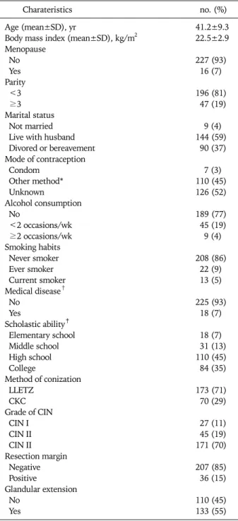
Table 1. Patients’ characteristics of 243 patients
Charateristics no. (%)
Age (mean±SD), yr 41.2±9.3
Body mass index (mean±SD), kg/m2 22.5±2.9 Menopause
No 227 (93)
Yes 16 (7)
Parity
<3 196 (81)
≥3 47 (19)
Marital status
Not married 9 (4)
Live with husband 144 (59)
Divored or bereavement 90 (37)
Mode of contraception
Condom 7 (3)
Other method* 110 (45)
Unknown 126 (52)
Alcohol consumption
No 189 (77)
<2 occasions/wk 45 (19)
≥2 occasions/wk 9 (4)
Smoking habits
Never smoker 208 (86)
Ever smoker 22 (9)
Current smoker 13 (5)
Medical disease†
No 225 (93)
Yes 18 (7)
Scholastic ability‡
Elementary school 18 (7)
Middle school 31 (13)
High school 110 (45)
College 84 (35)
Method of conization
LLETZ 173 (71)
CKC 70 (29)
Grade of CIN
CIN I 27 (11)
CIN II 45 (19)
CIN II 171 (70)
Resection margin
Negative 207 (85)
Positive 36 (15)
Glandular extension
No 110 (45)
Yes 133 (55)
SD: standard deviation, LLETZ: large loop excision of transfor- mation zone, CKC: cold knife conization, CIN: cervical intraepi- thelial noeplasia
*Periodic abstinence, intrauterine device, oral pill, tubal steri- lization, and vasectomy, †Hypertension, diabetes mellitus, chronic liver disease, and thyroid disease, ‡The school from which the pa- tient graduated last
after conization were available, 2) patients whose first fol- low-up cytology and HR-HPV test were performed within 6 months after conization, and 3) patients whose follow-up pe- riod was longer than 12 months.
The detailed methods for cervical cytology, HR-HPV test with HC II, and conization (LLETZ and CKC) were described in our previous reports.23,24 HC II is the only HPV test ap- proved by the United States Food and Drug Administration and is a liquid hybridization assay designed to detect 13 high-risk HPV types (HPV type 16, 18, 31, 33, 35, 39, 45, 51, 52, 56, 58, 59, and 68). In our study, a RLU/PC ratio of 1 or higher was considered a positive result. The follow-up HR-HPV test and cytology was performed at 3-6 months after conization, after which the patients were followed-up every 3-6 months.
A logistic regression model and the Kaplan-Meier method were used to identify risk factors for persistent or recurrent cytological and pathological abnormalities after conization, and to determine the relative risk of persistence or recurrence.
Student’s t-test and Mann-Whitney U-test were used to eval- uate the differences in the mean and median values between groups, and Chi-squared test and Fisher’s exact test were used to evaluate the differences in the proportions. The differ- ences were regarded as significant when the p-value was less than 0.05 in the two-sided test. SPSS software for Windows (version 9.0; SPSS inc., Chicago, IL) was used for analysis of data.
RESULTS
A total of 243 patients met the inclusion criteria and were in- cluded in this study. For the 243 study patients, the mean age was 41.2 years (range, 23 to 75 years), and 16 were post- menopausal. The parity was 1 or 2 in 196 patients. LLETZ was performed in 173 patients, and CKC was performed in 70 patients. Following conization, the diagnosis was CIN I in 27 patients, CIN II in 45 patients, and CIN III in 171 patients.
Patient characteristics are listed in Table 1. The first follow-up visit after conization was within 6 months for all patients, and the median follow-up period was 24 months (range, 12 to 57 months).
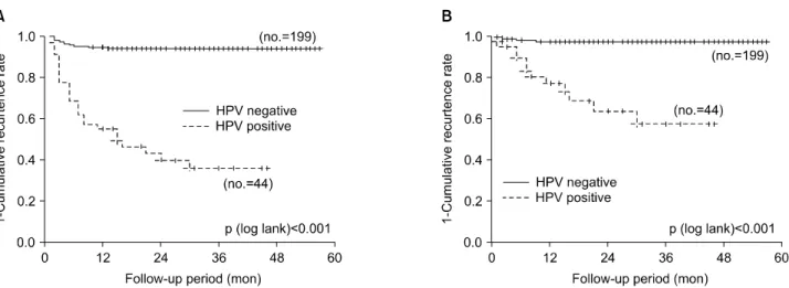
HR-HPV testing between 3 and 6 months after conization showed that 44 patients were HR-HPV positive and 199 were HR-HPV negative. Recurrent cytological abnormalities were found in 26 of the 44 HR-HPV positive patients, and in 12 of the 199 HR-HPV negative patients. Analysis showed that a positive HR-HPV result was a risk factor for recurrent cyto- logical abnormality (p<0.001, OR=22.51, 95% CI=9.74- 52.02) (Fig. 1).
The types of recurrent cytological abnormalities were ASCUS in 7 patients, ASCH in 8 patients, LSIL in 9 patients, and HSIL in 14 patients. Of these patients, 13 showed re- gression to normal cytology in subsequent follow-up tests, and 25 underwent colposcopy-directed biopsies of the cervix.
The biopsy results of those 25 patients showed that 9 had no dysplasia, while 16 had a recurrent pathological abnormality.
Recurrent pathological abnormalities were found in 12 of the 44 HR-HPV positive patients, and in 4 of the 199 HR-HPV
Fig. 1. Persistent or recurrent (A) cytological (left, p<0.001) or (B) pathological (right, p<0.001) abnormalities according to HR-HPV test re- sults between 3 and 6 months after LLETZ or CKC.
HR-HPV: high risk-human papilloma virus, LLETZ: large loop excision of transformation zone, CKC: cold knife conization.
negative patients. Analysis showed that a positive HR-HPV test result was a risk factor for recurrent pathological abnor- mality (p<0.001, OR=18.28, 95% CI=5.55-60.20). The types of recurrent pathological abnormalities were CIN I in 4 patients, CIN II in 2 patients, CIN III in 9 patients, and in- vasive carcinoma in 1 patient. Ten patients had repeat coniza- tions, and 6 had hysterectomies. The sensitivity, specificity, negative predictive value, and positive predictive values of the HR-HPV test results were 86%, 75%, 98%, and 27%, respectively.
The resection margin was positive in 36 patients and neg- ative in 207 patients. Recurrent cytological abnormalities were observed in 11 of 36 patients with positive resection margins, and in 27 of 207 patients with negative resection margins. Analysis showed that a positive resection margin was a risk factor for recurrent cytological abnormality (p=
0.01, OR=2.93, 95% CI=1.30-6.64). Recurrent pathological abnormalities occurred in 4 of 36 patients who were resection margin positive, and in 12 of 207 who were resection margin negative. Analysis found that a positive resection margin was not a risk factor for recurrent pathological abnormality (p=
0.268). There was no association between the HR-HPV test result and resection margin status (p=0.821).
Univariate analysis showed that age, body mass index, men- opausal status, parity, marital status, alcohol consumption, smoking habits, medical disease, scholastic ability, method of conization, grade of dysplasia, and glandular extension were not risk factors for recurrent cytological or pathological ab- normalities.
DISCUSSION
Our data showed that a positive HR-HPV test result between 3 and 6 months after conization was a significant risk factor for recurrent cytological or pathological abnormality for CIN.
The study also found there was no recurrent disease 10 months after conization in HR-HPV negative patients after conization (Fig. 1). In terms of patient management, the study data suggest that HR-HPV positive patients should undergo frequent and meticulous surveillance, while HR-HPV neg- ative patients do not require such high-level surveillance.
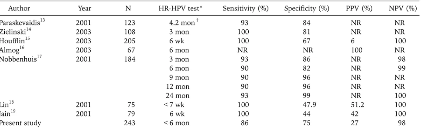
There is increasing evidence that HR-HPV testing after con- ization is important for detecting persistent or recurrent disease.1,13-22 The 2001 ASCCP guidelines state that HR-HPV testing is acceptable for post treatment surveillance.25 Post-conization HR-HPV testing is useful for detecting not only persistent disease but also recurrent disease. The sensi- tivity, specificity, positive and negative predictive values of HR-HPV testing for detecting persistent or recurrent disease after conization have been reported in several studies (Table 2).13-19 In particular, the negative predictive value was found to be very high in all studies.
Depending on the study, patients have been tested for HR-HPV at different times, including immediately after con- ization,21 within 6 months after conization,13-15,18,19,22 or at 6 months after conization (Table 2).16,17 Nobbenhuis et al. re- ported that results were similar at both 3 and 6 months after conization (Table 2).17 The 2001 American Society for Colposcopy and Cervical Pathology (ASCCP) guidelines rec- ommend that testing be performed at least 6 months after treatment to provide sufficient time for clearance of the HPV infection, and that it can be performed at 12 months after treatment unless a patient has risk factors for persistent/re- current CIN, such as a large lesion or endocervical exten- sion.25 While this may be a reasonable guideline under some circumstances, such a delay in testing may have a negative im- pact in cases where there is residual high grade CIN or in- vasive carcinoma after conization. In the present study, HR-HPV tests were performed between 3 and 6 months after conization, and the median time interval from conization to
Table 2. Studies which have examined the association between HR-HPV test results and persistent or recurrent CIN after conization
Author Year N HR-HPV test* Sensitivity (%) Specificity (%) PPV (%) NPV (%)
Paraskevaidis13 2001 123 4.2 mon† 93 84 NR NR
Zielinski14 2003 108 3 mon 100 81 NR NR
Houfflin15 2003 205 6 wk 100 67 6 100
Almog16 2003 67 6 mon NR NR 100 NR
Nobbenhuis17 2001 184 3 mon 93 86 NR 98
6 mon 90 82 NR 99
9 mon 90 96 NR NR
12 mon 90 96 NR NR
24 mon 93 99 NR 100
Lin18 2001 75 <7 wk 100 47.9 51.2 100
Jain19 2001 79 6 wk 100 44 42 100
Present study 243 <6 mon 86 75 27 98
HR-HPV: high risk-human papilloma virus, CIN: cervical intraepithelial neoplasia, PPV: positive predictive value, NPV: negative pre- dictive value
*Time interval from conization to HR-HPV test, †Mean time interval from conization to HR-HPV test
recurrence was 5 months (range, 1 to 30 months).
The present study indicates that HR-HPV testing between 3 and 6 months after conization is important for predicting the risk of disease persistence or recurrence. In addition, such testing can assist in designing patient management, since HR-HPV negative patients should undergo routine surveil- lance, while HR-HPV positive patients should undergo fre- quent and meticulous surveillance.
REFERENCES
1. Kucera E, Sliutz G, Czerwenka K, Breitenecker G, Leodolter S, Reinthaller A. Is high-risk human papillomavirus infection as- sociated with cervical intraepithelial neoplasia eliminated after conization by large-loop excision of the transformation zone?
Eur J Obstet Gynecol Reprod Biol 2001; 100: 72-6.
2. Nagai Y, Maehama T, Asato T, Kanazawa K. Persistence of hu- man papillomavirus infection after therapeutic conization for CIN 3: is it an alarm for disease recurrence? Gynecol Oncol 2000; 79: 294-9.
3. Soutter WP, de Barros Lopes A, Fletcher A, Monaghan JM, Duncan ID, Paraskevaidis E, et al. Invasive cervical cancer after conservative therapy for cervical intraepithelial neoplasia.
Lancet 1997; 349: 978-80.
4. Husseinzadeh N, Shbaro I, Wesseler T. Predictive value of cone margins and post-cone endocervical curettage with residual disease in subsequent hysterectomy. Gynecol Oncol 1989; 33:
198-200.
5. Jones HW 3rd, Buller RE. The treatment of cervical intra- epithelial neoplasia by cone biopsy. Am J Obstet Gynecol 1980;
137: 882-6.
6. Lapaquette TK, Dinh TV, Hannigan EV, Doherty MG, Yandell RB, Buchanan VS. Management of patients with positive mar- gins after cervical conization. Obstet Gynecol 1993; 82: 440-3.
7. Lin H, Chang HY, Huang CC, Changchien CC. Prediction of disease persistence after conization for microinvasive cervical carcinoma and cervical intraepithelial neoplasia grade 3. Int J Gynecol Cancer 2004; 14: 311-6.
8. Livasy CA, Maygarden SJ, Rajaratnam CT, Novotny DB.
Predictors of recurrent dysplasia after a cervical loop electro- cautery excision procedure for CIN-3: a study of margin, endo-
cervical gland, and quadrant involvement. Mod Pathol 1999;
12: 233-8.
9. Lu CH, Liu FS, Tseng JJ, Ho ES. Predictive factors for residual disease in subsequent hysterectomy following conization for CIN III. Gynecol Oncol 2000; 79: 284-8.
10. Moore BC, Higgins RV, Laurent SL, Marroum MC, Bellitt P.
Predictive factors from cold knife conization for residual cer- vical intraepithelial neoplasia in subsequent hysterectomy. Am J Obstet Gynecol 1995; 173: 361-6; discussion 6-8.
11. Paterson-Brown S, Chappatte OA, Clark SK, Wright A, Maxwell P, Taub NA, et al. The significance of cone biopsy resection margins. Gynecol Oncol 1992; 46: 182-5.
12. Phelps JY 3rd, Ward JA, Szigeti J 2nd, Bowland CH, Mayer AR.
Cervical cone margins as a predictor for residual dysplasia in post-cone hysterectomy specimens. Obstet Gynecol 1994; 84:
128-30.
13. Paraskevaidis E, Koliopoulos G, Alamanos Y, Malamou-Mitsi V, Lolis ED, Kitchener HC. Human papillomavirus testing and the outcome of treatment for cervical intraepithelial neoplasia.
Obstet Gynecol 2001; 98: 833-6.
14. Zielinski GD, Rozendaal L, Voorhorst FJ, Berkhof J, Snijders PJ, Risse EJ, et al. HPV testing can reduce the number of follow-up visits in women treated for cervical intraepithelial neoplasia grade 3. Gynecol Oncol 2003; 91: 67-73.
15. Houfflin Debarge V, Collinet P, Vinatier D, Ego A, Dewilde A, Boman F, et al. Value of human papillomavirus testing after conization by loop electrosurgical excision for high-grade squ- amous intraepithelial lesions. Gynecol Oncol 2003; 90: 587-92.
16. Almog B, Gamzu R, Bornstein J, Levin I, Fainaru O, Niv J, et al.
Clinical and economic benefit of HPV-load testing in follow-up and management of women postcone biopsy for CIN2-3. Br J Cancer 2003; 89: 109-12.
17. Nobbenhuis MA, Meijer CJ, van den Brule AJ, Rozendaal L, Voorhorst FJ, Risse EK, et al. Addition of high-risk HPV testing improves the current guidelines on follow-up after treatment for cervical intraepithelial neoplasia. Br J Cancer 2001; 84:
796-801.
18. Lin CT, Tseng CJ, Lai CH, Hsueh S, Huang KG, Huang HJ, et al.
Value of human papillomavirus deoxyribonucleic acid testing after conization in the prediction of residual disease in the sub- sequent hysterectomy specimen. Am J Obstet Gynecol 2001;
184: 940-5.
19. Jain S, Tseng CJ, Horng SG, Soong YK, Pao CC. Negative pre-
dictive value of human papillomavirus test following conization of the cervix uteri. Gynecol Oncol 2001; 82: 177-80.
20. Bar-Am A, Gamzu R, Levin I, Fainaru O, Niv J, Almog B.
Follow-up by combined cytology and human papillomavirus testing for patients post-cone biopsy: results of a long-term fol- low-up. Gynecol Oncol 2003; 91: 149-53.
21. Negri G, Gampenrieder J, Vigl EE, Haitel A, Menia E, Mian C.
Human papilloma virus typing at large loop excision of the transformation zone of the cervix uteri. Anticancer Res 2003;
23: 4289-92.
22. Chua KL, Hjerpe A. Human papillomavirus analysis as a prog- nostic marker following conization of the cervix uteri. Gynecol Oncol 1997; 66: 108-13.
23. Park JY, Lee SM, Yoo CW, Kang S, Park SY, Seo SS. Risk factors
predicting residual disease in subsequent hysterectomy follow- ing conization for cervical intraepithelial neoplasia (CIN) III and microinvasive cervical cancer. Gynecol Oncol 2007; 107:
39-44.
24. Park JY, Lee KH, Dong SM, Kang S, Park SY, Seo SS. The asso- ciation of pre-conization high-risk HPV load and the persis- tence of HPV infection and persistence/recurrence of cervical intraepithelial neoplasia after conization. Gynecol Oncol 2008;
108: 549-54.
25. Wright TC Jr, Cox JT, Massad LS, Carlson J, Twiggs LB, Wilkinson EJ. 2001 consensus guidelines for the management of women with cervical intraepithelial neoplasia. Am J Obstet Gynecol 2003; 189: 295-304.