Evaluation and Treatment of the Acute Cerebral Infarction with Convexal Subarachnoid Hemorrhage
Min Hyung Lee, Sang Uk Kim, Dong Hoon Lee, Young Il Kim, Chul Bum Cho, Seung Ho Yang, Il Sup Kim, Jae Taek Hong, Jae Hoon Sung, Sang Won Lee
Department of Neurosurgery, St. Vincent's Hospital, College of Medicine, The Catholic University of Korea, Suwon, Korea
Non-traumatic convexal subarachnoid hemorrhage (CSAH) is a comparatively infrequent with various vascular and nonvascular causes, it rarely occurs concomitant to acute ischemic stroke. We report a case of a 59-year-old woman, visited emergency room with right side subjective weakness spon- taneously. Magnetic resonance diffusion-weighted images revealed an acute infarction of anterior cerebral arterial territory. Computed tomographic an- giography showed a left frontal CSAH without any vascular lesions. And other laboratory studies were non-specific. We treated with dual anti- platelet drugs (cilostazole [Otsuka Pharmaceutical Co., Ltd. tokyo, Japan]
and Aspirin [Bayer Pharma AG., Leverkusen, Germany]). She has done well for a follow-up period. (5 months) This case demonstrates the CSAH with acute infarction is rare but need to work up to identify the etiology and antiplatelet dugs are taken into account for treatments.
J Cerebrovasc Endovasc Neurosurg.
2016 September;18(3):271-275 Received : 27 December 2015 Revised : 1 September 2016 Accepted : 12 September 2016 Correspondence to Sang Uk Kim
Department of Neurosurgery, St. Vincent's Hospital, The Catholic University College of Medicine, 93 Jungbu-daero, Paldal-gu, Suwon 16247, Korea
Tel : 82-31-249-8163 Fax : 82-31-245-5208 E-mail : [email protected]
ORCID : http://orcid.org/0000-0002-2117-8022
This is an Open Access article distributed under the terms of the Creative Commons Attribution Non- Commercial License (http://creativecommons.org/li- censes/by-nc/3.0) which permits unrestricted non- commercial use, distribution, and reproduction in any medium, provided the original work is properly cited.
Keywords Convexal subarachnoid hemorrhage, Ischemic stroke, Antiplatelet drug, Computed tomographic angiography
INTRODUCTION
Non-traumatic convexal subarachnoid hemorrhage (CSAH) observed at the convexity of the brain is a relatively uncommon entity with various vascular and nonvascular causes.: cerebral venous thrombosis (CVT),1) reversible cerebral vasoconstriction syndrome (RCVS),4) vascular malformations, vasculitides,12) infectious aneur- ysms,10) Moyamoya disease or syndrome,22) severe car- otid atherosclerosis,6) posterior reversible encephalop- athy syndrome (PRES),24) cerebral amyloid angiopathy (CAA),5) and nonvascular disorders, such as primary and secondary brain neoplasms7) or abscess.18)
Cerebral infarcts in the territory of the anterior cere- bral artery (ACA) are reported to comprise 0.5-3% of all ischemic strokes9)11) and few studies have specifi-
cally assessed the clinical characteristics of stroke pa- tients with ACA infarction.2) We report a case of ACA cerebral infarction with spontaneous CSAH lead to stenosis of the ACA. It is a infrequent case, but is worth consideration that the management and evalua- tion of these patients.
CASE REPORT
A 59-year-old female patient visited emergency room with right side subjective weakness spontaneously. She had a clinical history of hypertension and other past medical history was unremarkable. There was no his- tory of head trauma. Initial systolic and diastolic blood pressure was 140 mmHg / 80 mmHg. Routine laboratory finding is non-specific. She complained that right side
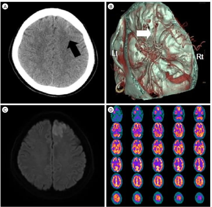
A B
C D
Fig. 1. (A) Computed tomography scan of the patient showing subarachnoid hemorrhage (black arrow) on left frontal convexity. (B) CT angiography shows that both A2 segments were mild focal stenosis (white arrow). (C) Diffusion-weighted images shows a high intensity area in the territory of left anterior cerebral artery. (D) Brain SPECT with Tc-99 m HMPAO shows a small area of decreased perfusion at left frontal area. CT = computed thmography; SPECT = single-photon emission computed tomography; HMPAO = hexam- ethylpropylene amine oxime; Lt = left; Rt = Right.
subjective weakness and mild sensory numbness.
However, The National Institute of Health Stroke Scale (NIHSS) was 0. Initial computed tomography angiography at one and half hours after symptom on- set demonstrated subarachnoid hemorrhage (SAH) lo- calized in the left frontal convexity and mild focal
stenosis at both A2 segments. Also the Magnetic reso- nance diffusion-weighted images (DWI) revealed an acute infarction of anterior cerebral arterial territory (Fig. 1).
We could not find any vascular lesion on computed tomographic angiography (CTA) and magnetic reso-
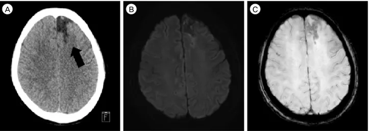
A B C
Fig. 2. (A) Computed tomography shows that low intensity of previous ACA infarction territory (black arrow) and no high intensity presenting SAH on left frontal convexity. (B) and (C) there is no evidence of acute infarction in DWI and hemorrhagic transformation in SWI. ACA = anterior cerebral artery; SAH = subarachnoid hemorrhage; DWI = diffusion-weighted images; SWI = susceptibility weighted imaging.
nance angiography (MRA) and treated with dual anti- platelet drugs (cilostazole [Otsuka Pharmaceutical Co., Ltd. tokyo, Japan] 50 mg bid/day plus aspirin [Bayer Pharma AG., Leverkusen, Germany] 100 mg) and hy- dration for 7 days. Other work up for evaluation of acute cerebral infarction was echocardiogram and hol- ter monitoring. But there was no abnormal finding ex- cept a few APB on holter monitoring. The patient's neurological finding was improved on day 2. She has done well for a follow-up period and her Modified Rankin Scale was 0 at day 5. The follow-up evalua- tion was performed at out-patient clinic after 5 months. CTA shows that the SAH was washed out and encephalomalatic change in left ACA territory.
MRI also reveals that no evidences of acute infarction in DWI and hemorrhagic transformation in suscepti- bility weighted imaging (SWI) (Fig. 2).
DISCUSSION
Although the prevalence of non-traumatic convexal SAH is reported to be 7.5% of all spontaneous SAH patients,13) little has been conscious of concerning the incidence of CSAH accompanying with acute infarction among many different etiologies of CSAH. Cuvinciuc et al. demostrated the image protocol to cover the wide
spectum of entities potentially responsible for CSAH that brain CTA with a paired channel at both arterial and venous phases and MR imaging including GRE T2 sequences, FLAIR, DWI, MRA 3D TOF, contrast- enhanced venogram, and pre- and postgadoliniumT1- weighted imaging.3)
However, We evaluated the CTA and MR imaging cluding T2, DWI, SWI and 3D TOF MRA. these work-up tests were enough to distinguish the etiolgies of CSAH.
CTA and MRA are useful to determine the vascular causes of CSAH such as vascular malformations, RCVS, vasulitides, high-grade stenosis, moyamoya disease, and septic aneurysms. In addition, MR imging is as- certainable diagnositic tools between dural and cort- ical CVT, and non-vascular causes such as, CAA, PRES, neoplasm, abscess and cavernoma.
In this case, ACA territory infarction seemed to oc- cur hemodynamically because of stenotic lesion on A2 segment and hypoperfusion on brain SPECT with Tc-99 m HMPAO. Nakajima et al. assumed that the accompanying an infarction and non traumatic CSAH might demonstrate that hemodynamic insufficiency because of arterial stenosis or occlusion get to the crit- ical point. For instance, after an acute infarction due to occlusive disease by the embolic or hemodynamic mechanism, ensuing dynamic changes of intracranial
perfusion pressure might bring about the CSAH.14) Also Cuvinciuc et al. explained that the main mecha- nism of CSAH because of chronic arterial occlusive diseases is considered to be the rupture of dilated vulnerable compensatory pial vessels.3)
Antiplatelet treatment is suggested for patients who have suffered the infarction. A large randominzed con- trolled trial studied Clopidogrel and Aspirin in High-Risk Patients with Acute Nondisabling Cerebrovascular Events (CHANCE) was finished in china. The result revealed that antiplatelet double therapy with clopi- dogrel plus aspirin was better than aspirin alone for reducing the risk of recurrent stroke and not increase the risk of bleeding among patients with high risk transient ischemic attack (TIA) or minor infarction23) Due to our patient's acute hemorrhage on the frontal convexity, we were concerned the hemorrhagic trans- formation and rebleeding because dual antiplatelet therapy (clopidogrel plus aspirin) was associated with a significant trend to increase moderate bleeding.17) So we treated with cilostazol plus aspirin successfully.
The patient's symptom was improved and there was no evidence of rebleeding on following studies. Tan et al. reported that cilostazol, alone or with aspirin, de- crease recurrence of ischemic stroke significantly, post- stroke intracranial hemorrhage, and extracranial bleed- ing in patients with a prior ischemic stroke as com- pared with other antiplatelet treatments.21) Cilostazol, a selective inhibitor of cyclic nucleotidephosphodies- terase 3, increases activated intracellular cyclic adenosine monophosphate (cAMP) concentrations and thus in- hibits platelet aggregation.19) Cilostazol is a known di- rect arterial vasodilator,15) and has antiatherosclerotic effect,8) which can strengthen the endothelial barrier20) and additionally may play a role in neuroprotection.16)
CONCLUSION
Acute infarction with spontaneous convexal SAH is rare but it is worth to work up to identify the etiol- ogy of CSAH using the CTA or MRA and MR image
including T2, DWI, SWI. And we suggest that dual antiplatelet dugs (cilostazole and aspirin) are taken in- to account for treatments that improved the neuro- logic symptoms and reduced the concerns about stroke recurrence and rebleeding.
Disclosure
The authors report no conflict of interest concerning the materials or methods used in this study or the findings specified in this paper.
REFERENCES
1. Arévalo-Lorido JC, Carretero-Gómez J. Cerebral venous thrombosis with subarachnoid hemorrhage: a case report.
Clin Med Res. 2015 Mar;13(1):40-3.
2. Arboix A, García-Eroles L, Sellarés N, Raga A, Oliveres M, Massons J. Infarction in the territory of the anterior cerebral artery: clinical study of 51 patients. BMC Neurol.
2009 Jul;9:30.
3. Cuvinciuc V, Viguier A, Calviere L, Raposo N, Larrue V, Cognard C, et al. Isolated acute nontraumatic cortical subarachnoid hemorrhage. AJNR Am J Neuroradiol. 2010 Sep;31(8):1355-62.
4. Ducros A, Boukobza M, Porcher R, Sarov M, Valade D, Bousser MG. The clinical and radiological spectrum of reversible cerebral vasoconstriction syndrome. A prospective series of 67 patients. Brain. 2007 Dec;130(Pt 12):3091-101.
5. Finelli PF. Cerebral amyloid angiopathy as cause of con- vexity SAH in elderly. Neurologist. 2010 Jan;16(1):37-40.
6. Geraldes R, Santos C, Canhão P. Atraumatic localized convexity subarachnoid hemorrhage associated with acute carotid artery occlusion. Eur J Neurol. 2011 Feb;18(2):e28-9.
7. Hentschel S, Toyota B. Intracranial malignant glioma presenting as subarachnoid hemorrhage. Can J Neurol Sci. 2003 Feb;30(1):63-6.
8. Ito H, Uehara K, Matsumoto Y, Hashimoto A, Nagano C, Niimi M, et al. Cilostazol inhibits accumulation of triglyceride in aorta and platelet aggregation in choles- terol-fed rabbits. PLoS One. 2012;7(6):e39374.
9. Kang SY, Kim JS. Anterior cerebral artery infarction:
stroke mechanism and clinical-imaging study in 100 patients. Neurology. 2008 Jun;70(24 Pt 2):2386-93.
10. Kannoth S, Iyer R, Thomas SV, Furtado SV, Rajesh BJ, Kesavadas C, et al. Intracranial infectious aneurysm:
presentation, management and outcome. J Neurol Sci.
2007 May;256(1-2):3-9.
11. Kazui S, Sawada T, Naritomi H, Kuriyama Y, Yamaguchi T. Angiographic evaluation of brain infarction limited to the anterior cerebral artery territory. Stroke. 1993 Apr;24(4):
549-53.
12. Kumar R, Wijdicks EF, Brown RD Jr, Parisi JE, Hammond CA. Isolated angiitis of the CNS presenting as sub- arachnoid haemorrhage. J Neurol Neurosurg Psychiatry.
1997 Jun;62(6):649-51.
13. Kumar S, Goddeau RP Jr, Selim MH, Thomas A, Schlaug G, Alhazzani A, et al. Atraumatic convexal subarachnoid hemorrhage: clinical presentation, imaging patterns, and etiologies. Neurology. 2010 Mar;74(11):893-9.
14. Nakajima M, Inatomi Y, Yonehara T, Hirano T, Ando Y.
Nontraumatic convexal subarachnoid hemorrhage con- comitant with acute ischemic stroke. J Stroke Cerebrovasc Dis. 2014 Jul;23(6):1564-70.
15. Nakamura K, Ikomi F, Ohhashi T. Cilostazol, an inhibitor of type 3 phosphodiesterase, produces endothelium-in- dependent vasodilation in pressurized rabbit cerebral penetrating arterioles. J Vasc Res. 2006;43(1):86-94.
16. Nonaka Y, Tsuruma K, Shimazawa M, Yoshimura S, Iwama T, Hara H. Cilostazol protects against hemor- rhagic transformation in mice transient focal cerebral is- chemia-induced brain damage. Neurosci Lett. 2009 Mar;452 (2):156-61.
17. O'Donnell MJ, Hankey GJ, Eikelboom JW. Antiplatelet therapy for secondary prevention of noncardioembolic ischemic stroke: a critical review. Stroke. 2008 May;39(5):
1638-46.
18. Rhode V, van Oosterhout A, Mull M, Gilsbach JM.
Subarachnoid haemorrhage as initial symptom of multiple brain abscesses. Acta Neurochir (Wien). 2000;142(2):205-8.
19. Sudo T, Tachibana K, Toga K, Tochizawa S, Inoue Y, Kimura Y, et al. Potent effects of novel anti-platelet aggregatory cilostamide analogues on recombinant cyclic nucleotide phosphodiesterase isozyme activity. Biochem Pharmacol. 2000 Feb;59(4):347-56.
20. Sugiura Y, Morikawa T, Takenouchi T, Suematsu M, Kajimura M. Cilostazol strengthens the endothelial bar- rier of postcapillary venules from the rat mesentery in situ. Phlebology. 2014 Oct;29(9):594-9.
21. Tan L, Margaret B, Zhang JH, Hu R, Yin Y, Cao L, et al. Efficacy and Safety of Cilostazol Therapy in Ischemic Stroke: A Meta-analysis. J Stroke Cerebrovasc Dis. 2015 May;24(5):930-8.
22. Wan M, Han C, Xian P, Yang WZ, Li DS, Duan L.
Moyamoya disease presenting with subarachnoid hemor- rhage: Clinical features and neuroimaging of a case series. Br J Neurosurg. 2015;29(6):804-10.
23. Wang Y, Pan Y, Zhao X, Li H, Wang D, Johnston SC, et al. Clopidogrel With Aspirin in Acute Minor Stroke or Transient Ischemic Attack (CHANCE) Trial: One-Year Outcomes. Circulation. 2015 Jul;132(1):40-6.
24. Yoon SD, Cho BM, Oh SM, Park SH, Jang IB, Lee JY.
Clinical and radiological spectrum of posterior reversible encephalopathy syndrome. J Cerebrovasc Endovasc Neurosurg.
2013 Sep;15(3):206-13.