Brief Report
Vol. 29, No. 1, 2017 111
Received July 29, 2015, Revised December 31, 2015, Accepted for publication January 6, 2016
Corresponding author: Nack In Kim, Department of Dermatology, Kyung Hee University Medical Center, 23 Kyungheedae-ro, Dongdaemun-gu, Seoul 02447, Korea. Tel: 82-2-958-8501, Fax: 82-2-969-6538, E-mail: [email protected]
This is an Open Access article distributed under the terms of the Creative Commons Attribution Non-Commercial License (http://creativecommons.org/
licenses/by-nc/4.0) which permits unrestricted non-commercial use, distribution, and reproduction in any medium, provided the original work is properly cited.
Copyright © The Korean Dermatological Association and The Korean Society for Investigative Dermatology
https://doi.org/10.5021/ad.2017.29.1.111
Immune Parameters of Korean Patients with Psoriasis Treated with Low-Dose Cyclosporine
Jeong Hwee Choi, Eun-Jae Shin, Min Kyung Shin, Nack In Kim
Department of Dermatology, College of Medicine, Kyung Hee University, Seoul, Korea
Dear Editor:
Cyclosporine A (CsA) is an immunosuppressive drug that interferes with T-cell receptor signaling at a critical stage zof T-cell activation. CsA has also been reported to exert effects on B and natural killer (NK) cells1,2. It is well-known that low-dose CsA improves the clinical signs and symp- toms of psoriasis, atopic dermatitis, pyoderma gangreno- sum, and chronic idiopathic urticaria3. Earlier, psoriasis was generally regarded as a disease of keratinocytes; the inflammatory response was thought to be secondary.
Later, psoriasis came to be considered an autoimmune dis- ease in which T lymphocytes play central roles. T cells are almost certainly involved in the initiation of psoriatic lesions. Activated T cells of the dermal-epidermal junction are thought to drive the hyperplastic proliferative response by elaborating T helper cell 1 cytokines, including tumor necrosis factor (TNF)-α, interferon (IFN)-γ, and various ILs4. The purpose of the present study was to examine the effect of low-dose CsA on the immune systems of Korean patients with psoriasis, by counting the numbers of T, B, and NK cells.
In total, 67 patients clinically diagnosed with histopatho- logically confirmed psoriasis vulgaris were selected from January 2009 to June 2014. Clinical data (age, sex, dura- tion of disease, duration of CsA treatment, and the
“psoriatic area and severity index” [PASI]) were collected.
The exclusion criteria were any history of the use of an- other immunosuppressant within 6 months before treat- ment, concomitant use of other immunosuppressants, any hematological disorder, and any other chronic illness.
Informed consent was obtained from all patients. This
study was approved by the Kyung Hee University Medical Center institutional review board (KMC IRB 1522-14). We collected venous blood samples from all patients (from 11 prior to CsA treatment and from 56 at least 1 month after such treatment, at 100∼200 mg/day). For T cells, CD3, CD4, and CD8 served as cell-surface markers. For B and NK cells, CD19 and CD16/56 served respectively. We used the Becton Dickinson SimultestTM (Becton Dickinson, CA, USA) IMK-Lymphocyte kit to calculate the proportions of mature human leukocytes in erythrocyte-free whole blood. Each sample was introduced into the flow cytometer.
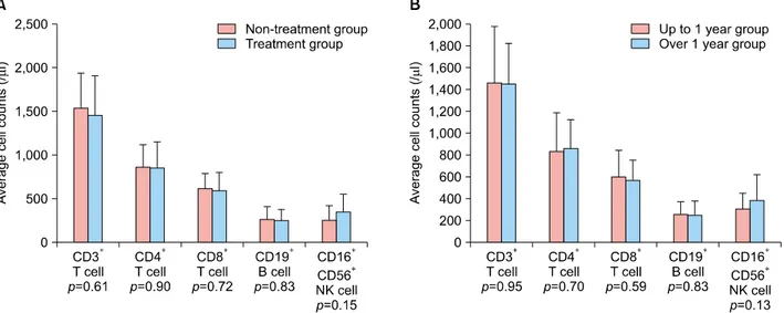
The clinical characteristics of all patients are shown in Table 1. No statistically significant difference was noted in any of mean age, duration of disease, or baseline average PASI score, between the non-treatment and treatment groups. In the treatment group, the average PASI score de- creased significantly from baseline (13.18±6.79) to after treatment (7.56±5.81) (p<0.000). The mean counts of CD3+, CD4+, CD8+ cells and CD19+ cells in the treat- ment group were lower than those in the non-treatment group. The mean count of CD16+CD56+ cells was higher in the treatment group. However, all of the difference was not statistically significant (Fig. 1A). Twenty-six patients were treated with low-dose CsA for less than 1 year, whereas 30 patients were treated for more than 1 year.
The mean counts of CD3+, CD8+ cells and CD19+ cells in the patients treated for more than 1 year were lower than those in the patients treated for up to 1 year. The mean counts of CD4+ cells and CD16+CD56+ cells were higher in the patients treated for more than 1 year, but the difference was not significant (Fig. 1B).
Brief Report
112 Ann Dermatol
Table 1. Demographic and clinical findings
Characteristic Non-treatment group Treatment group p-value
Mean age (yr) 34 (18∼61) 39 (17∼65) 0.24
Male:female ratio 1.7:1 2.3:1 NA
Mean duration of disease (mo) 9.2 (1∼25) 12.4 (2∼45) 0.31
Average PASI (mean±SD) 11.4±7.65 (baseline) 13.18±6.79 (baseline) 0.44
7.56±5.81 (after treatment) <0.000
Mean duration of CsA therapy (mo) No treatment 18.9 (1∼58) NA
SD: standard deviation, NA: not applicable, PASI: psoriasis area and severity index, CsA: cyclosporine.
Fig. 1. (A) Average differences in cell counts between the two groups, by cell type. (B) Differences in average cell counts during the cyclosporine dosing period. NK: natural killer.
After the efficacy of CsA in treating psoriasis was first re- ported in 1979, subsequent studies showed that low doses of the drug yielded acceptable results even in patients with severe recalcitrant psoriasis5. CsA is the first im- munosuppressive drug to directly target T cells. Helper T cells are the principal targets, but T-suppressor cells may also be affected. CsA forms a complex with cyclophilin;
the complex inhibits calcineurin phosphatase. Consequently, the enzyme is unable to phosphorylate “nuclear factor of activated T cells,” which thus cannot enter the nucleus to promote transcription of genes encoding cytokines such as interleukin (IL)-2, IFN-γ, granulocyte macrophage colony stimulating factor (GM-CSF), IL-3, IL-4, TNF-α6. As a re- sult, CsA depletes lymphocyte and macrophage numbers in the epidermis and dermis, and inhibits activation of T, B, and NK cells1-3.
Conflicting reports on the effects of low-dose CsA on T cells have appeared. Brandt et al.7 found that low-dose CsA increased the peripheral regulatory T-cell sub- population (CD4+CD25+). However, Miroux et al.8 re- ported that low-dose CsA inhibited regulatory T-cell
function. Takashima and Morita9 analyzed numbers and phenotypes of T cells, T cell activation by mitogen stim- ulation, and in vivo responsiveness to a foreign antigen.
Despite the fact that the lesions of psoriasis improved sig- nificantly in response to low-dose CsA treatment, no sig- nificant suppression was detected in any of the T cell ac- tivities and CD3+, CD4+, CD8+ cell populations. In our present study, CD3+, CD4+, and CD8+ cell numbers re- mained unchanged significantly. Also, the mean post-treat- ment PASI decreased significantly after CsA treatment.
Thus, we suggest that partial inhibition of T-cell activity may be adequate to improve the PASI, and systemic sup- pression in T-cell activity may not necessarily accompany the beneficial effect that is seen with CsA in psoriasis.
Turning to B cells, several reports have found that CsA suppresses B-cell activation1. However, in our present study, CD19+ cell numbers decreased only slightly. This may be because we used a low dose of CsA. The reported effects of CsA on NK cells numbers and functions have been poorly understood. Morteau et al.2 found that CsA reduced NK cell degranulation and IFN-γ production.
Brief Report
Vol. 29, No. 1, 2017 113 Wang et al.10 reported that CsA augmented NK cell cyto-
toxicity against leukemia cell lines. In our present study, CD16+CD56+ numbers increased slightly. Further studies on the effect of CsA on NK cells are warranted.
The limitations of our study are that we could not com- pare individual cell counts before and after CsA treatment, and we did not analyze the CD4+CD25+ T-cell sub- population. Further studies on the CD4+CD25+ T cell subpopulation are required.
Our present study shows that low-dose CsA treatment did not significantly affect the numbers of T, B, or NK cells, but significantly decreased the PASI of Korean patients with psoriasis. We suggest that low-dose CsA should be viewed as an immunomodulatory, thus not an immuno- suppressive, treatment.
REFERENCES
1. Wicker LS, Boltz RC Jr, Matt V, Nichols EA, Peterson LB, Sigal NH. Suppression of B cell activation by cyclosporin A, FK506 and rapamycin. Eur J Immunol 1990;20:2277-2283.
2. Morteau O, Blundell S, Chakera A, Bennett S, Christou CM, Mason PD, et al. Renal transplant immunosuppression impairs natural killer cell function in vitro and in vivo. PLoS One 2010;5:e13294.
3. Amor KT, Ryan C, Menter A. The use of cyclosporine in dermatology: part I. J Am Acad Dermatol 2010;63:925-946;
quiz 947-948.
4. Cerman AA, Bozkurt S, Sav A, Tulunay A, Elbaşi MO, Ergun
T. Serum leptin levels, skin leptin and leptin receptor ex- pression in psoriasis. Br J Dermatol 2008;159:820-826.
5. Swimberghe S, Ghislain PD, Daci E, Allewaert K, Den- haerynck K, Hermans C, et al. Clinical, quality of life, patient adherence, and safety outcomes of short-course (12 weeks) treatment with cyclosporine in patients with severe psoriasis (the practice study). Ann Dermatol 2013;25:28-35.
6. Giese T, Zeier M, Schemmer P, Uhl W, Schoels M, Dengler T, et al. Monitoring of NFAT-regulated gene expression in the peripheral blood of allograft recipients: a novel pers- pective toward individually optimized drug doses of cyclo- sporine A. Transplantation 2004;77:339-344.
7. Brandt C, Pavlovic V, Radbruch A, Worm M, Baumgrass R.
Low-dose cyclosporine A therapy increases the regulatory T cell population in patients with atopic dermatitis. Allergy 2009;64:1588-1596.
8. Miroux C, Moralès O, Carpentier A, Dharancy S, Conti F, Boleslowski E, et al. Inhibitory effects of cyclosporine on human regulatory T cells in vitro. Transplant Proc 2009;
41:3371-3374.
9. Takashima A, Morita A. Genomic, phenotypic, and func- tional analyses of T cells in patients with psoriasis under- going systemic cyclosporin A treatment. J Invest Dermatol 1991;96:376-382.
10. Wang H, Grzywacz B, Sukovich D, McCullar V, Cao Q, Lee AB, et al. The unexpected effect of cyclosporin A on CD56+
CD16- and CD56+CD16+ natural killer cell subpopulations.
Blood 2007;110:1530-1539.