Mixed Carcinoma as an Independent Prognostic Factor in Submucosal Invasive Gastric Carcinoma
Mixed carcinoma shows a mixture of glandular and signet ring/poorly cohesive cellular histological components and the prognostic significance of each component is not fully understood. This study aimed to investigate the significance of the poorly cohesive cellular histological component as a risk factor for lymph node metastasis and to examine the diagnostic reliability of endoscopic biopsy. Clinicopathologic characteristics of 202 patients who underwent submucosal invasive gastric carcinoma resection with lymph node dissection in 2005-2012 were reviewed. Mixed carcinoma accounted for 27.2% (56/202) of cases. The overall prevalence of lymph node metastasis was 17.3% (35/202). Lymphatic invasion (P < 0.001), family history of carcinoma (P = 0.025), tumor size (P = 0.004), Lauren classification (P = 0.042), and presence of any poorly cohesive cellular histological component (P = 0.021) positively correlated with the lymph node metastasis rate on univariate analysis. Multivariate analyses revealed lymphatic invasion, family history of any carcinoma, and the presence of any poorly cohesive cellular histological component to be significant and independent factors related to lymph node metastasis. Review of preoperative biopsy slides showed that preoperative biopsy demonstrated a sensitivity of 63.6% and a specificity of 100% in detecting the presence of the poorly cohesive cellular histological component, compared with gastrectomy specimens. The presence of any poorly cohesive cellular histological component was an independent risk factor associated with lymph node metastasis in submucosal invasive gastric carcinoma. Endoscopic biopsy had limited value in predicting the presence and proportion of the poorly cohesive cellular histologic component due to the heterogeneity of mixed carcinoma.
Keywords: Early Gastric Cancer; Lymph Node Metastasis; Biopsy; Mixed Carcinoma;
Gastrointestinal Endoscopy Hyung Kyu Park,1 Kyung-Yung Lee,2
Moon-Won Yoo,2 Tae Sook Hwang,1 and Hye Seung Han1
1Department of Pathology, Konkuk University Medical Center, Konkuk University School of Medicine, Seoul, Korea; 2Department of Surgery, Konkuk University Medical Center, Konkuk University School of Medicine, Seoul, Korea Received: 19 October 2015
Accepted: 9 March 2016 Address for Correspondence:
Hye Seung Han, MD
Department of Pathology, Konkuk University Medical Center, Konkuk University School of Medicine, 120-1 Neungdong-ro, Gwangjin-gu, Seoul 05030, Korea
E-mail: [email protected]
http://dx.doi.org/10.3346/jkms.2016.31.6.866 • J Korean Med Sci 2016; 31: 866-872
INTRODUCTION
Traditionally, surgical resection with lymph node (LN) dissec- tion was considered the only curative therapeutic option for early gastric carcinoma (EGC). However, surgical resection is associated with its own mortality and morbidity (1). In addition, even after successful surgery, a patient’s quality of life could be significantly reduced due to surgical complications (2).
Endoscopic mucosal resection (EMR) and endoscopic sub- mucosal dissection (ESD) were introduced for organ-sparing, minimally invasive endoscopic removal of benign and early malignant lesions in the gastrointestinal tract (3). These endo- scopic resections have little effect on the quality of life. Howev- er, as these procedures do not include LN dissection, the indi- cation for endoscopic resection is limited to EGC with an ignor- able risk of LN metastasis. Predicting the possibility of LN me- tastasis is important to determine the appropriateness of endo- scopic resection for EGC (4,5).
Initially, the indication for endoscopic resection was limited
to small, intestinal type intramucosal lesions without ulceration (6,7). With the accumulation of statistical data and advances in the technique, numerous attempts have been made to extend the indications for endoscopic resection, and attention has been focused on the clinicopathologic risk factors of LN metastasis.
Mixed carcinoma is a newly defined histologic type of gastric carcinoma in the 2010 World Health Organization (WHO) clas- sification. This type of carcinoma shows a mixture of morpho- logically discrete glandular and signet ring/poorly cohesive cel- lular (PCC) histological components (8). The prognostic rele- vance of LN metastasis and of the proportion of each compo- nent in mixed carcinoma has not been fully understood. In ad- dition, previous reports related to the risk of LN metastasis in histologically heterogeneous gastric carcinoma have concen- trated on mixed carcinoma, consisting of differentiated and un- differentiated components based on the Japanese classification.
The aim of this study was to investigate the significance of the proportion of the PCC histological component as a risk factor for LN metastasis in submucosal invasive gastric carcinoma and Oncology & Hematology
to investigate the diagnostic reliability of endoscopic biopsy.
MATERIALS AND METHODS Patients
A total of 202 patients who had undergone gastrectomy with LN dissection for submucosal invasive EGC at Konkuk University Medical Center between 2005 and 2012 were studied. Clinico- pathological parameters including age, sex, family history of cancer, past medical history, follow-up data, and gross tumor type were retrieved from hospital databases. The preoperative biopsy slides were available for only 165 patients. The remain- ing 37 patients were diagnosed and referred from another hos- pital without proper biopsy slides. All available slides from pre- operative biopsy and resection were reviewed, and the tumors were classified according to the 2010 WHO classification (8).
We counted and recorded the numbers of the preoperative bi- opsied pieces. If any PCC histological components were identi- fied, the proportions of PCC histological components were also evaluated. Additionally, depth of invasion, presence of lympho- vascular and perineural invasion, and LN metastasis were also re-evaluated in the resection specimens.
Depth of invasion was recorded in three different ways. First, we measured the maximum depth of tumor invasion from the muscularis mucosa. Second, we recorded it according to the Japanese Classification of Gastric Carcinoma (JCGC) (9). The JCGC subclassifies submucosal invasion as SM1 (tumor inva- sion is less than 500 μm from the muscularis mucosa) or SM2 (tumor invasion is 500 μm or deeper from the muscularis mu- cosa). Third, we classified it into three groups (sm1, sm2, and sm3) based on the maximum depth of tumor invasion into the upper third (sm1), middle third (sm2), and lower third (sm3) of the submucosa when possible.
Statistical analysis
Continuous variables were tested for normality of distribution using the Kolmogorov-Smirnov test. Continuous variables show- ing normal distribution are described as the mean and standard deviation (SD), and were compared using the unpaired t-test.
Continuous variables that did not show normal distribution are described as the median and interquartile range (IQR), and were compared using the Mann-Whitney U-test. Categorical variables are described as the n (%) and were compared using the chi-square test or Fisher’s exact test. Multivariate analysis was performed using multiple logistic regression testing. Analy- sis of receiver operating characteristic (ROC) curves, logistic re- gression test analysis, intraclass correlation, and the Bland-Alt- man plot were applied to evaluate diagnostic accuracy. A P val- ue of < 0.05 (two-tailed) was considered to indicate statistical significance. All statistical analyses were conducted using SPSS v. 19.0 (SPSS Inc., Chicago, IL, USA) and dBSTAT v. 5 (dBSTAT
Co., Chuncheon, Korea).
Ethics statement
This study was approved by the institutional review board of Konkuk University Medical Center (KUH1210041). Informed consents were waived by the board.
RESULTS
Clinicopathologic features in this study
The mean age of patients was 61.4 years (range: 30-85). One hundred thirty-one (64.9%) patients were male, and 71 (35.1%) were female. The median tumor size was 3.0 (2.08-4.5) cm and the incidence of LN metastasis was 17.3% (35/202). The histo- logic types of submucosal invasive carcinomas according to the 2010 WHO classification were tubular adenocarcinomas in 119 cases (58.9%), mixed carcinomas in 56 cases (27.7%), poorly cohesive carcinomas in 18 cases (8.9%), medullary carcinomas in 7 cases (3.5%), and mucinous carcinomas in 2 cases (1.0%).
The proportion of the PCC histological component in mixed carcinomas ranged from 2% to 95%. A family history of cancer was identified in 20.4% (40/202) of the patients: 24 gastric carci- nomas, 2 urothelial carcinomas of the bladder, 2 non-small cell carcinomas of the lung, 2 hepatocellular carcinomas, 1 cholan- giocarcinoma, 1 thyroid papillary carcinoma, 2 colorectal ade- nocarcinomas, 1 nasopharyngeal carcinoma, 1 laryngeal carci- noma, 3 pancreatic carcinomas, 3 ductal carcinomas of the bre- ast, and 1 non-Hodgkin lymphoma. Eight patients had more than one cancer patient in their family.
The risk factors for LN metastasis
The relationship between each clinicopathologic parameter and the risk of LN metastasis is summarized in Table 1.
Univariate analysis revealed that the presence of LN metas- tasis had a statistically significant correlation with tumor size (P = 0.004), family history of cancer (P = 0.025), Lauren classifi- cation (P = 0.026), lymphatic invasion (P < 0.001), and presence of any PCC histological components (P = 0.021). Age (P = 0.352), sex (P = 0.293), venous invasion (P = 0.873), and perineural in- vasion (P = 0.464) did not show statistical significance. The depth of invasion was deeper in the group with LN metastasis, but the difference was not sufficient to show statistical significance (P = 0.059). The rate of LN metastasis was higher in the mixed carcinoma group (26.8%) than in the pure poorly cohesive car- cinoma group (16. 7%) or the poorly differentiated tubular car- cinoma group (12.5%), but the differences were insufficient to show statistical significance (P = 0.533 and P = 0.327). Because the presence of any PCC histologic component showed statisti- cal significance, we tried to find the optimal cutoff point of the percentage of the PCC histologic components using ROC curve analysis. However, we failed to find a statistically significant and
clinically important cutoff point.
Multivariate analysis revealed that family history of cancer (P = 0.034), lymphatic invasion (P < 0.001), and presence of any PCC histological components (P = 0.015) are significant and independent factors associated with the presence of LN metastasis (Table 2). Tumor size showed no statistical signifi- cance. Lauren classification was excluded from multivariate analysis due to a strong correlation (P < 0.001) with the pres- ence of any PCC histological components.
The diagnostic value of endoscopic biopsy
Preoperative biopsy specimens were available in 81.7% (165/202) of the patients. In detecting the PCC component, preoperative
biopsy showed a sensitivity of 63.6%, a specificity of 100%, a false positive rate of 0%, and a false negative rate of 36.3%. The medi- an of the number of biopsied pieces was 4 (IQR: 3-5, range: 1-10).
The number of biopsied pieces showed no statistically significant correlation with the detection of PCC components (P = 0.573).
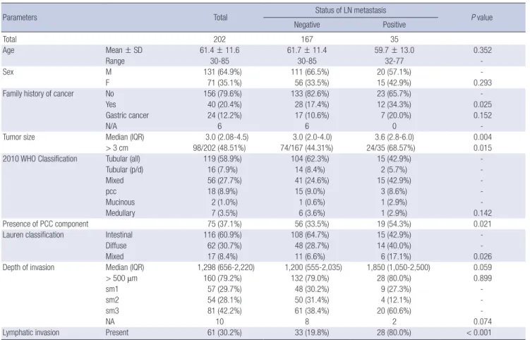
In predicting the exact percentage of the PPC components, preoperative biopsy showed an intraclass correlation coefficient of 0.586, which indicates a poor correlation. The relationship between the percentages in the biopsy and resection specimens is demonstrated in a scatter plot and a Bland-Altman plot (Fig. 1).
DISCUSSION
Mixed carcinoma is a newly described histologic type of gastric carcinoma in the 2010 WHO classification (8) (Fig. 2). In the 2000 WHO classification, the histologic type of gastric carcino- ma is determined based on the predominant histologic pattern (10). In the 2010 WHO classification, carcinomas showing a mix- ture of morphologically discrete glandular and signet ring/PCC histological components are defined as mixed carcinoma (8).
In this study, mixed carcinoma accounted for 27.2% (56/202) of all patients. This result suggests that the mixed carcinoma is not a rare type of gastric carcinoma, but a relatively common and Table 1. Relationship between clinicopathologic parameters and lymph node metastasis in 202 submucosal invasive gastric carcinomas: results of univariate analysis
Parameters Total Status of LN metastasis
P value
Negative Positive
Total 202 167 35
Age Mean ± SD 61.4 ± 11.6 61.7 ± 11.4 59.7 ± 13.0 0.352
Range 30-85 30-85 32-77 -
Sex M 131 (64.9%) 111 (66.5%) 20 (57.1%) -
F 71 (35.1%) 56 (33.5%) 15 (42.9%) 0.293
Family history of cancer No 156 (79.6%) 133 (82.6%) 23 (65.7%) -
Yes 40 (20.4%) 28 (17.4%) 12 (34.3%) 0.025
Gastric cancer 24 (12.2%) 17 (10.6%) 7 (20.0%) 0.152
N/A 6 6 0 -
Tumor size Median (IQR) 3.0 (2.08-4.5) 3.0 (2.0-4.0) 3.6 (2.8-6.0) 0.004
> 3 cm 98/202 (48.51%) 74/167 (44.31%) 24/35 (68.57%) 0.015
2010 WHO Classification Tubular (all) 119 (58.9%) 104 (62.3%) 15 (42.9%) -
Tubular (p/d) 16 (7.9%) 14 (8.4%) 2 (5.7%) -
Mixed 56 (27.7%) 41 (24.6%) 15 (42.9%) -
pcc 18 (8.9%) 15 (9.0%) 3 (8.6%) -
Mucinous 2 (1.0%) 1 (0.6%) 1 (2.9%) -
Medullary 7 (3.5%) 6 (3.6%) 1 (2.9%) 0.142
Presence of PCC component 75 (37.1%) 56 (33.5%) 19 (54.3%) 0.021
Lauren classification Intestinal 116 (60.9%) 108 (64.7%) 15 (42.9%) -
Diffuse 62 (30.7%) 48 (28.7%) 14 (40.0%) -
Mixed 17 (8.4%) 11 (6.6%) 6 (17.1%) 0.026
Depth of invasion Median (IQR) 1,298 (656-2,220) 1,200 (555-2,035) 1,850 (1,050-2,500) 0.059
> 500 μm 160 (79.2%) 132 (79.0%) 28 (80.0%) 0.899
sm1 57 (29.7%) 48 (30.2%) 9 (27.3%) -
sm2 54 (28.1%) 50 (31.4%) 4 (12.1%) -
sm3 81 (42.2%) 61 (38.4%) 20 (60.6%) -
NA 10 8 2 0.074
Lymphatic invasion Present 61 (30.2%) 33 (19.8%) 28 (80.0%) < 0.001
LN, lymph node; SD, standard deviation; N/A, not available; IQR, interquartile range; p/d, poorly differentiated; pcc, poorly cohesive carcinoma; PCC component, signet ring/
poorly cohesive cellular histological component.
Table 2. Multivariate analysis of independent clinicopathologic factors associated with lymph node metastasis and their significance
Variables Odds ratio 95% CI
P value Lower Upper
Lymphatic invasion 32.257 10.181 102.201 < 0.001 Presence of PCC component 3.615 1.290 10.128 0.015 Family history of cancer 2.866 1.080 7.604 0.034 CI, confidence interval; PCC component, signet ring/poorly cohesive cellular histologi- cal component.
clinically important type of gastric carcinoma.
In the present study, we found that tumor size, Lauren classi- fication, and lymphatic invasion have a relationship with LN
metastasis. These results are in close agreement with those of numerous authors (11-14). On the other hand, the depth of in- vasion has been reported as an independent risk factor for LN
PCC components in resection (%)
PCC components in biopsy (%)
0 20 40 60 80 100 120
120 100 80 60 40 20 0
Scatter plot
Average of means
Mean difference
0 20 40 60 80 100 120
80 60 40 20 0 -20 -40 -60 -80
Bland-Altman plot
Mean+2SD
Mean-2SD Mean
A B
Fig. 1. Relationship between the proportions of the poorly cohesive cellular histological component in biopsy and resection specimens.
Fig. 2. A representative case of mixed carcinoma. (A-C) Between gland forming moderately differentiated tubular adenocarcinoma components (arrows), signet ring/poorly co- hesive cellular histological components are present in lamina propria (arrow heads). (D) The signet ring/poorly cohesive cellular histological components show characteristic in- tracytoplasmic mucin vacuole, which pushes the nucleus to the cell periphery. Magnification: (A) × 40; (B-C) × 200; (D) × 600.
A
C
B
D
metastasis by many previous studies, in contrast to the present study, which failed to find a correlation. In a review of the previ- ous studies, we found that most studies were based on the JCGC.
The JCGC divides tumors according to the depth of submucosal invasion into two groups: the SM1 group includes tumors which invade less than 500 μm from the muscularis mucosa and the SM2 group includes tumors which invade 500 μm or deeper from the muscularis mucosa (9). According to the JCGC, 79.2%
(160/202) of patients were classified as the SM2 group in the present study. Perhaps the proportion in the SM2 group was too high to show a statistical difference.
Interestingly, family history of cancer was found to be a sig- nificant and independent risk factor associated with LN metas- tasis. This is the first study that statistically evaluated the rela- tionship between family history of cancer and the risk of LN metastasis in gastric carcinoma, but similar findings have been reported in gastrointestinal and other carcinoma entities. Ku- pelian et al. (15) reported that prostate cancer patients with a family history of prostate carcinoma show worse clinical out- comes. Hata et al. (16) reported that a family history of gastric cancer is a risk factor for colorectal neoplasia. Minami et al. (17) reported that a family history of gastric cancer increases mor- tality in gastric cancer patients under 60 years old. Finally, Wong et al. (18) reported that esophageal cancer patients with a fami- ly history of esophageal cancer show a higher tendency toward LN metastasis. Yu et al. (19) reported that family history of can- cer was not statistically correlated with sex, age, or the histolog- ic type of gastric carcinoma. However, they did not evaluate the relationship between family history of cancer and the risk of LN metastasis. Considering that LN metastasis is one of the most important prognostic factors, it would be not surprising if there is an association between family history of cancer and LN me- tastasis. Maybe there are still unknown genetic factors that can help the lymphatic invasion but this question was out of our reach in this study.
The presence of any PCC histological component appeared as an independent risk factor associated with LN metastasis in submucosal invasive gastric carcinoma. Furthermore, the pro- portion of the PCC histological component was not in correla- tion with the risk of LN metastasis. To our knowledge, there were no previous reports on this subject based on the histologic type of the 2010 WHO classification. There are several similar stud- ies, but they are based on the histologic type of the JCGC report- ed similar results. The JCGC defined mixed carcinoma as a mix- ture of differentiated (well or moderately differentiated tubular adenocarcinoma and papillary adenocarcinoma) and undiffer- entiated (poorly differentiated adenocarcinoma, signet-ring cell carcinoma, and mucinous adenocarcinoma) histological components (9). Accordingly, the mixed carcinoma of the JCGC classification is different from the mixed carcinoma of the 2010 WHO classification, and only overlaps partly.
Many previous studies based on the JCGC reported that the presence of an undifferentiated histological component is a risk factor for LN metastasis (20-23). However, the importance of the proportion of the undifferentiated histologic component is still debated. One previous report suggested that there is no sig- nificant difference in LN metastasis between differentiated-pre- dominant mixed carcinoma and undifferentiated-predominant mixed carcinoma (21). But another report suggested that undif- ferentiated-predominant mixed carcinoma has the highest risk of LN metastasis (22). In addition, some previous reports sug- gest that mixed carcinoma has an even higher risk of LN metas- tasis than purely undifferentiated carcinoma (22,24). In accor- dance with previous studies, the rate of LN metastasis of the mixed carcinoma was higher than that of the purely poorly co- hesive carcinoma or the poorly differentiated tubular carcino- ma in our study. However, the difference was insufficient to show statistical significance. Further study would be needed to ad- dress this question.
Maybe it is evident that the presence of an undifferentiated histological component is a risk factor for LN metastasis in JCGC.
But in practice, diagnoses are made solely based on WHO clas- sification in many countries including Korea. The question is how we can apply the result of previous studies to WHO classi- fication. In WHO classification, the diagnosis is made based on the predominant histologic pattern of the tumor. If there is mi- nor portion of poorly differentiated tubular adenocarcinoma in moderately differentiated tubular adenocarcinoma, we may be able to make the diagnosis as ‘tubular adenocarcinoma, mod- erately to poorly differentiated’. But if there is minor portion of signet-ring/PCC histological components in tubular adenocar- cinoma, we only are able to make the diagnosis as ‘mixed carci- noma’. Therefore, we have to find out the significance of the PCC histologic components and the meaning of mixed carcinoma in predicting the risk factors and deciding the therapeutic op- tions. We think our study have solved this problem.
Preoperative endoscopic forceps biopsy showed a sensitivity of only 63.6% in detecting the presence of the PCC histologic component, and the number of the biopsied pieces showed no significant correlation. Moreover, it showed a poor correlation in predicting the proportion of PCC histologic components.
It is not uncommon to find a heterogeneous histologic com- ponent in the resection specimen, which was not identified in the forceps biopsy specimen (25). Several previous studies have reported the incidence of histologic discrepancies between for- ceps biopsied specimens and resected specimens as 2.3%-11.9%
(26). Tumor location at the mid-stomach and easy friability were reported as risk factors for histologic discrepancy (27).
There are several explanations for our findings. First, mixed carcinomas tend to be glandular in the intramucosal compo- nent and to grow diffusely in the deeper portion (28). Second, the distribution of the PCC histological components is hetero-
geneous and the presence of the PCC histological component in the biopsy specimen largely depends on the biopsy sites. How- ever, in this study, the number of biopsied pieces showed no significant correlation. We supposed that maybe the problem is not the number of biopsied pieces, but the number of biopsied sites. Further prospective study would be needed to address this question. Third, a part of mixed carcinoma is derived from intestinal type carcinoma in the course of the tumorigenesis (29).
As we mentioned above, there are no previous studies on mixed carcinoma based on the 2010 WHO classification. It would be possible to speculate, however, from the studies based on the Japanese classification. Several previous studies have suggested that histologically heterogeneous carcinoma shows more ag- gressive features, including larger tumor size, deeper invasion, ulcer formation, lymphatic invasion, and LN metastasis (21,23, 25,30). There was also a suggestion that complete and curative resection rates are significantly lower in this type of carcinoma, mainly due to positive resection margins (20). The long-term outcome of the histologically heterogeneous carcinoma remains controversial. Shimizu et al. reported that this type of carcino- ma shows a poorer 5-year survival rate after curative gastrecto- my and LN dissection (21). In contrast, another report suggest- ed that there are no differences in the survival rate between pure- ly differentiated type carcinoma and histologically heterogene- ous carcinoma after curative resection (24,25).
In conclusion, we suggest that a family history of cancer and the presence of any PCC histological components are significant and independent risk factors for LN metastasis. We also suggest that endoscopic biopsy has limited value in predicting the pres- ence and proportion of the PCC histological component. This result reminds us that endoscopic resection is not only a thera- peutic option, but also a diagnostic option. We should consider the possibility of additional resection before endoscopic resection.
DISCLOSURE
The authors have no potential conflicts of interest to disclose.
AUTHOR CONTRIBUTION
Study design: Han HS. Data acquisition, analysis and interpre- tation: Park HK, Lee KY, Yoo MW. Drafting of the manuscript:
Park HK, Han HS. Review of manuscript for important intellec- tual content: Lee KY, Yoo MW, Hwang TS, Han HS. Approval of the final manuscript and submission: all authors.
ORCID
Hyung Kyu Park http://orcid.org/0000-0002-5972-3516 Kyung-Yung Lee http://orcid.org/0000-0002-5288-8772
Moon-Won Yoo http://orcid.org/0000-0003-0346-9042 Tae Sook Hwang http://orcid.org/0000-0002-3602-9257 Hye Seung Han http://orcid.org/0000-0002-3591-9995 REFERENCES
1. Yi HW, Kim SM, Kim SH, Shim JH, Choi MG, Lee JH, Noh JH, Sohn TS, Bae JM, Kim S. Complications leading reoperation after gastrectomy in patients with gastric cancer: frequency, type, and potential causes. J Gas- tric Cancer 2013; 13: 242-6.
2. Jeon YW, Han SI, Jeon CE, Kim JJ, Park SM. Quality of life in patients with stomach cancer after operation. J Korean Gastric Cancer Assoc 2004; 4:
27-31.
3. ASGE Technology Committee, Kantsevoy SV, Adler DG, Conway JD, Diehl DL, Farraye FA, Kwon R, Mamula P, Rodriguez S, Shah RJ, et al. Endoscop- ic mucosal resection and endoscopic submucosal dissection. Gastroin- test Endosc 2008; 68: 11-8.
4. Gotoda T. Endoscopic resection of early gastric cancer: the Japanese per- spective. Curr Opin Gastroenterol 2006; 22: 561-9.
5. Lee JH, Kim JG, Jung HK, Kim JH, Jeong WK, Jeon TJ, Kim JM, Kim YI, Ryu KW, Kong SH, et al. Clinical practice guidelines for gastric cancer in Ko- rea: an evidence-based approach. J Gastric Cancer 2014; 14: 87-104.
6. Hiki Y, Shimao H, Mieno H, Sakakibara Y, Kobayashi N, Saigenji K. Modi- fied treatment of early gastric cancer: evaluation of endoscopic treatment of early gastric cancers with respect to treatment indication groups. World J Surg 1995; 19: 517-22.
7. Japanese Gastric Cancer Association. Japanese gastric cancer treatment guidelines 2010 (ver. 3). Gastric Cancer 2011; 14: 113-23.
8. Lauwers GY, Carneiro F, Graham DY, Curado MP, Franceschi S, Mont- gomery E. Tumours of the stomach. In: Bosman FT, Carneiro F, Hruban RH, Theise ND, editors. WHO Classification of Tumours of the Digestive System. 4th ed. Lyon: IARC Press, 2010, p48-58.
9. Japanese Gastric Cancer Association. Japanese classification of gastric carcinoma: 3rd English edition. Gastric Cancer 2011; 14: 101-12.
10. Fenoglio-Preiser C, Carneiro F, Correa P, Guilford P, Lambert R, Megraud F, Munoz N, Powell SM, Rugge M, Sasako M, et al. Tumours of the stom- ach. In: Hamilton SR, Aaltonen LA, editors. Pathology and Genetics of Tumours of the Digestive System: WHO Classification of Tumours. 3rd ed. Lyon: IARC Press, 2000, p37-68.
11. Gotoda T, Sasako M, Ono H, Katai H, Sano T, Shimoda T. Evaluation of the necessity for gastrectomy with lymph node dissection for patients with submucosal invasive gastric cancer. Br J Surg 2001; 88: 444-9.
12. Hirasawa T, Gotoda T, Miyata S, Kato Y, Shimoda T, Taniguchi H, Fujisaki J, Sano T, Yamaguchi T. Incidence of lymph node metastasis and the fea- sibility of endoscopic resection for undifferentiated-type early gastric can- cer. Gastric Cancer 2009; 12: 148-52.
13. Kunisaki C, Takahashi M, Nagahori Y, Fukushima T, Makino H, Takagawa R, Kosaka T, Ono HA, Akiyama H, Moriwaki Y, et al. Risk factors for lymph node metastasis in histologically poorly differentiated type early gastric cancer. Endoscopy 2009; 41: 498-503.
14. Kurihara N, Kubota T, Otani Y, Ohgami M, Kumai K, Sugiura H, Kitajima M. Lymph node metastasis of early gastric cancer with submucosal inva- sion. Br J Surg 1998; 85: 835-9.
15. Kupelian PA, Kupelian VA, Witte JS, Macklis R, Klein EA. Family history of
prostate cancer in patients with localized prostate cancer: an indepen- dent predictor of treatment outcome. J Clin Oncol 1997; 15: 1478-80.
16. Hata K, Shinozaki M, Toyoshima O, Toyoshima A, Matsumoto S, Saisho T, Tsurita G. Impact of family history of gastric cancer on colorectal neopla- sias in young Japanese. Colorectal Dis 2013; 15: 42-6.
17. Minami Y, Kawai M, Fujiya T, Suzuki M, Noguchi T, Yamanami H, Kakuga- wa Y, Nishino Y. Family history, body mass index and survival in Japanese patients with stomach cancer: a prospective study. Int J Cancer 2015; 136:
411-24.
18. Wong VC, Ko JM, Qi RZ, Li PJ, Wang LD, Li JL, Chan YP, Chan KW, Stan- bridge EJ, Lung ML. Abrogated expression of DEC1 during oesophageal squamous cell carcinoma progression is age- and family history-related and significantly associated with lymph node metastasis. Br J Cancer 2011;
104: 841-9.
19. Yu J, Fu B, Zhao Q. Family history of malignant neoplasm and its relation with clinicopathologic features of gastric cancer patients. World J Surg Oncol 2013; 11: 201.
20. Shim CN, Chung H, Park JC, Lee H, Shin SK, Lee SK, Lee YC. Early gastric cancer with mixed histology predominantly of differentiated type is a dis- tinct subtype with different therapeutic outcomes of endoscopic resec- tion. Surg Endosc 2015; 29: 1787-94.
21. Shimizu H, Ichikawa D, Komatsu S, Okamoto K, Shiozaki A, Fujiwara H, Murayama Y, Kuriu Y, Ikoma H, Nakanishi M, et al. The decision criterion of histological mixed type in “T1/T2” gastric carcinoma--comparison be- tween TNM classification and Japanese Classification of Gastric Cancer.
J Surg Oncol 2012; 105: 800-4.
22. Hanaoka N, Tanabe S, Mikami T, Okayasu I, Saigenji K. Mixed-histologic- type submucosal invasive gastric cancer as a risk factor for lymph node metastasis: feasibility of endoscopic submucosal dissection. Endoscopy 2009; 41: 427-32.
23. Lee JH, Choi IJ, Han HS, Kim YW, Ryu KW, Yoon HM, Eom BW, Kim CG, Lee JY, Cho SJ, et al. Risk of lymph node metastasis in differentiated type mucosal early gastric cancer mixed with minor undifferentiated type his- tology. Ann Surg Oncol 2015; 22: 1813-9.
24. Kozuki T, Yao T, Nakamura S, Matsumoto T, Tsuneyoshi M. Differences in p53 and cadherin-catenin complex expression between histological sub- types in diffusely infiltrating gastric carcinoma. Histopathology 2002; 41:
56-64.
25. Min BH, Kim KM, Park CK, Lee JH, Rhee PL, Rhee JC, Kim JJ. Outcomes of endoscopic submucosal dissection for differentiated-type early gastric cancer with histological heterogeneity. Gastric Cancer 2015; 18: 618-26.
26. Joo M, Kim KM. Histologic discrepancy between gastric biopsy and re- section specimen in the era of endoscopic treatment for early gastric can- cer. Korean J Gastroenterol 2014; 64: 256-9.
27. Shim CN, Kim H, Kim DW, Chung HS, Park JC, Lee H, Shin SK, Lee SK, Lee YC. Clinicopathologic factors and outcomes of histologic discrepan- cy between differentiated and undifferentiated types after endoscopic resection of early gastric cancer. Surg Endosc 2014; 28: 2097-105.
28. Carneiro F, Machado JC, Nabais S, Santos CM, Sobrinho Simões M. Mixed carcinoma of the stomach: a clinicopathological entity. Histopathology 2003; 43: 94-5.
29. Honda T, Tamura G, Endoh Y, Nishizuka S, Kawata S, Motoyama T. Ex- pression of tumor suppressor and tumor-related proteins in differentiat- ed carcinoma, undifferentiated carcinoma with tubular component and pure undifferentiated carcinoma of the stomach. Jpn J Clin Oncol 2005;
35: 580-6.
30. Zheng HC, Li XH, Hara T, Masuda S, Yang XH, Guan YF, Takano Y. Mixed- type gastric carcinomas exhibit more aggressive features and indicate the histogenesis of carcinomas. Virchows Arch 2008; 452: 525-34.