Copyrights © 2016 The Korean Society of Radiology
508
Case Report
pISSN 1738-2637 / eISSN 2288-2928 J Korean Soc Radiol 2016;75(6):508-511 https://doi.org/10.3348/jksr.2016.75.6.508
INTRODUCTION
Traumatic adrenal hemorrhage is rare because of the small size and retroperitoneal location of the adrenal glands, and because the adrenal gland is surrounded by the paraspinal muscles, the rib cage, and liver (1). Recently, because of the widespread and frequent use of computed tomography (CT), a high incidence of adrenal injury, up to 25%, has been detected in cases of severe abdominal trauma (2). In pediatric patients, isolated post-trau- matic adrenal injuries are infrequent and they are usually accom- panied by multi-organ injuries, which have been perceived as in- cidental findings (3).
The management of adrenal injuries generally depends on the concomitant injuries, while isolated adrenal injury is usually managed conservatively or may require transcatheter emboliza- tion (4). Particularly in pediatric patients, conservative manage- ment is generally performed, even in cases of adrenal bleeding requiring transfusion (5).
We report a case of pediatric adrenal hemorrhage treated with transcatheter embolization.
CASE REPORT
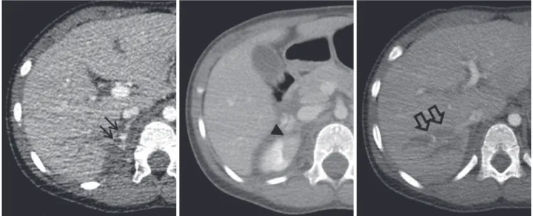
A 5-year-old boy fell from a height of about 5 m. Upon admis- sion to the emergency room of our hospital, he did not complain of any symptoms, except for headache and some bruises on his face and back. Plain radiography of the skull and CT of the brain revealed right occipital and parietal bone fractures without intra- cranial hemorrhage. Chest CT showed multifocal lung contu- sions. Abdominal CT demonstrated a small right suprarenal and retroperitoneal hematoma with contrast extravasation into the right adrenal gland as well as multifocal lacerations in the right lobe of the liver and segmental renal infarction in the anterosu- perior aspect of the right kidney (Fig. 1). Laboratory tests re- vealed a slightly decreased hemoglobin level of 11.2 g/dL (mean, 12.5 g/dL) and slightly elevated liver enzymes. He was alert and
Endovascular Treatment of a Post-Traumatic Adrenal Hemorrhage in a Pediatric Patient: A Case Report
소아 환자에서 외상성 부신 출혈에 대한 중재적 시술: 증례 보고
Dong Gun Kim, MD, Hyun Seok Jung, MD*
Department of Radiology, Busan Paik Hospital, Inje University College of Medicine, Busan, Korea
Adrenal hemorrhage following blunt trauma is a rare occurrence. We report here the case of a 5-year-old child with adrenal hemorrhage, which developed as a result of an accidental fall. Embolization treatment of adrenal hemorrhage was success- fully performed. To the best of our knowledge, this is the first report of adrenal hemorrhage occurring in a child which was treated with transcatheter embolization.
Index terms Adrenal Glands Hemorrhage
Embolization, Therapeutic Child
Trauma
Received April 30, 2016 Revised June 9, 2016 Accepted June 22, 2016
*Corresponding author: Hyun Seok Jung, MD Department of Radiology, Busan Paik Hospital, Inje University College of Medicine, 75 Bokji-ro, Busanjin-gu, Busan 47392, Korea.
Tel. 82-51-890-6549 Fax. 82-51-896-1085 E-mail: [email protected]
This is an Open Access article distributed under the terms of the Creative Commons Attribution Non-Commercial License (http://creativecommons.org/licenses/by-nc/3.0) which permits unrestricted non-commercial use, distri- bution, and reproduction in any medium, provided the original work is properly cited.
509
Dong Gun Kim, et al
jksronline.org J Korean Soc Radiol 2016;75(6):508-511 his vital signs were stable, except for a markedly increased pulse rate of 124/min. The following several reasons led us to perform transcatheter embolization therapy: 1) a definite active bleeding focus in the right adrenal gland; 2) possibility of additional active bleeding; 3) the patient was assigned the major trauma category according to the injury severity score (ISS) of more than 15 (med- ical score to assess trauma severity) (6); 4) unpredictable status, including vital signs of the pediatric patient.
Access was achieved via the right common femoral artery puncture with a 4-Fr vascular sheath (Terumo, Tokyo, Japan) and a 21-G puncture needle under ultrasonographic guidance. Non- selective aortography was performed with a 4-Fr pig-tail catheter (Cook Inc., Bloomington, IN, USA), which revealed no demon- strable abnormality. A 4-Fr Cobra catheter (Terumo, Tokyo, Ja- pan) was used for selective right renal arteriography to demon- strate the arterial supply of the adrenal gland. There was no arte- rial supply to the adrenal gland from the right renal artery. Addi- tional selective right inferior phrenic artery (RIPA) arteriography was performed in the same way, and it revealed that the superior, middle, and inferior adrenal arteries originated from the RIPA with extravasation of contrast medium (Fig. 2). We primarily embolized the superior and middle adrenal arteries with a mix- ture of N-butyl cyanoacrylate (Histoacryl; B.Braun, Tuttlingen, Germany) and lipiodol (Guerbet, Paris, France) at a dilution of 1:4. On follow-up RIPA arteriography, contrast extravasation into the adrenal gland was still detected via the middle and inferior
adrenal arteries. Additional embolization was performed via the proximal branch of the superior and middle adrenal arteries.
There was no evidence of definite extravasation of contrast medi- um, but subtle parenchymal staining in the right adrenal gland was detected on additional follow-up RIPA arteriography. To prevent delayed hemorrhage and to obtain a temporary embolic effect, we performed embolization via the common trunk of the RIPA with a gelatin sponge (Alicon, Zhejiang, China). During the procedure, a pseudoaneurysm in the adrenal parenchyma was newly detected via the inferior adrenal artery. We performed additional embolization of the inferior adrenal artery with a mix- ture of N-butyl cyanoacrylate and lipiodol at a dilution of 1:4.
Embolization treatment of adrenal hemorrhage was successfully performed and active bleeding was no longer detected (Fig. 3).
After 1 week, follow-up abdominal CT demonstrated no active bleeding in the right adrenal gland. Furthermore, the patient re- mained clinically well on the out-patient follow-up examination one month later.
DISCUSSION
Traumatic adrenal injury rarely occurs in the pediatric or the general population (5). Several studies have reported that the in- cidence of traumatic adrenal injury in the pediatric population was 0.03–4.95% (7), 3–5% (5), and 7.5% (8), which increased with the use of multi-detector CT (2).
Fig. 1. Contrast-enhanced computed tomography demonstrating a retroperitoneal hematoma with contrast extravasation into the right adrenal gland (solid arrows), segmental renal infarction in the right kidney (arrowhead), and multifocal liver lacerations (open arrows).
510
Endovascular Treatment of a Post-Traumatic Adrenal Hemorrhage in a Pediatric Patient
jksronline.org
J Korean Soc Radiol 2016;75(6):508-511 The following three possible mechanisms for the occurrence of
adrenal hemorrhage have been proposed: 1) Compression of the adrenal gland against the spinal column; 2) An acute increase in the intra-adrenal venous pressure due to compression of the infe-
rior vena cava. This may explain why the right adrenal gland is more commonly affected; and 3) Shearing of small vessels that perforate the adrenal capsule, resulting from secondary decelera- tion forces (9).
The management of adrenal hemorrhage may be either surgi- cal or conservative according to the extent of the injury and the patient’s hemodynamic stability (10). The modern management of blunt adrenal trauma is usually non-operative (2). Emboliza- tion treatment is occasionally reported in patients with an isolat- ed adrenal gland injury (5). Several previous studies have report- ed that several pediatric patients with traumatic adrenal hemorrhage were successfully cured with conservative treatment (3, 7). In our case, we decided to perform transcatheter emboliza- tion therapy with consideration for a definite active bleeding fo- cus, possibility of additional active bleeding, trauma severity, and unpredictable status including vital signs of pediatric patient. In our literature search, we could not identify any pediatric patient who was treated with transcatheter embolization for post-trau- matic adrenal hemorrhage.
Embolization can be performed with various agents, including coil, glue, and gelatin sponge. Embolic agents must be applied on a case by case basis, considering the clinical purpose, preference of the radiologists, and accessibility of the agent (9). In our case, we preferred N-butyl cyanoacrylate glue for primary superior and middle adrenal arterial embolization because the vessels are of small-caliber and relatively tortuous. After we embolized the Fig. 2. RIPA arteriography demonstrating the superior (solid arrow),
middle (arrowhead), and inferior (open arrow) adrenal arteries origi- nating from the RIPA and extravasation of contrast medium.
RIPA = right inferior phrenic artery
Fig. 3. Follow-up RIPA arteriography (A) revealing adrenal parenchymal staining via the inferior adrenal artery (arrows), but extravasation of contrast medium was unclear. During preventive embolization via the common trunk of the RIPA (B), a pseudoaneurysm (arrowheads) was de- tected. Additional embolization was performed. Final angiography (C) revealing complete embolization of the pseudoaneurysm in the adrenal parenchyma. There is no further evidence of active bleeding.
RIPA = right inferior phrenic artery
A B C
511
Dong Gun Kim, et al
jksronline.org J Korean Soc Radiol 2016;75(6):508-511 proximal branch of the superior and middle adrenal arteries, we used a gelatin sponge, a temporary agent for additional preven- tive embolization. It is inevitable that we would have to embolize via the common trunk of the RIPA because a pseudoaneurysm was unfortunately discovered in the adrenal parenchyma. How- ever, the contralateral adrenal gland was so intact that the proce- dure was performed without any special complications. The pa- tient was healthy after 1 month despite right adrenal infarction.
In conclusion, transcatheter embolization may be a treatment option for post-traumatic adrenal hemorrhage in pediatric pa- tients.
REFERENCES
1. Dinc H, Sims¸ek A, Ozyavuz R, Ozgür GK, Gümele HR. Endo- vascular treatment of massive retroperitoneal haemor- rhage due to inferior adrenal artery injury. A case report.
Acta Radiol 2002;43:326-328
2. Agrawal N, Rao S, Zellweger R, Knight T. Adrenal pseudoa- neurysm due to blunt trauma. Indian J Surg 2013;75(Sup- pl 1):155-157
3. Gabal-Shehab L, Alagiri M. Traumatic adrenal injuries. J
Urol 2005;173:1330-1331
4. To’o KJ, Duddalwar VA. Imaging of traumatic adrenal inju- ry. Emerg Radiol 2012;19:499-503
5. Roupakias S, Papoutsakis M, Mitsakou P. Blunt adrenal gland trauma in the pediatric population. Asian J Surg 2011;
34:103-110
6. Copes WS, Champion HR, Sacco WJ, Lawnick MM, Keast SL, Bain LW. The Injury Severity Score revisited. J Trauma 1988;28:69-77
7. Sivit CJ, Ingram JD, Taylor GA, Bulas DI, Kushner DC, Eichel- berger MR. Posttraumatic adrenal hemorrhage in children:
CT findings in 34 patients. AJR Am J Roentgenol 1992;158:
1299-1302
8. Choi SJ, Kim JE, Ryu Il, Kim JJ, Choi HY. MDCT findings of traumatic adrenal injury in children. J Korean Soc Radiol 2011;64:191-195
9. Fowler AM, Burda JF, Kim SK. Adrenal artery embolization:
anatomy, indications, and technical considerations. AJR Am J Roentgenol 2013;201:190-201
10. Rammelt S, Mucha D, Amlang M, Zwipp H. Bilateral adre- nal hemorrhage in blunt abdominal trauma. J Trauma 2000;
48:332-335
소아 환자에서 외상성 부신 출혈에 대한 중재적 시술: 증례 보고
김동건 · 정현석*
외상 후 부신 출혈은 드물게 발생한다. 본 저자들은 외상 후에 부신 출혈이 있었던 5살 남자아이에 대해 동맥색전술을 이 용해 치료한 사례를 보고하며 이 보고는 소아에서 동맥색전술로 치료한 외상성 부신 출혈의 첫 증례이다.
인제대학교 의과대학 부산백병원 영상의학과