J Korean Soc Radiol 2016;74(6):351-360 http://dx.doi.org/10.3348/jksr.2016.74.6.351
INTRODUCTION
The use of breast magnetic resonance imaging (MRI) is in- creasing for screening and diagnosis of breast cancer. MRI has greater sensitivity than other imaging modalities for the screen- ing of women at higher risk of breast cancer, with more than half of lesions only detected on MRI (1, 2). MRI can identify prima- ry cancer in suspected occult breast cancer patients (3). In ad- dition, breast MRI most accurately depicts the extent of breast cancer (4-6). Although the clinical relevance of additional lesions is still controversial, preoperative MRI identifies more synchro- nous, ipsilateral or contralateral breast cancers than mammog- raphy and ultrasound (7-9).
However, breast MRI has limited specificity (10). If a suspi- cious lesion is detected, biopsy is mandatory to avoid unneces- sary surgery. In addition, biopsy under MRI guidance is required for suspected occult lesions on mammography or ultrasound.
The American College of Radiology requires performance of
MRI-guided intervention or contact with available referral site when performing breast MRI (11). The MRI guideline of the Eu- ropean Society of Breast Imaging also emphasizes the necessity of offering MRI-guided intervention at a site performing breast MRI (12). The number of medical centers providing breast MRI is growing in Korea, and more MRI-guided breast biopsies are required. However, few reports describe MRI-guided breast bi- opsy in Korea (13, 14). In this article, we describe preprocedur- al considerations and technique, and correlation of radiologic and pathologic findings in MRI-guided breast biopsy.
PREPROCEDURAL CONSIDERATIONS FOR MRI-GUIDED BREAST BIOPSY
Second-Look Studies
Second-look studies should be considered for all women with suspicious lesions on MRI. If the lesion is delineated with mam- mography or ultrasound, biopsy under imaging guidance using
Breast Magnetic Resonance Imaging-Guided Biopsy
유방 자기공명유도 조직검사Bo La Yun, MD
1, Sun Mi Kim, MD
1*, Mijung Jang, MD
1, Nariya Cho, MD
2, Woo Kyung Moon, MD
2, Hak Hee Kim, MD
31Department of Radiology, Seoul National University Bundang Hospital, Seongnam, Korea
2Department of Radiology, Seoul National University Hospital, Seoul, Korea
3Department of Radiology, Asan Medical Center, University of Ulsan College of Medicine, Seoul, Korea
Despite the high sensitivity of breast magnetic resonance imaging (MRI), pathologic confirmation by biopsy is essential because of limited specificity. MRI-guided biopsy is required in patients with lesions only seen on MRI. We review preprocedural consider- ations and the technique of MRI-guided biopsy, challenging situations and trouble- shooting, and correlation of radiologic and pathologic findings.
Index terms Breast Cancer Biopsy, Needle
MR Guided Interventional Procedures
Received January 4, 2016 Revised March 3, 2016 Accepted March 23, 2016
*Corresponding author: Sun Mi Kim, MD Department of Radiology, Seoul National University Bundang Hospital, 82 Gumi-ro 173beon-gil, Bundang-gu, Seongnam 13620, Korea.
Tel. 82-31-787-7617 Fax. 82-31-787-4011 E-mail: [email protected]
This is an Open Access article distributed under the terms of the Creative Commons Attribution Non-Commercial License (http://creativecommons.org/licenses/by-nc/3.0) which permits unrestricted non-commercial use, distri- bution, and reproduction in any medium, provided the original work is properly cited.
these modalities is preferred. MRI-guided biopsy is more expen- sive and difficult for the patient. However, the reported correla- tion rate of second-look ultrasound ranges from 23–57%; MRI- only lesions require an MRI-guided biopsy (15-18).
Patient Preparation
Informed consent should be obtained before the biopsy. Pa- tients with a known contraindication for MRI or gadolinium administration should not have an MRI-guided biopsy. Patients with allergy to gadolinium or local anesthetics are also not suit- able. Bleeding risk due to use of aspirin or anticoagulants or an underlying coagulation disorder is a relative contraindication for biopsy, requiring careful consideration of the risks and ben- efits. The patient should be able to remain prone during the bi- opsy for a minimum of 60 minutes. The possibility of a nonvisu- alized target lesion should be discussed with the patient. Even with successful visualization, the cancellation of a biopsy is some- times necessary because of unforeseen safety issues. Complica-
tions such as hematoma, infection, and skin injury should also be discussed with the patient. For patients with breast implants, rupture is a possible complication of the biopsy.
MRI-GUIDED BREAST BIOPSY PROCEDURE
Table 1 summarizes the biopsy procedure.
Positioning
The patient lies in a prone position, and a dedicated interven- tional breast coil is used. Either a grid or a pillar and post system is generally used. We focus on the grid system, which is most widely used (Fig. 1). The breast within the coil is compressed by the grid. Compression pressure is adjusted for stable immobiliza- tion while preserving blood flow. The approach to a posterior mass is often difficult, requiring that sufficient breast tissue be contained in the grid to maintain distance from the chest wall.
Table 1. Summary of Breast Biopsy MRI Procedure
Procedure Check Point
Patient positioning Attach fiducial marker
Precontrast T1-weighted fat-saturation images (axial and sagittal) Check location of target and grid
Postcontrast T1-weighted fat-saturation images (axial and sagittal) Calculate distance from grid and fiducial marker using worksheet Skin preparation, local anesthesia, and insertion of introducer with obturator
Postcontrast T1-weighted fat-saturation images (axial and sagittal) Repeat until adequate location achieved Change obturator to VAB device and perform biopsy
Post-biopsy images Repeat biopsy if inadequate sample retrieval
Marker clip insertion Post-marker-insertion image with MRI or mammography
VAB = vacuum assisted biopsy
Fig. 1. MRI-guided biopsy grid system.
Introducer stylet
Obturator
Introducer sheath
Needle guide
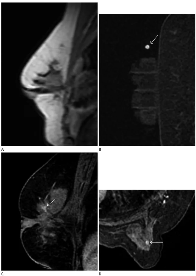
Fig. 2. MRI-guided biopsy kit contains introducer stylet, obturator, in- troducer sheath, and needle guide.
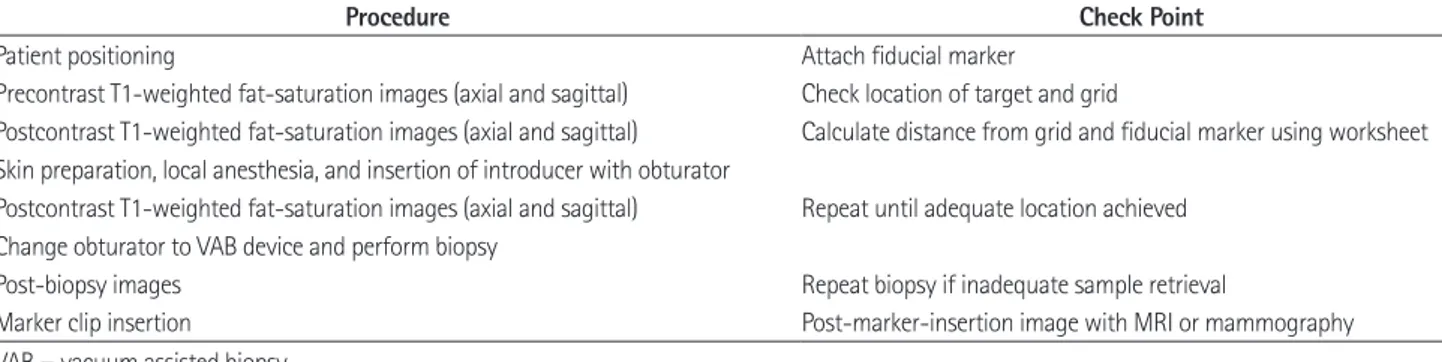
Fig. 3. MRI-guided VAB procedure.
After localizing image (A), precontrast images with fiducial marker (arrow) (B) are obtained. Sagittal and axial postcontrast images (C, D) are ob- tained to identify target location (arrow).
VAB = vacuum assisted biopsy
A B
C D
Fig. 3. MRI-guided VAB procedure.
After location of introducer sheath and obturator, sagittal and axial images (E, F) are obtained to confirm the position before lesion sampling. The target lesion (arrowhead) and obturator (arrow) are well demonstrated in these images. After tissue sampling, additional sagittal and axial images (G, H) are obtained to confirm adequate biopsy location and marker (arrow) placement. The target lesion was confirmed as ductal carcinoma in situ.
VAB = vacuum assisted biopsy
E F
H G
Fixing a fiducial marker to the grid enhances target localization.
Equipment
MRI-guided vacuum assisted biopsy (VAB) has advantages over standard core biopsy. The larger core size results in decrea- sed sampling error and compensates for tissue shifting during needle placement (19). The European consensus group recom- mend a minimum 11-gauge probe size (20). The MRI-guided biopsy kit consists of an introducer stylet, obturator, introducer sheath, and needle guide. The needle guide, a cube-shaped plas- tic block with multiple holes for the biopsy device (Fig. 2), main- tains the VAB device perpendicular to the grid.
Image Acquisition and Lesion Localization
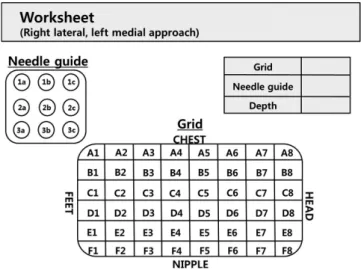
Fig. 3 shows the MRI-guided biopsy sequence. A precontrast T1-weighted fat-saturation sequence is obtained to determine whether the target lesion is within the grid. If the target lesion is too small for detection on the precontrast image, anatomic land- marks are helpful for identification. If the precontrast image in- dicates that the lesion is inaccessible, the patient’s position should be adjusted. After planning the proper approach route, post- contrast T1-weighted fat-saturation images are obtained. The thickness and in-plane resolution, which are similar to that of diagnostic imaging for accurate needle positioning, make local- ization easier. Sagittal and axial plane images are obtained. Al- ternatively, axial or sagittal plane images with perpendicular re- construction can be used for lesion localization depending on the physician’s preference. The entry site on the grid and depth from the skin to the lesion are measured using the fiducial mark-
er as a reference. A worksheet provided by the manufacturer is helpful in manual localization (Fig. 4). The thickness of the nee- dle guide should be included in the calculation. A computer-as- sisted diagnostic system that improves accuracy and speed of the procedure is also commercially available. About 8–13% of MRI- guided biopsy target lesions are not visible at the time of biopsy (21-23). If the target is not visible on the first postcontrast im- age, it is sometimes identified on a delayed image. Overpressure by the grid may interfere with breast perfusion and should be checked. A subtraction image also aids in lesion identification (Fig. 5). If the target lesion is still not visible, short-term follow- up is recommended at about 6 months.
Biopsy Procedure
After standard skin preparation, local anesthesia is adminis- tered. A small nick in the skin facilitates smooth entry by the VAB device. The introducer stylet within its sheath is inserted through the needle guide to the measured depth. A twisting mo- tion is helpful to avoid skin tenting and tissue displacement. The stylet is removed and replaced by the obturator. T1-weighted fat-saturation images are then obtained to confirm the depth and position of the introducer (24, 25). In the case of insufficient depth, the introducer sheath can be advanced after reinsertion of the stylet; if advanced past the target lesion, the introducer is gradually withdrawn. In general, the optimal position is in the center of the target. Directional sampling can be performed with a peripheral location of the needle. When the introducer position is verified as correct, the obturator is exchanged with a VAB de- vice. The European consensus group recommends no less than 24 samples for an 11-gauge or equivalent volume if a larger probe is used (20). Liberman (26) reported that an 11-gauge VAB de- vice collects 100 mg, and a 9-gauge VAB device collects 200 mg.
T1-weighted fat-saturation images are obtained immediately after the biopsy to evaluate adequacy. Image assessment can be difficult due to contrast washout, background enhancement, hemorrhage, and air, but careful review using anatomic land- marks improves the evaluation accuracy. An additional biopsy can be performed if the target sample is insufficient. Once the target sampling is acceptable, marker clip should be placed through the introducer sheath. A post-clip-insertion image may be obtained with MRI or mammography. The importance of marker clip insertion should never be underestimated. The mark- Fig. 4. Example of worksheet.
er clip facilitates mammographic or ultrasound-guided localiza- tion in place of MRI-guided localization for subsequent exci- sional biopsy. Moreover, if the entire target lesion is removed by VAB, the marker clip is the only way to identify the biopsy site.
The breast is compressed for at least 15 minutes after the biopsy.
Complications and Management
Major complications requiring surgical intervention seldom occur with MRI-guided biopsy. Complication rates are less than 5%. Bleeding and hematoma formation are most common, and
can be controlled by compression. Other rare complications are skin laceration, vasovagal syncope, and infection. Termination of the procedure due to a complication is rare (19, 27-36).
CHALLENGING SITUATIONS AND TROUBLESHOOTING
Targeting deep-seated lesions is prone to chest wall injury. To avoid this, traction on the breast tissue as much as possible, coil padding removal, and biopsy toward the anterior side are some Fig. 5. A 35-year-old woman with ipsilateral breast cancer surgery 9 months previously.
Postoperative MRI shows a new, round, fast and washout-enhancing mass (A). The patient underwent MRI-guided biopsy. The target lesion is not identified on the postcontrast sagittal image (B). On the subtraction image (C), a subtle enhancing mass is well-delineated (arrow). Repeat MRI (D, E) shows introducer sheath with obturator (arrowhead) in correct position, with the tip at the target lesion (arrow). The enhancing mass was pathologically confirmed as reactive hyperplasia in intramammary lymph node.
D E
A B C
solutions (33). A posteromedial target location is the most diffi- cult to access. If the patient is small, a lateral approach from the contralateral opening of the breast coil can make a deep poste- rior location more accessible (Fig. 6). A thin breast is another challenge for MRI-guided biopsy. A generous amount of anes- thetic agent helps to increase breast thickness, and a reverse com- pression paddle is also useful (24, 37). VAB devices with smaller apertures or blunt tips can be used for targets in thin breasts and
also those near the skin (Table 2).
RADIOLOGIC-PATHOLOGIC CORRELATION
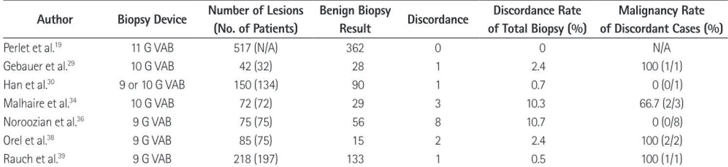
As with all other image-guided biopsy techniques, MRI-guid- ed biopsy results should be evaluated for radiologic-pathologic concordance. MRI-guided biopsy has no corresponding evalua- tion method such as specimen imaging in stereotactic biopsy or real-time monitoring in ultrasound-guided biopsy; therefore, ra- diologic-pathologic concordance requires caution. The positive predictive value of a lesion detected by MRI with subsequent MRI-guided biopsy is 16–61%; a radiologist should be aware that for radiologic-pathologic correlation, the positive predictive value is affected by the prevalence of breast cancer in a patient popula- tion (19, 30, 33, 36, 38-40). A six-month follow-up is recom- mended for a benign concordant biopsy result (41). The rate of radiology-pathology discordance is not high (0% to 10.7%); but the mean proportion of malignancies in discordant cases is 37.5%, and surgical excision is recommended for discordant le- sions (Table 3) (19, 29, 30, 34, 36, 38, 39).
The atypical ductal hyperplasia (ADH) upgrade rate at sur- gery is reportedly 25–38% (19, 30, 38, 40, 42, 43). In ductal car- cinoma in situ (DCIS), the upgrade rate ranges from 5–24% (19, 30, 38, 44). The underestimation rates for ADH and DCIS on MRI-guided biopsy are slightly higher than those for stereotac- tic biopsy (21% and 11%, respectively) (45, 46) or ultrasound- guided biopsy (23.3% and 13.8%, respectively) (47). Atypical lobular hyperplasia and lobular carcinoma in situ also have a high upgrade rate (27%) (40). Despite a limited number of stud- ies in the underestimation rate for other high-risk lesions such as radial scars and papillomas, surgical excision for all such le- sions using MRI-guided biopsy is recommended (30, 38, 40).
Grid Breast biopsy coil
Fig. 6. Lateral approach for posteromedial located target.
Table 2. Challenging Situations and Troubleshooting
Problem Solution
Nonvisible target Decrease compression pressure Obtain delayed image Subtle enhancement target Subtraction image
Deep-seated target Traction on breast as much as possible Remove biopsy coil padding
Posteromedial located target Lateral approach from contralateral side of breast
Thin breast Generous amount of anesthetic agent Reverse compression paddle Use small aperture or blunt tip device
Table 3. Radiology-Pathology Discordance and Malignancy Rate in Discordant Cases Author Biopsy Device Number of Lesions
(No. of Patients)
Benign Biopsy
Result Discordance Discordance Rate of Total Biopsy (%)
Malignancy Rate of Discordant Cases (%)
Perlet et al.19 11 G VAB 517 (N/A) 362 0 0 N/A
Gebauer et al.29 10 G VAB 42 (32) 28 1 2.4 100 (1/1)
Han et al.30 9 or 10 G VAB 150 (134) 90 1 0.7 0 (0/1)
Malhaire et al.34 10 G VAB 72 (72) 29 3 10.3 66.7 (2/3)
Noroozian et al.36 9 G VAB 75 (75) 56 8 10.7 0 (0/8)
Orel et al.38 9 G VAB 85 (75) 15 2 2.4 100 (2/2)
Rauch et al.39 9 G VAB 218 (197) 133 1 0.5 100 (1/1)
N/A = not available, VAB = vacuum assisted biopsy
CONCLUSION
Breast MRI is an important screening and diagnostic tool, but the limited specificity requires biopsy confirmation. MRI- only lesions that are occult on mammography and ultrasound require routine evaluation by MRI-guided biopsy. Radiologists who perform the procedure understand best the indications, preprocedural considerations, imaging protocols, biopsy tech- niques, and possible complications of MRI-guided VAB. Pa- tients should be informed about the demanding nature of the procedure due to prolonged immobilization, the possibility of cancellation of the procedure, and the need for imaging follow- up despite a benign biopsy result. Appropriate patient manage- ment based on radiologic-pathologic correlation is emphasized.
REFERENCES
1. Kuhl C, Weigel S, Schrading S, Arand B, Bieling H, König R, et al. Prospective multicenter cohort study to refine man- agement recommendations for women at elevated familial risk of breast cancer: the EVA trial. J Clin Oncol 2010;28:
1450-1457
2. Berg WA, Zhang Z, Lehrer D, Jong RA, Pisano ED, Barr RG, et al. Detection of breast cancer with addition of annual screening ultrasound or a single screening MRI to mam- mography in women with elevated breast cancer risk. JAMA 2012;307:1394-1404
3. Morris EA, Schwartz LH, Dershaw DD, van Zee KJ, Abramson AF, Liberman L. MR imaging of the breast in patients with occult primary breast carcinoma. Radiology 1997;205:437- 440
4. Boetes C, Mus RD, Holland R, Barentsz JO, Strijk SP, Wobbes T, et al. Breast tumors: comparative accuracy of MR imag- ing relative to mammography and US for demonstrating extent. Radiology 1995;197:743-747
5. Gavenonis SC, Roth SO. Role of magnetic resonance imag- ing in evaluating the extent of disease. Magn Reson Imag- ing Clin N Am 2010;18:199-206, vii-viii
6. Mann RM. The effectiveness of MR imaging in the assess- ment of invasive lobular carcinoma of the breast. Magn Re- son Imaging Clin N Am 2010;18:259-276, ix
7. Pediconi F, Catalano C, Roselli A, Padula S, Altomari F, Mori-
coni E, et al. Contrast-enhanced MR mammography for evaluation of the contralateral breast in patients with diag- nosed unilateral breast cancer or high-risk lesions. Radiolo- gy 2007;243:670-680
8. Wang SY, Long JB, Killelea BK, Evans SB, Roberts KB, Silber A, et al. Preoperative breast magnetic resonance imaging and contralateral breast cancer occurrence among older women with breast cancer. J Clin Oncol 2016;34:321-328 9. Iacconi C, Galman L, Zheng J, Sacchini V, Sutton EJ, Der-
shaw D, et al. Multicentric cancer detected at breast MR imaging and not at mammography: important or not? Ra- diology 2016;279:378-384
10. Peters NH, Borel Rinkes IH, Zuithoff NP, Mali WP, Moons KG, Peeters PH. Meta-analysis of MR imaging in the diag- nosis of breast lesions. Radiology 2008;246:116-124 11. American College of Radiology. ACR practice parameter
for the performance of contrast-enhanced magnetic reso- nance imaging (MRI) of the breast. 2014. Available at:
http://www.acr.org/~/media/ACR/Documents/PGTS/guide- lines/MRI_Breast.pdf. Accessed May 4, 2016
12. Mann RM, Kuhl CK, Kinkel K, Boetes C. Breast MRI: guide- lines from the European Society of Breast Imaging. Eur Ra- diol 2008;18:1307-1318
13. Jung HN, Han BK, Ko EY, Shin JH. Initial experience with magnetic resonance-guided vacuum-assisted biopsy in Ko- rean women with breast cancer. J Breast Cancer 2014;17:
270-278
14. Choi HY, Kim SM, Jang M, Yun BL, Kim SW, Kang E, et al.
MRI-guided intervention for breast lesions using the free- hand technique in a 3.0-T closed-bore MRI scanner: feasi- bility and initial results. Korean J Radiol 2013;14:171-178 15. Abe H, Schmidt RA, Shah RN, Shimauchi A, Kulkarni K, Sen-
nett CA, et al. MR-directed (“Second-Look”) ultrasound ex- amination for breast lesions detected initially on MRI: MR and sonographic findings. AJR Am J Roentgenol 2010;194:
370-377
16. LaTrenta LR, Menell JH, Morris EA, Abramson AF, Dershaw DD, Liberman L. Breast lesions detected with MR imaging:
utility and histopathologic importance of identification with US. Radiology 2003;227:856-861
17. Meissnitzer M, Dershaw DD, Lee CH, Morris EA. Targeted ul- trasound of the breast in women with abnormal MRI find-
ings for whom biopsy has been recommended. AJR Am J Roentgenol 2009;193:1025-1029
18. Lee SH, Kim SM, Jang M, Yun BL, Kang E, Kim SW, et al. Role of second-look ultrasound examinations for MR-detected lesions in patients with breast cancer. Ultraschall Med 2015;36:140-148
19. Perlet C, Heywang-Kobrunner SH, Heinig A, Sittek H, Cas- selman J, Anderson I, et al. Magnetic resonance-guided, vacuum-assisted breast biopsy: results from a European multicenter study of 538 lesions. Cancer 2006;106:982-990 20. Heywang-Köbrunner SH, Sinnatamby R, Lebeau A, Lebrecht
A, Britton PD, Schreer I; Consensus Group. Interdisciplinary consensus on the uses and technique of MR-guided vacu- um-assisted breast biopsy (VAB): results of a European con- sensus meeting. Eur J Radiol 2009;72:289-294
21. Hefler L, Casselman J, Amaya B, Heinig A, Alberich T, Koelbl H, et al. Follow-up of breast lesions detected by MRI not biopsied due to absent enhancement of contrast medium.
Eur Radiol 2003;13:344-346
22. Brennan SB, Sung JS, Dershaw DD, Liberman L, Morris EA.
Cancellation of MR imaging-guided breast biopsy due to lesion nonvisualization: frequency and follow-up. Radiology 2011;261:92-99
23. Perlet C, Heinig A, Prat X, Casselman J, Baath L, Sittek H, et al. Multicenter study for the evaluation of a dedicated biopsy device for MR-guided vacuum biopsy of the breast.
Eur Radiol 2002;12:1463-1470
24. Liberman L, Morris EA, Dershaw DD, Thornton CM, Van Zee KJ, Tan LK. Fast MRI-guided vacuum-assisted breast biopsy:
initial experience. AJR Am J Roentgenol 2003;181:1283- 1293
25. Price ER. Magnetic resonance imaging-guided biopsy of the breast: fundamentals and finer points. Magn Reson Imag- ing Clin N Am 2013;21:571-581
26. Liberman L. Percutaneous image-guided core breast biopsy.
Radiol Clin North Am 2002;40:483-500, vi
27. Bahrs SD, Hattermann V, Preibsch H, Hahn M, Staebler A, Claussen CD, et al. MR imaging-guided vacuum-assisted breast biopsy: reduction of false-negative biopsies by short- term control MRI 24-48 h after biopsy. Clin Radiol 2014;69:
695-702
28. Crystal P, Sadaf A, Bukhanov K, McCready D, O’Malley F,
Helbich TH. High-risk lesions diagnosed at MRI-guided vacuum-assisted breast biopsy: can underestimation be predicted? Eur Radiol 2011;21:582-589
29. Gebauer B, Bostanjoglo M, Moesta KT, Schneider W, Schlag PM, Felix R. Magnetic resonance-guided biopsy of suspi- cious breast lesions with a handheld vacuum biopsy device.
Acta Radiol 2006;47:907-913
30. Han BK, Schnall MD, Orel SG, Rosen M. Outcome of MRI- guided breast biopsy. AJR Am J Roentgenol 2008;191:1798- 1804
31. Hauth EA, Jaeger HJ, Lubnau J, Maderwald S, Otterbach F, Kimmig R, et al. MR-guided vacuum-assisted breast biopsy with a handheld biopsy system: clinical experience and results in postinterventional MR mammography after 24 h.
Eur Radiol 2008;18:168-176
32. Kuhl CK, Morakkabati N, Leutner CC, Schmiedel A, Wardel- mann E, Schild HH. MR imaging--guided large-core (14- gauge) needle biopsy of small lesions visible at breast MR imaging alone. Radiology 2001;220:31-39
33. Liberman L, Bracero N, Morris E, Thornton C, Dershaw DD.
MRI-guided 9-gauge vacuum-assisted breast biopsy: initial clinical experience. AJR Am J Roentgenol 2005;185:183- 193
34. Malhaire C, El Khoury C, Thibault F, Athanasiou A, Petrow P, Ollivier L, et al. Vacuum-assisted biopsies under MR guid- ance: results of 72 procedures. Eur Radiol 2010;20:1554- 1562
35. Meeuwis C, Veltman J, van Hall HN, Mus RD, Boetes C, Barentsz JO, et al. MR-guided breast biopsy at 3T: diag- nostic yield of large core needle biopsy compared with vacuum-assisted biopsy. Eur Radiol 2012;22:341-349 36. Noroozian M, Gombos EC, Chikarmane S, Georgian-Smith
D, Raza S, Denison CM, et al. Factors that impact the du- ration of MRI-guided core needle biopsy. AJR Am J Roent- genol 2010;194:W150-W157
37. Jackman RJ, Marzoni FA Jr. Stereotactic histologic biopsy with patients prone: technical feasibility in 98% of mam- mographically detected lesions. AJR Am J Roentgenol 2003;
180:785-794
38. Orel SG, Rosen M, Mies C, Schnall MD. MR imaging-guid- ed 9-gauge vacuum-assisted core-needle breast biopsy:
initial experience. Radiology 2006;238:54-61
39. Rauch GM, Dogan BE, Smith TB, Liu P, Yang WT. Outcome analysis of 9-gauge MRI-guided vacuum-assisted core nee- dle breast biopsies. AJR Am J Roentgenol 2012;198:292- 299
40. Heller SL, Elias K, Gupta A, Greenwood HI, Mercado CL, Moy L. Outcome of high-risk lesions at MRI-guided 9-gauge vacuum-assisted breast biopsy. AJR Am J Roentgenol 2014;202:237-245
41. Li J, Dershaw DD, Lee CH, Kaplan J, Morris EA. MRI follow- up after concordant, histologically benign diagnosis of breast lesions sampled by MRI-guided biopsy. AJR Am J Roentgenol 2009;193:850-855
42. Liberman L, Holland AE, Marjan D, Murray MP, Bartella L, Morris EA, et al. Underestimation of atypical ductal hyper- plasia at MRI-guided 9-gauge vacuum-assisted breast bi- opsy. AJR Am J Roentgenol 2007;188:684-690
43. Strigel RM, Eby PR, Demartini WB, Gutierrez RL, Allison KH, Peacock S, et al. Frequency, upgrade rates, and characteris-
tics of high-risk lesions initially identified with breast MRI.
AJR Am J Roentgenol 2010;195:792-798
44. Lee JM, Kaplan JB, Murray MP, Mazur-Grbec M, Tadic T, Sti- mac D, et al. Underestimation of DCIS at MRI-guided vacu- um-assisted breast biopsy. AJR Am J Roentgenol 2007;189:
468-474
45. Eby PR, Ochsner JE, DeMartini WB, Allison KH, Peacock S, Lehman CD. Frequency and upgrade rates of atypical duc- tal hyperplasia diagnosed at stereotactic vacuum-assisted breast biopsy: 9-versus 11-gauge. AJR Am J Roentgenol 2009;192:229-234
46. Jackman RJ, Burbank F, Parker SH, Evans WP 3rd, Lechner MC, Richardson TR, et al. Stereotactic breast biopsy of nonpalpable lesions: determinants of ductal carcinoma in situ underestimation rates. Radiology 2001;218:497-502 47. Lee SH, Kim EK, Kim MJ, Moon HJ, Yoon JH. Vacuum-assist-
ed breast biopsy under ultrasonographic guidance: analysis of a 10-year experience. Ultrasonography 2014;33:259-266
유방 자기공명유도 조직검사
윤보라
1· 김선미
1* · 장미정
1· 조나리야
2· 문우경
2· 김학희
3유방 자기공명영상은 높은 예민도를 갖지만, 낮은 특이도로 인해 조직검사를 통한 병리 확진이 필수적이다. 다른 유방 영상 검사에서는 발견되지 않고 오직 자기공명영상에서만 보이는 병변을 진단하려면 자기공명영상 유도하 조직생검술을 시행해 야 한다. 유방 자기공명유도 조직검사의 사전 고려사항과 시술방법, 어려운 상황에 대처하는 방법과 마지막으로 영상-병리 소견 연관의 시행에 대해 고찰하고자 한다.
1분당서울대학교병원 영상의학과, 2서울대학교병원 영상의학과, 3울산대학교 의과대학 서울아산병원 영상의학과