Introduction
Although there are currently many anti-glaucomatous drugs formulated without preservatives, preservative agent such as benzalkonium chloride (BAK) is still frequently used in intraocular pressure lowering eyedrops. Preserv- atives used in topical eye drops may cause ocular surface disorders, including superficial punctate keratitis, corneal
erosion, conjunctival allergy, conjunctival injection, and anterior chamber inflammation.1-3 While the toxic effects of BAK could be negligible for a short-term exposure, chronic or repeated eye drop use can have dose-dependent toxic effects. After repeated instillations, BAK penetrates healthy eyes and is detected in both ocular surface structures and deeper tissues, such as the trabecular meshwork (TM) and optic nerve.4 Eye drop preservatives may cause long-term trabecular degeneration and increased outflow resistance.5,6 Therefore, investigating protective agents against BAK-in- duced TM damage may improve the treatment and preven- tion of glaucoma.
Ribonuclease 5 which is the fifth member of RNase and is also called angiogenin (ANG), is known to undergo nuclear
섬유주세포에서 MMP-1과 α-SMA에 대한 식물생명공학 기법으로 합성한 안지오제닌의 효과
Effect of Plant-derived Angiogenin Fusion Protein’s on Matrix Metalloproteinase-1 and Alpha Smooth Muscle Actin in Trabecular Meshwork Cells
정재훈
1,2, 고기성
3, 이정환
3Jae Hoon Jeong, MD, PhD1,2, Kisung Ko, MD3, Jeong Hwan Lee, MD3
건양대학교병원 안과1, 건양대학교 명곡안연구소2, 중앙대학교 의과대학 의과학교실3
1Departement of Ophthalmology, Konyang University Hospital, Daejeon, Korea
2Myunggok Eye Research Institute, Konyang University, Daejeon, Korea
3Department of Medical Science, Chung-Ang University College of Medicine, Seoul, Korea
Purpose: Benzalkonium chloride (BAK), commonly used in glaucoma treatment, is an eye drop preservative with dose-dependent toxicity. Previous studies have observed the multi-functional benefits of angiogenin (ANG) against glaucoma. In our study, we developed a plant-derived ANG fusion protein and evaluated its effect on the trabecular meshwork (TM) damage induced by BAK in human TM cell lines.
Methods: Plant-derived ANG (ANG-FcK) was synthesized by fusing immunoglobulin G’s Fc region and KDEL to conventional recombinant human ANG (Rh-ANG) purified from transgenic tobacco plants. Expression patterns of matrix metalloproteinase-1 (MMP-1) and alpha smooth muscle actin (a-SMA) in human TM cell lines were evaluated by immunocytochemical analysis and at the levels of protein and mRNA. We used BAK to look for toxic changes, and ANG-FcK and Rh-ANG for the protective effects.
Results: In human TM cell lines, BAK induced more prominent expressions of MMP-1 and α-SMA. ANG co-treatment augmented the MMP-1 expression, however lessened the α-SMA expression. Expression were showed similar pattern between the ANG-FcK and Rh-ANG.
Conclusions: Plant-derived ANG-FcK showed similar effects as Rh-ANG in the BAK-induced degenerative TM cell lines, and that effect might be related to the anti-fibrosis mechanism, suggesting that plant-derived ANG-FcK could be a promising glaucoma treatment.
Key words: Angiogenin; Benzalkonium chloride; Fibrosis; Molecular farming; Trabecular meshwork
Received: 2020. 4. 19. Revised: 2020. 5. 24.
Accepted: 2020. 6. 6.
Corresponding Author: Jae Hoon Jeong, MD, PhD
Department of Ophthalmology, Konyang University Hospital, #158 Gwanjeodong-ro, Seo-gu, Daejeon 35365, Korea
Tel: +82-42-600-8965, Fax: +82-42-600-9251 E-mail: [email protected]
translocation and stimulate rRNA transcription to perform its various activities.7 ANG is associated with cancer and neurological disease through angiogenesis and activating gene expression that suppresses apoptosis.8-10 ANG is highly concentrated in normal tear fluid that has pooled overnight and helps maintain corneal avascularity. It is suggesting that ANG plays a physiological role which is separate from its angiogenic role under normal ocular surface conditions.11 In addition, it has been reported that ANG can be a candi- date survival booster for transformed human TM cell lines with possible mechanisms such as activated Akt-mediated signals for nitric oxide production, and TM remodeling by regulating the production of matrix metalloproteinase (MMP)-1 and -3, and rho-kinase.12
However, recombinant human ANG (Rh-ANG) is expen- sive to apply in large quantities to research, even though ANG has a protective effect against the toxicity of BAK in TM. Therefore, we developed a plant-derived ANG fusion protein using molecular farming technique and evaluated its effect in TM cells. Molecular farming uses plant trans- formation to generate transgenic plants that can produce therapeutically valuable proteins.13 Plant production systems provide the opportunity to generate antibodies on a very large scale. In addition, molecular farming using a trans- genic plant expression system may prove to be a less expen- sive, large-scale alternative for existing fermentation-based production systems.14
The possibility of exploiting plants for the production of recombinant functional full-size antibody was first demon- strated in transgenic tobacco with its successful expression in 1989.15 Since that time, the biopharmaceutical industry has produced recombinant proteins for use as protective subunit vaccines, diagnostic and therapeutic antibodies, hormones, cytokines and many other proteins for medical or industrial application in plants.16 The purpose of the pres- ent study was attempted to introduce molecular farming technology to the ophthalmology field, and to explore the ANG’s possibility in protecting TM cell lines.
Materials and Methods
Plant-derived ANG-FcK protein development
cDNA fragments encoding the human ANG fused Fc region of immunoglobulin G-tagged endoplasmic reticu- lum retention signal, KDEL (ANG-FcK), was cloned into a pBI121 plant expression vector. The gene was inserted with the alfalfa mosaic virus untranslated leader sequence (AMV) of RNA4 under the control of the cauliflower mosa- ic virus 35S promoter into the vector. The ANG-FcK gene expression cassette was transferred as a HindIII-EcoRI fragment into the plant binary vector pBI121. Agrobacteri- um-mediated plant transformation was conducted using the vector to generate transgenic tobacco (Nicotiana tabacum) lines expressing ANG-FcK.
Transgenic plant leaf tissue (100 mg) was homogenized in 300 μL of 1× phosphate buffer saline (PBS). Plant extracts were resolved by 12.5% sodium dodecyl sulfate polyacryla- mide gel electrophoresis (SDS-PAGE), and transferred to a nitrocellulose membrane (Millipore, Bedford, MA, USA).
The membrane was incubated in blocking solution (5%
[w/v] skim milk [Fluka] in 1× TBS, 0.05% [v/v] Tween 20 [TBST]), followed by anti-ANG antibody (1:250, Abcam Inc., Cambridge, MA, USA) as the primary antibody and anti-mouse immunoglobulin G2a Fc fragment (1:3,000, Jackson ImmunoResearch Labs, West Grove, PA, USA) conjugated to horseradish peroxidase as the secondary an- tibody to detect ANG-FcK. The signal was detected using SuperSignal (Pierce, Rockford, IL, USA) chemilumines- cence substrate. Rh-ANG (R&D Systems, Minneapolis, MN, USA) was used as a positive control.
To purify plant-derived ANG-FcK, tobacco plant leaves were homogenized using an HR2094 aluminum blender (Philips, Seoul, Korea) on ice with extraction buffer (37.5 mM Tris-HCl pH 7.5, 50 mM NaCl, 15 mM EDTA, 75 mM sodium citrate, and 0.2% sodium thiosulfate). After centrif- ugation at 8,800 × g for 30 minutes at 4°C, the suspension was filtered through a Miracloth (Biosciences, La Jolla, CA, USA) and its pH was adjusted to 5.1 by the addition of acetic acid (pH 2.4). The solution was centrifuged at 10,200
× g for 30 minutes at 4°C. The pH was brought to 7.0 by
the addition of 3 M Tris-HCl, and ammonium sulfate was added to 8% saturation. After centrifugation at 8,800 × g for 30 minutes at 4°C, the precipitate was discarded and ammonium sulfate was added to the supernatant to 40%
saturation. After overnight incubation at 4°C, the solution was centrifuged, the pellet was resuspended in extraction buffer to 1/10 of the original volume, and the final solution was then centrifuged at 10,200 × g for 30 minutes at 4°C.
The supernatant was filtered through a 0.45-mm filter and loaded onto a HiTrap Protein A column (Pharmacia, Uppsa- la, Sweden). Soluble protein extract was applied to a protein A column (GE Healthcare, Piscataway, NJ, USA). Elutes of plant-derived ANG-FcK protein were dialyzed against 1× PBS buffer. Aliquots were frozen in liquid nitrogen and stored at -80°C for the glycosylation analysis.
Culture of primary human TM cells and chemical treatment Primary human TM (HTM) cells were purchased from ScienCell Research Laboratories (Carlsbad, CA, USA).
HTMs were cultured in TM Cell Medium (Cat No. 6591) containing 2% FBS, TM cell growth supplement (Cat. No.
6592), and 1% penicillin/streptomycin solution in an incu- bator at 37°C, 5% CO2 and the medium was changed every 2 days. When cells grew to 95% confluence, they were pas- saged using standard trypsinization techniques.
For protein and RNA prep, HTM cells were cultured in six-well plates for 2 days. They were washed twice with PBS. The medium of confluent HTM cells was changed to serum-free TMEM 4 hours before treatment. Cells were treated with Rh-ANG (5 μg/mL) or Ang-FcK (5 μg/mL) for 23 hours, and then co-treated 0.001% BAK for 10 minutes.
Medium was replaced with serum-free TMEM, following by incubation at 37°C, 5% CO2 for 1 hour.
Cell viability assay
HTM cells were cultured in 96-well plates for 1 day. The medium of confluent HTM cells was changed to serum-free TMEM 4 hours before treatment. HTM cells were incu- bated in the presence or absence of Rh-ANG (5 μg/mL) or ANG-FcK (5 μg/mL) 23 hours after seeding. They were co-treated with 0.0001%, 0.0005%, 0.001%, and 0.002%
BAK for 10 minutes and media were changed to serum-free TMEM. Cell viability was assessed using 3-(4,5-dimeth- ylthiazol-2-yl)-2,5-diphenyltetrazolium bromide (MTT) assays. In brief, MTT was dissolved in PBS at 5 mg/mL.
MTT was added to each well (10 μL per 100 μL of medium), and plates were incubated at 37°C for 2 hours in dark con- ditions. The medium was replaced with 100 μL of dimethyl sulfoxide, and absorbance was measured at 570 nm using a Spectramax microplate photometer (Molecular Devices, Sunnyvale, CA, USA).
Immunocytochemical analyses of MMP-1 and α-SMA HTM cells were seeded at 5 × 10⁴ cells/cm2 on poly-l-ly- sine-coated 12-mm coverslips. HTM cells cultured for 2 days were washed twice with PBS and fixed with 4%
paraformaldehyde for 20 minutes. Non-specific binding was blocked using 10% normal goat blocking solution in PBS (pH 7.4) for 1 hour at room temperature, followed by overnight incubation with anti-MMP-1 or α-SMA antibody (1:100 in PBS containing 1% normal goat serum, 0.1% Tri- ton X-100). Slides were incubated with secondary antibody conjugated to Alexa Fluor™ 488 (green) or Alexa Fluor™
568 (red) (1:400 in PBS with 5% normal goat serum) for 2 hours at room temperature. Flat-mounts were placed on glass slides with the HTM cell layer facing up and mounted in Fluoroshield™ mounting medium with DAPI. The slides were examined using a confocal microscope (LSM700 META, Carl Zeiss, Jena, Germany).
Western blot analysis
Total cell lysates were prepared by lysing HTM with PRO- PREP (iNtRON Biotechnology, Seoul, Korea). The total protein concentration was measured using a spectrophotom- eter (ND-1000; NanoDrop Technologies, Inc., Wilmington, DE, USA). Individual protein samples were separated by SDS-PAGE and transferred to a polyvinylidene fluoride membrane (PVDF; Millipore Corporation). The membrane was blocked with 5% non-fat milk in tris-buffered saline (TBS; 50 mM Tris-HCl pH 7.5, 150 mM NaCl). Primary antibodies against MMP-1 (Bioworld Technology, Inc., St. Louis Park, MN, USA), β-actin (Sigma-Aldrich), and
α-SMA (EMD Millipore) diluted in TBS-T (1:1,000) con- taining 5% BSA were applied to the PVDF membrane and incubated overnight at 4°C. The secondary antibodies were diluted in TBS (1:2,000), applied to the membrane, and in- cubated for 1 hour at room temperature. After each step, the PVDF membrane was washed four times with TBS-T (0.1%
Tween 20 in TBS buffer) for 10 minutes. The protein signal was visualized using an Enhanced Chemiluminescence Western Blotting Detection Kit (Pierce Biotechnology, Inc.).
Quantitative real-time polymerase chain reaction (qRT- PCR)
Total RNA was extracted from cultured HTM cells using TRIzol reagent (Invitrogen-Gibco) according to the man- ufacturer’s instructions. After washing with 75% ethanol, the final RNA was eluted in 30 μL of distilled diethylpyro- carbonate-treated water. Total cDNA was synthesized using a cDNA synthesis kit (Takara Bio Inc., Otsu, Japan). qRT- PCR was performed using SYBR Premix Ex Taq (Takara).
SybrGreen fluorescence was quantified using the CFX96™
Real-Time PCR Detection System (BioRad, Hercules, CA, USA) and an appropriate standard curve was obtained from autonomous qRT-PCR assay reactions. Each sample and the positive control were analyzed by triplicate qRT-PCR.
Statistical analysis
Cells at passage 3-5 were used for all experiments. The
results of cell viability was expressed as means ± standard errors, and normality and equal variances in groups were tested. The mRNA expression was normalized to the ex- pression level of GAPDH and calculated using the follow- ing equation: Fold change = 2-ΔΔCT. Unpaired t-test was used to compare the data, and the probability level for statistical significance was set at 5%. Data were recorded and analyzed using SPSS for Windows, version 18.0 (SPSS Inc., Chicago, IL, USA).
Results
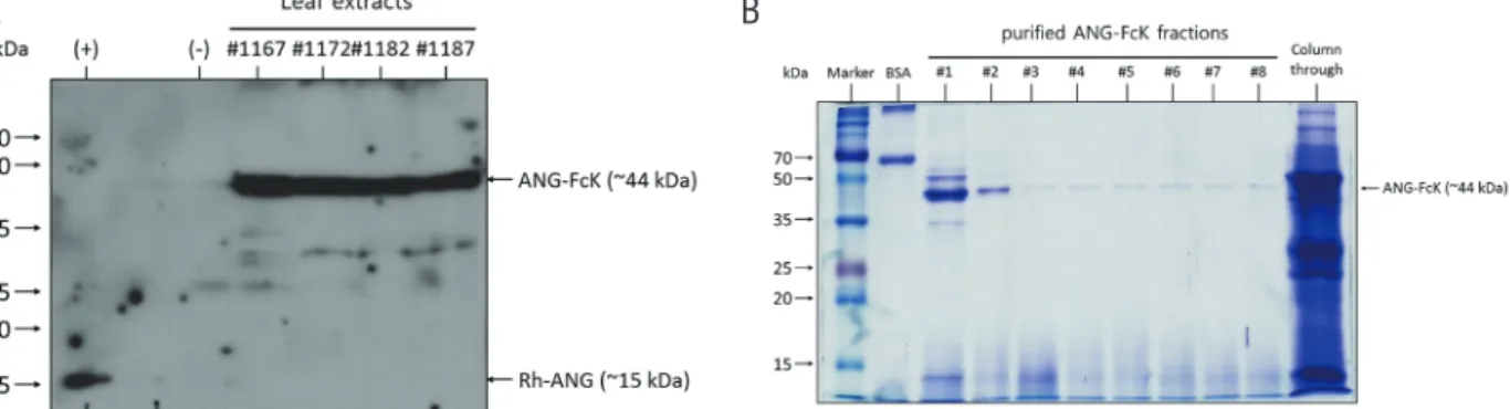
Expression and purification of ANG-FcK in transgenic plants Randomly selected transgenic plants were selected and their expression was confirmed (Fig. 1A). Western blot analysis with anti-human Fc fragment antibody was con- ducted to confirm the expression of ANG-FcK in plants.
Positive control, the Rh-ANG protein band was detected at approximately 15 kDa and the ANG-FcK was detected at approximately 44 kDa. No band was observed in the non-transgenic plant. ANG-FcK was purified from leaves harvested from transgenic tobacco plants. The protein A column purification yielded an average of 2 mg of plant-de- rived ANG-FcK per kg of fresh leaves from high protein expressing line. SDS-PAGE analysis of the purified ANG- FcK revealed one major band (44 kDa) (Fig. 1B).
Figure 1. Development of angiogenin fusion protein (ANG-FcK). (A) Expression of ANG-FcK in randomly selected transgenic plants. (+), positive control, recombinant human angiogenin (Rh-ANG); (-), non-transgenic tobacco plant leaf extract. #1167–1187, transgenic plant line number. (B) Sodium dodecyl sulfate polyacrylamide gel electrophoresis results for purified ANG-FcK. #1–8, purified protein fraction number; Column through, plant extracts passed through a column.
A B
Cellular toxicity induced by BAK in HTM cells
The MTT assay showed a cell viability decrease with BAK concentration, and this decrease of viability was even greater with concentration of 0.002%, reaching less than 40% (Fig. 2A). The lowest concentration (0.0001%) showed no difference with the control. BAK 0.001% with a cell via- bility decrease of about 70% was used for the other test, be-
cause our aim was making degeneration in TM by chronic exposure to low concentration of BAK. The morphological analysis of HTM cell showed that administration of BAK reduced TM cell density and cell shape (Fig. 2B). Both Rh- ANG and ANG-FcK had little effect on the cell viability in HTM cells.
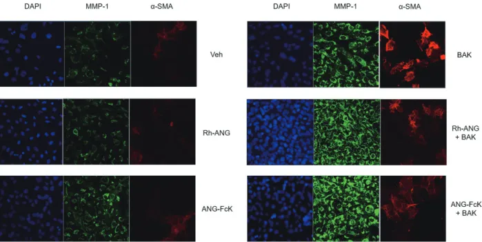
Figure 3. Immunocytochemical analyses of matrix metalloproteinase-1 (MMP-1) and alpha smooth muscle actin (a-SMA) in human trabecular meshwork cells. The expression of MMP-1 and α-SMA were increased by benzalkonium chloride (BAK) treatment; however, that were preserved by the single angiogenin (ANG) treatment. The expression of α-SMA was decreased by co-treatment with ANG compared to the single BAK treatment. All images were magnified 200 times. Veh = vehicle for sham-treated control; DAPI = diamidino phenylindol; Rh-ANG = recombinant human angiogenin; ANG-FcK = plant-derived angiogenin fusion protein.
Figure 2. Cellular toxicity induced by benzalkonium chloride (BAK). (A) In a 3-(4,5-dimethylthiazol-2-yl)-2,5-diphenyltetrazolium bro- mide (MTT) assay, the viability of human trabecular meshwork cells decreased with the BAK concentration, and this decrease reached about 70% for 0.001% BAK. (B) BAK reduced the human trabecular meshwork cell density and affected cell shape. Rh-ANG = recom- binant human angiogenin; ANG-FcK = plant-derived angiogenin fusion protein.
A B
Effects of BAK and ANG on MMP-1 and α-SMA in HTM cells Single ANG treatment showed no significant change how- ever, increased MMP-1 and α-SMA expression were obvious in response to the BAK treatment by immunocytochemistry (Fig. 3). The combination of ANG and BAK induced more MMP-1 expression and lesser α-SMA expression compared to the single BAK treatment. Expression pattern of MMP- 1 and α-SMA in response to the ANG-FcK was similar to that of Rh-ANG with or without BAK. Expression levels of MMP-1 at the protein and mRNA were significantly aug- mented in BAK treatment groups compared to single ANG and control groups, and the expression in combination ANG with BAK was higher than the single BAK treatment group, but it was not significant statistically (Fig. 4A, B). Expres- sion of α-SMA were significantly increased in BAK treat- ment groups compared to single ANG and control groups at the protein and mRNA levels. The expression of α-SMA in the single BAK treatment was about 2.2 times (p < 0.01) and that in the combination treatment of ANG and BAK were 1.5 to 1.8 times (p < 0.01) more than control, and the dif- ferences between single BAK and combination with ANG were significant (p < 0.01) also (Fig. 4A, C).
Discussion
In this study, we developed ANG-FcK, using molecular farming technique, and examined its defenses against the toxicity of BAK exposure on TM cell lines. Expressions of MMP-1 and α-SMA were increased through BAK treat- ment in human TM cell lines, and co-treatment with ANG induced more MMP-1 expression and significantly less remarkable α-SMA expression compared to the single BAK treatment, suggesting that ANG prevents fibrosis. Treat- ment of the ANG-FcK in the TM cell lines showed similar response to that of Rh-ANG, indicating that ANG-FcK has important practical applications as a protecting substance.
The isolation and purification of proteins from plant biomass is one of salient factors in the production of ther- apeutic proteins by transgenic plants on the molecular lev- el.13 Fusion of Fc region of immunoglobulin G and KDEL brought on greater molecular weight of the ANG-FcK Figure 4. Western blot analysis and quantitative real-time poly-
merase chain reaction (qRT-PCR) of matrix metalloproteinase-1 (MMP-1) and alpha smooth muscle actin (a-SMA) in human trabecular meshwork cells. (A) Expression levels of MMP-1 and α-SMA were augmented by benzalkonium chloride (BAK), and especially, α-SMA expression was notable in the single BAK treat- ment at the protein level. (B) MMP-1 expression levels were higher after BAK treatment than in the control group and the single angiogenin (ANG) treatment group. Levels of MMP-1 expression were increased by co-treatment with ANG, although there was no significant difference. (C) Levels of α-SMA also were elevated after BAK treatment than in the control group and the single ANG treat- ment group. Elevated expression of α-SMA, in contrast, was inhib- ited by co-treatment with ANG. Error bars represent standard errors of the mean. Rh-ANG = recombinant human angiogenin; ANG-FcK
= plant-derived angiogenin fusion protein. *p < 0.01; #p < 0.05.
A
B
C
(44 kDa) over Rh-ANG (15 kDa), and that might have im- proved yields by enhancing protein stability and facilitating purification. Purifying plant-derived ANG-FcK yielded an average of 2 mg per kg of fresh leaves. With a cost of ap- proximately 1,000 U.S. dollars per 250 μg of Rh-ANG, 1 kg of transgenic plants is worth approximately 8,000 U.S. dol- lars of conventional protein. To the best of our knowledge, this is the first study to apply molecular farming techniques in ophthalmology, and our production of recombinant ANG may be beneficial to this field.
Increased MMP-1 and α-SMA expressions in human TM cell lines have been observed in response to the BAK treat- ment, and these findings imply that BAK causes epithelial mesenchymal transition-like phenomenon and myofibro- blast like phenotype change in TM. This epithelial mesen- chymal transition like phenomenon results in the abundant expression of fibronectin and activation of motility in TM cells, and the cells switch to a myofibroblast-like phenotype which strengthens simultaneously both their actin cytoskel- eton and their directly associated extracellular matrix.
Overall, the changes cause an increase in TM rigidity and resistance to aqueous humor outflow.17,18
Combination use of ANG and BAK have been showed more MMP-1 and significantly less noticeable α-SMA ex- pression compared to the single BAK treatment, and these findings support that ANG protects from the fibrosis and myofibroblast like phenotype changes induced by BAK.
The protective mechanism of ANG could be explained by the regulation of MMPs that initiate the turnover of extracellular matrix in the TM, and necessary to maintain outflow facility.19,20 In human TM cells, MMP-1 was up- regulated by the BAK exposure and the levels of MMP- 1 expression were elevated with ANG. These results were in close agreement with previous study showed that MMP- 1 expression were defective in primary glaucomatous TM cells and augmented by ANG treatment over the course of time.14 MMP activation by ANG may induce cytoskeletal changes in TM cells, to probably defend against the expres- sion of α-SMA as for a fibrogenic marker21,22 induced by BAK.
Our study has its limitations. First, we used TM cells
from a commercial source and did not characterize them thoroughly. There is strong evidence in the field that the TM cells from this commercial source are not a pure pop- ulation.23 Second, effects of BAK on the TM and ANG’s protective mechanism have not been demonstrated and in vivo studies are needed for more comprehensive data when managing glaucoma. Third, extra in vitro experiments are necessary to investigate the protective mechanism of ANG against BAK. Previous studies showed that ANG may ac- tivate Akt-mediated signals for nitric oxide production and TM remodeling by regulating MMP and rho-kinase.12 Fi- nally, further research on ANG’s effect on retinal ganglion cells may clarify its function and improve its clinical effec- tiveness. Because glaucoma is an ocular neurodegenerative disease characterized by the progressive death of retinal ganglion cells, the importance of ANG enrichment in nor- mal motor neurons has been observed in studies on amyo- trophic lateral sclerosis, a fetal neurodegenerative disease.24
Conclusions
In conclusion, ANG’s protective effect on TM may involve an anti-fibrotic function against the exposure to single toxic substance such as BAK. Plant-derived ANG-FcK’s protec- tive effect is similar to that of Rh-ANG, and it is a promis- ing candidate for an alternative eye drop additive. Future studies should focus on ANG’s detailed defense mechanism and potential applications in glaucoma management.
Acknowledgement
This report was supported by the National Research Foun- dation of Korea (2017R1C1B5018031).
References
1. Baudouin C. Detrimental effect of preservatives in eye- drops: implications for the treatment of glaucoma. Acta Ophthalmol 2008;86:716-26.
2. Noecker R, Miller KV. Benzalkonium chloride in glau- coma medications. Ocul Surf 2011;9:159-62.
3. Rosin LM, Bell NP. Preservative toxicity in glau- coma medication: clinical evaluation of benzalkonium
chloride-free 0.5% timolol eye drops. Clin Ophthalmol 2013;7:2131-5.
4. Brignole-Baudouin F, Desbenoit N, Hamm G, et al. A new safety concern for glaucoma treatment demonstrated by mass spectrometry imaging of benzalkonium chloride distribution in the eye, an experimental study in rabbits.
PLoS One 2012;7:e50180.
5. Baudouin C, Denoyer A, Desbenoit N, et al. In vitro and in vivo experimental studies on trabecular meshwork degen- eration induced by benzalkonium chloride (an American Ophthalmological Society thesis). Trans Am Ophthalmol Soc 2012;110:40-63.
6. Chang C, Zhang AQ, Kagan DB, et al. Mechanisms of benzalkonium chloride toxicity in a human trabecular meshwork cell line and the protective role of preservative- free tafluprost. Clin Exp Ophthalmol 2015;43:164-72.
7. Gao X, Xu Z. Mechanisms of action of angiogenin. Acta Biochim Biophys Sin (Shanghai) 2008;40:619-24.
8. Li S, Yu W, Hu GF. Angiogenin inhibits nuclear translo- cation of apoptosis inducing factor in a Bcl-2-dependent manner. J Cell Physiol 2012;227:1639-44.
9. Steidinger TU, Standaert DG, Yacoubian TA. A neuropro- tective role for angiogenin in models of Parkinson’s dis- ease. J Neurochem 2011;116:334-41.
10. Tello-Montoliu A, Patel JV, Lip GY. Angiogenin: a review of the pathophysiology and potential clinical applications.
J Thromb Haemost 2006;4:1864-74.
11. Sack RA, Conradi L, Krumholz D, et al. Membrane array characterization of 80 chemokines, cytokines, and growth factors in open- and closed-eye tears: angiogenin and other defense system constituents. Invest Ophthalmol Vis Sci 2005;46:1228-38.
12. Kim KW, Park SH, Oh DH, et al. Ribonuclease 5 coor- dinates signals for the regulation of intraocular pressure and inhibits neural apoptosis as a novel multi-functional anti-glaucomatous strategy. Biochim Biophys Acta 2016;1862:145-54.
13. Jamal A, Ko K, Kim HS, et al. Role of genetic factors and environmental conditions in recombinant protein produc-
tion for molecular farming. Biotechnol Adv 2009;27:914- 23.
14. Gomord V, Sourrouille C, Fitchette AC, et al. Production and glycosylation of plant-made pharmaceuticals: the anti- bodies as a challenge. Plant Biotechnol J 2004;2:83-100.
15. Hiatt A, Cafferkey R, Bowdish K. Production of antibod- ies in transgenic plants. Nature 1989;342:76-8.
16. Ma JK, Barros E, Bock R, et al. Molecular farming for new drugs and vaccines. Current perspectives on the pro- duction of pharmaceuticals in transgenic plants. EMBO Rep 2005;6:593-9.
17. Takahashi E, Inoue T, Fujimoto T, et al. Epithelial mesen- chymal transition-like phenomenon in trabecular mesh- work cells. Exp Eye Res 2014;118:72-9.
18. Tamm ER. Functional morphology of the outflow path- ways of aqueous humor and their changes in open angle glaucoma. Ophthalmologe 2013;110:1026-35.
19. Bradley JM, Kelley MJ, Zhu X, et al. Effects of mechani- cal stretching on trabecular matrix metalloproteinases.
Invest Ophthalmol Vis Sci 2001;42:1505-13.
20. Bradley JM, Vranka J, Colvis CM, et al. Effect of matrix metalloproteinases activity on outflow in perfused human organ culture. Invest Ophthalmol Vis Sci 1998;39:2649- 58.
21. Ko MK, Tan JC. Contractile markers distinguish struc- tures of the mouse aqueous drainage tract. Mol Vis 2013;19:2561-70.
22. Pattabiraman PP, Maddala R, Rao PV. Regulation of plas- ticity and fibrogenic activity of trabecular meshwork cells by Rho GTPase signaling. J Cell Physiol 2014;229:927- 42.
23. Keller KE, Bhattacharya SK, Borras T, et al. Consensus recommendations for trabecular meshwork cell isolation, characterization and culture. Exp Eye Res 2018;171:164- 73.
24. Kieran D, Sebastia J, Greenway MJ, et al. Control of motoneuron survival by angiogenin. J Neurosci 2008;28:14056-61.
국문초록
섬유주세포에서 MMP-1과 α-SMA에 대한 식물생명공학 기법으로 합성한 안지오제닌의 효과
목적: 안압하강제 및 안약 보존제로서 널리 이용되는 벤잘코니움은 사용량에 비례하는 독성을 나타낼 수 있다. 안지오제닌의 다양한 기능이 녹내장 치료에 이용될 수 있다는 기존 보고들에 근거하여, 벤잘코니움으로 손상된 사람 섬유주세포에서 식물생명공학 기법으로 합성한 안지오제닌의 효과를 알아보고자 한다.
대상과 방법: 상용화된 재조합 사람 안지오제닌(Rh-ANG)에 면역글로불린 G의 Fc 부분과 KDEL을 결합하여 식물로부터 안지오제닌(ANG-FcK)을 합성하였다. 면역화학 분석을 통해 단백질과 mRNA 수준에서 섬유주세포에서의 MMP-1과 α-SMA 발현에 대해 확인하였다. 독성 자극으로 벤잘코니움을 이용하였고, 두 가지 안지오제닌(Rh-ANG, ANG-FcK)을 보호물질로 이용하였다.
결과: 벤잘코니움에 노출된 섬유주세포에서 MMP-1과 α-SMA 발현이 유의하게 증가하였다. 안지오제닌을 벤잘코니움과 병용 투여한 결과 MMP-1 발현은 증가하는 양상이었으나, α-SMA 발현은 벤잘코니움 단독 노출에 비해 유의하게 감소하는 양상으로 나타났다. 두 가지 안지오제닌(Rh-ANG, ANG-FcK)의 발현 양상은 큰 차이가 없었다.
결론: 식물생명공학 기법으로 합성한 안지오제닌의 섬유주세포에 대한 효과는 상용화된 안지오제닌과 비슷하였고, 항섬유화 기전과 연관이 있을 것으로 보이며, 녹내장 치료에 이용될 수 있는 가능성을 시사한다.