ABSTRACT
Purpose: Campylobacter species are currently the most common cause of bacterial
gastroenteritis. In Lebanon, Campylobacter infection occurrence is underdiagnosed owing to the lack of specific culture and rapid test kits, particularly among children. This study aimed to evaluate the prevalence, laboratory findings, and clinical characteristics of Campylobacter infection in hospitalized children with acute gastroenteritis in South Lebanon.
Methods: We conducted a 6-month retrospective cohort study between January and June 2018, including 291 children aged between 1 month and 12 years, who were admitted to a tertiary healthcare center in South Lebanon. The medical files of the patients were reviewed to retrieve the required clinical information, including clinical and laboratory data.
Results: The prevalence of campylobacteriosis agents in pediatric patients with acute gastroenteritis is 12.02%. Patients infected with Campylobacter had more severe acute gastroenteritis than Campylobacter-negative patients and often presented with high-grade fever, diarrhea episodes more than six times per day, diarrhea lasting for more than five days, and dehydration. Indeed, children with high-grade fever (≥38.5°C) were five times more likely to test positive for Campylobacter than those with low-grade fever. In addition, the results showed a higher Vesikari score for the majority of Campylobacter-positive patients with severe acute gastroenteritis compared to a moderate profile for Campylobacter-negative patients.
Conclusion: The present study findings highlight that Campylobacter infection is frequent among children with acute gastroenteritis. Therefore, the detection of Campylobacter should be carried out for the diagnosis of human gastroenteritis in Lebanon, along with the detection of routine enteropathogens.
Keywords: Campylobacteriosis; Campylobacter; Gastroenteritis; Children; Diarrhea; Lebanon
Original Article
Received: Oct 15, 2020 Revised: Jan 14, 2021 Accepted: Mar 6, 2021 Correspondence to Ali El Roz
Rammal Hassan Rammal Research Laboratory, PhyToxE Research Group, Faculty of Sciences (V), Lebanese University, Nabatieh P.O. Box 6573/14, Lebanon.
E-mail: [email protected]
*These two authors contributed equally to this work as co-first authors.
Copyright © 2021 by The Korean Society of Pediatric Gastroenterology, Hepatology and Nutrition
This is an open-access article distributed under the terms of the Creative Commons Attribution Non-Commercial License (https://
creativecommons.org/licenses/by-nc/4.0/) which permits unrestricted non-commercial use, distribution, and reproduction in any medium, provided the original work is properly cited.
ORCID iDs Ghassan Ghssein
https://orcid.org/0000-0002-9313-1418 Rana Awada
https://orcid.org/0000-0001-8737-6892 Ali Salami
https://orcid.org/0000-0003-3343-4035 Hisham F. Bahmad
https://orcid.org/0000-0003-3799-2595 Ali Awad
https://orcid.org/0000-0001-5911-8642 Wissam H. Joumaa
https://orcid.org/0000-0002-1272-2719 Ali El Roz
https://orcid.org/0000-0003-4198-5371
Ghassan Ghssein ,1,2,* Rana Awada ,1,3,* Ali Salami ,4 Hisham F. Bahmad ,5 Ali Awad ,3 Wissam H. Joumaa ,1 and Ali El Roz 1,3
1 Rammal Hassan Rammal Research Laboratory, PhyToxE Research Group, Faculty of Sciences, Lebanese University, Nabatieh, Lebanon
2 Department of Laboratory Sciences, Faculty of Nursing and Health Sciences, Islamic University of Lebanon, Khalde, Lebanon
3Department of Biology, Faculty of Sciences, Lebanese University, Hadath, Lebanon
4 Department of Mathematics, Faculty of Sciences, Lebanese University, Nabatieh, Lebanon
5 Arkadi M. Rywlin M.D. Department of Pathology and Laboratory Medicine, Mount Sinai Medical Center, Miami Beach, FL, USA
Prevalence, Laboratory Findings and Clinical Characteristics of
Campylobacteriosis Agents among
Hospitalized Children with Acute
Gastroenteritis in Lebanon
Conflict of Interest
The authors have no financial conflicts of interest.
INTRODUCTION
Campylobacteriosis-causing agents are the most common cause of bacterial gastroenteritis in developed countries [1], with an incidence rate of 64.8 per 100,000 population in Europe [2]. According to the United States (US) Centers for Disease Control and Prevention, it is estimated that around 1.3 million cases of campylobacteriosis are found every year in the US alone, with an annual economic loss of up to $ 6.8 billion [3]. Campylobacteriosis is an intestinal infection caused by Campylobacter species [4] and presents with common gastrointestinal symptoms, including abdominal pain, diarrhea, fever, nausea, and vomiting.
This infection can be accompanied by clinical manifestations that are fatal in children, the elderly, and immunocompromised individuals [5].
Campylobacteriosis in humans usually occurs after ingestion of water or food contaminated with bacteria, especially from poultry, which represents the highest percentage (50–70%) of cases [6]. Once ingested, Campylobacter prompts a toxin-releasing inflammatory response in the colon, accompanied by infiltration of the intestinal mucosa with neutrophils and lymphocytes [7]. Clinical signs of the disease range from mild self-limiting enteritis with watery diarrhea to bloody mucoidal diarrhea in cases of severe acute campylobacteriosis [8]. Diagnosing this disease usually requires direct stool culture on Campylobacter specific media or polymerase chain reaction (PCR)-based methods [9]. Currently, since no vaccination is available to protect against this enteropathogen (as in Rotavirus), the only way to hinder the spread of campylobacteriosis is via hygiene and biosecurity [10].
Campylobacter species are spiral, microaerobic, and gram-negative bacteria belonging to the 16S rRNA superfamily VI. The genus includes 17 species and six subspecies, although most are thought to be nonpathogenic [11]. Campylobacter species are sometimes isolated from symptomatic patients and healthy individuals, with Campylobacter jejuni being the most commonly isolated species among children in the US [12]. In Lebanon, Campylobacter infection in both children and adults is underestimated owing to the lack of specific culture and rapid test kits. In addition, the extensive use of antibiotics in Lebanon increases the occurrence of resistant bacteria [13]. To the best of our knowledge, few studies have been published in Lebanon on the prevalence, laboratory findings, and clinical characteristics of campylobacteriosis in children. In our study, we aimed to evaluate these parameters in hospitalized children with acute gastroenteritis in South Lebanon.
MATERIALS AND METHODS
Ethics approval and consent to participate
All experimental protocols followed in this study were approved by the ethics committee of the Sheikh Ragheb Harb University Hospital (SRHUH) (Approval No. AER2017). Ethical clearance was obtained as per the norms and in accordance with relevant guidelines and regulations of the tertiary healthcare center. Recruitment was performed randomly after obtaining written informed consent for participation from the patients' caregivers, in accordance with the Declaration of Helsinki.
Patient selection
During a six-month period, from January to June 2018, we collected clinical, demographic, and laboratory data from hospitalized Lebanese children (aged 1 month to 12 years), admitted to the pediatric department of a tertiary care center located in South Lebanon.
The patients included in this study were hospitalized children with acute gastroenteritis, i.e., vomiting and/or diarrhea. The exclusion criteria were children with chronic diarrhea,
immunodeficiency, malnutrition, and multiple malformations, as these might negatively affect the length of hospitalization and severity of the disease, and hence, disrupt the analysis of our results.
Clinical variables
Upon admission, data were collected as follows:
• Demographic data included age, sex, date of admission, status of breastfeeding, type of drinking water, family size of patients, and use of antibiotics prior to hospitalization.
• Clinical data comprised signs and symptoms of acute gastroenteritis, such as fever, diarrhea (frequency and duration), vomiting (frequency and duration); dehydration, and presence of blood and mucus in stool; stool texture (classified based on a combination of clinical experience and scientific evidence based on the Bristol stool chart); duration of hospitalization; and calculation of the index of severity “Vesikari Score” (VSS) [14].
• Laboratory findings included blood levels of white blood cells, red blood cells, hemoglobin, hematocrit, sugars, and C-reactive protein (CRP) and absolute neutrophil count (ANC).
Laboratory methods
Stool samples were freshly acquired from patients and directly analyzed within one hour for the presence of infectious agents. Rotavirus, adenovirus, and Entamoeba histolytica kit tests (CerTest; CerTest Biotec, Zaragoza, Spain) were used to detect possible enteropathogens.
As an additional diagnostic tool, which is not routinely used in the diagnosis of acute gastroenteritis in hospitals, stool samples were cultured for the detection of Campylobacter.
Campylobacter culture
Campylobacter stool cultures were performed on the selective medium “charcoal cefoperazone desoxycholate agar” (Liofilchem, Roseto degli Abruzzi, Italy) containing cefoperazone and amphotericin B (32 mg/L and 10 mg/L, respectively). After sample streaking, the plates were incubated micro-aerobically (6% O2) at 37°C for 48 hours. Suspect colonies (small, gray, and translucent colonies [12 mm in diameter] with a metallic sheen and butter-like consistency) were identified at the Campylobacter genus level using oxidase and catalase tests and the Gram stain technique (gram-negative rods, oxidase positive, and catalase positive).
Sample size and power of the study
Sample size calculations were performed to calculate the prevalence rate based on the following assumptions:
• An effect size-g-of 6%. The value of the constant proportion was set conservatively to 11.1% for the prevalence rate of Campylobacter infection in children in the region of North Lebanon [15].
• An alpha, type 1 error, balanced on each side, set to 5%.
• A type 2 error of 20%, yielding a statistical power of 80%.
Using the G*Power 3.1.9.2 software [16], the exact binomial distribution yielded 255 subjects as the minimum required sample size to satisfy the above assumptions.
Statistical analysis
Descriptive statistics were carried out and reported as numbers and percentages for categorical variables, whereas mean and standard deviation (±) were used for continuous variables.
Patients' clinical characteristics were tabulated and comparisons between the two groups
(Campylobacter-positive vs. Campylobacter-negative patients) were made using the Mann–Whitney test for continuous variables. To evaluate whether a significant difference was present between the categorical variables, the chi-squared test was used. We determined the associations between Campylobacter cases (yes/no) as the dependent variable and sex, breastfeeding, daycare, fever, and antibiotic use before hospitalization as independent variables using logistic regression analyses. Normality was tested using the Kolmogorov–Smirnov test. Statistical analysis was performed using SPSS Statistics for Windows, Version 22.0 (IBM Co., Armonk, NY, USA). The level of significance was set at p<0.05 for all statistical analyses.
RESULTS
Socio-demographic and clinical characteristics of patients
This study included 291 hospitalized children with acute gastroenteritis aged between 1 month and 12 years. Of these, 169 (58%) were male and 122 (42%) were female. The age distribution of our study participants was as follows: 3.43% aged 1–3 months, 19.58% aged 4–11 months, 24.74%
aged 12–23 months, 15.46% aged 24–35 months, 10.99% aged 36–47 months, 5.49% aged 48–59 months, and 20.27% aged >60 months, which was equivalent to 5 years of age (Table 1).
Examination of the stool specimens revealed that 35 out of 291 samples (12.02%) were positive for Campylobacter spp., while the rest were positive for non-Campylobacter enteropathogens, including viruses (such as rotavirus and adenovirus), parasites (such as Entamoeba histolytica), and other bacteria. The percentages of Campylobacter-positive patients among hospitalized males and females were similar (12.4% vs. 11.5%, respectively). With regard to age distribution, the highest rates of infection were observed in the 36–47 month group (25%), with no statistical difference observed between the groups.
The clinical characteristics of the patients are summarized in Table 2. Our results revealed statistically significant differences between the study groups with respect to fever,
Table 1. Socio-demographic characteristics of patients
Demographic Campylobacteriosis Total gastroenteritis p-value
Yes No
Age (mo) 0.067
1–3 2 (5.71) 8 (3.13) 10
4–11 8 (22.86) 49 (19.14) 57
12–23 4 (11.43) 68 (26.56) 72
24–35 2 (5.71) 43 (16.79) 45
36–47 8 (22.86) 24 (9.38) 32
48–59 2 (5.71) 14 (5.47) 16
≥60 9 (25.72) 50 (19.53) 59
Sex 0.806
Female 14 (40.00) 108 (42.19) 122
Male 21 (60.00) 148 (57.81) 169
Breastfeeding* 0.798
No 3 (60.00) 52 (54.17) 55
Yes 2 (40.00) 44 (45.83) 46
Daycare* 0.102
No 1 (20.00) 55 (57.29) 56
Yes 4 (80.00) 41 (42.71) 45
Mean family size 4.20±0.45 4.43±1.19 4.42 0.927
Values are presented as number (%) or mean±standard deviation.
*Missing data.
stool texture, diarrhea duration, and dehydration, among other variables. Interestingly, Campylobacter-positive patients presenting with high-grade fever (≥38.5°C) comprised a significantly higher percentage (52.38%) than those with low-grade fever (<38.5°C) (17.86%), and Campylobacter-negative patients mainly had low-grade fever (p=0.001). Regarding stool texture, the majority of the Campylobacter-positive patients had mucoidal (56.52%) diarrhea, whereas most of the Campylobacter-negative patients had watery diarrhea (50.66%) (p<0.001).
Furthermore, the results showed that a significantly higher percentage of Campylobacter- positive patients passed stool >6 times per day (22.22%) or 4–5 times a day (66.67%).
Whereas, 38.66% and 54.64% of Campylobacter-negative patients passed stool 1–3 times or 4–5 times a day, respectively (p=0.002). Almost 25.93% of patients in the Campylobacter-positive group experienced diarrhea for ≥5 days, while barely 2.58% of patients in the Campylobacter-
Table 2. Clinical characteristics of patients
Clinical characteristic Campylobacteriosis Total gastroenteritis p-value
Yes No
Stool texture*,† <0.001
Mucoidal 13 (56.52) 40 (26.32) 53
Watery 0 (0.00) 77 (50.66) 77
Bloody 8 (34.78) 32 (21.05) 40
Semi-solid 0 (0.00) 3 (1.97) 3
Bloody mucoidal 2 (8.70) 0 (0.00) 2
Fever* 0.001
Low-grade fever (<38.5°C) 5 (17.86) 110 (52.38) 115
High-grade fever (≥38.5°C) 23 (82.14) 100 (47.62) 123
Abdominal pain* 0.077
No 0 (0.00) 19 (14.39) 19
Yes 19 (100.00) 113 (85.61) 132
Diarrhea 0.456
No 0 (0.00) 4 (1.56) 4
Yes 35 (100.00) 252 (98.44) 287
Diarrhea episodes per day* 0.002
1–3 3 (11.11) 75 (38.66) 78
4–5 18 (66.67) 106 (54.64) 124
≥6 6 (22.22) 13 (6.70) 19
Diarrhea duration (d)* <0.001
1–4 20 (74.07) 189 (97.42) 209
≥5 7 (25.93) 5 (2.58) 12
Vomiting 0.767
No 15 (42.86) 103 (40.23) 118
Yes 20 (57.14) 153 (59.77) 173
Vomiting episodes per day* 0.815
0 15 (53.57) 103 (47.69) 118
1 3 (10.71) 30 (13.89) 33
2–4 10 (35.72) 83 (38.42) 93
Vomiting duration (d)* 0.134
0 15 (53.57) 103 (47.68) 118
1 6 (21.43) 87 (40.28) 93
2 6 (21.43) 22 (10.19) 28
≥3 1 (3.57) 4 (1.85) 5
Severity of dehydration (%)* 0.004
No 13 (38.23) 160 (64.26) 173
1–5 21 (61.77) 82 (32.93) 103
≥6 0 (0.00) 7 (2.81) 7
Antibiotic before hospitalization 0.002
No 34 (97.14) 188 (73.44) 222
Yes 1 (2.86) 68 (26.56) 69
Values are presented as number (%).
*Missing data. †Stool texture was classified based on a combination of clinical experience and scientific evidence based on the Bristol stool chart.
negative group showed a significant difference (p<0.001). Lastly, it is noteworthy that while dehydration represented a major presenting clinical sign in Campylobacter-positive patients (61.77%), the majority of patients did not have dehydration (64.26%) (p=0.004).
The laboratory and clinical findings are outlined in Table 3. Our results revealed that the mean body temperature of Campylobacter-positive patients was significantly higher by almost 1°C than that of Campylobacter-negative patients (39.23±0.90 vs. 38.28±0.99; p<0.001).
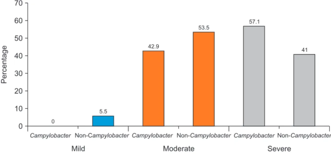
As expected, following the analysis of the previously mentioned clinical characteristics (diarrhea, vomiting, dehydration, and body temperature), the VSS showed a severe profile of acute gastroenteritis in Campylobacter-positive patients (11.37±2.76) compared to a moderate profile of acute gastroenteritis in Campylobacter-negative patients (9.92±2.50) (p=0.004).
The severity of gastroenteritis based on the VSS
As shown in Fig. 1, no mild gastroenteritis cases were observed in patients infected with Campylobacter. In contrast, those patients showed moderate and severe profiles with a higher percentage of severe cases (57.1%) compared to Campylobacter-negative patients (41.0%).
Table 3. Clinical and laboratory data of patients Clinical and laboratory
characteristic Campylobacteriosis Total gastroenteritis p-value
Yes No
Frequency (%) 35 (12.0) 256 (88.0) 291 -
Body temperature (°C) 39.23±0.90 38.28±0.99 38.39±1.02 <0.001
Vesikari score 11.37±2.76 9.92±2.50 10.09±2.57 0.004
Duration of hospitalization (hr) 66.31±18.30 64.78±20.83 64.90±20.61
Laboratory findings 0.885
PLT (×103 per mm3) 234.00±103.59 283.15±80.34 280.71±81.71 0.343
ANC (×102 per mm3) 51.62±19.71 62.21±16.07 61.41±16.55 0.026
HGB (g/dL) 11.45±0.83 11.48±1.11 11.48±1.09 0.847
BS (mg/dL) 85.00±19.54 92.71±20.73 91.81±20.52 0.426
CRP (mg/L) 33.32±24.01 27.36±28.72 27.83±28.35 0.180
HCO3 (mEq/L) 17.57±2.07 17.19±2.98 17.24±2.86 0.948
BUN (mg/dL) 9.71±3.19 11.68±5.39 11.47±5.22 0.438
Creatinine (mg/dL) 0.26±0.06 0.99±3.87 0.92±3.65 0.788
Values are presented as number (%) or mean±standard deviation.
PLT: platelets, ANC: absolute neutrophil count, HGB: hemoglobin, BS: blood sugar, CRP: C-reactive protein, BUN:
blood urea nitrogen.
Campylobacter 70
60 50 40 30 20 10
Percentage
Mild Moderate Severe
0
Non-CampylobacterCampylobacter Non-CampylobacterCampylobacterNon-Campylobacter 0
5.5
42.9
53.5
57.1
41
Fig. 1. The severity of gastroenteritis in Campylobacter-negative and Campylobacter-positive patients based on the Vesikari score system (Severity rating scales: mild, <7; moderate, 7–10; severe, ≥11).
Laboratory characteristics of patients
Infectious markers such as ANC and CRP levels were also measured in the patients' peripheral blood (Table 3). Interestingly, Campylobacter-positive patients were found to have significantly lower ANC than Campylobacter-negative patients (p=0.026). In contrast, children with campylobacteriosis had higher levels of CRP than Campylobacter-negative patients, though the difference was not statistically significant (p=0.180). As for other laboratory findings, there were no significant differences between the two groups.
Prevalence and predictors of Campylobacter infection
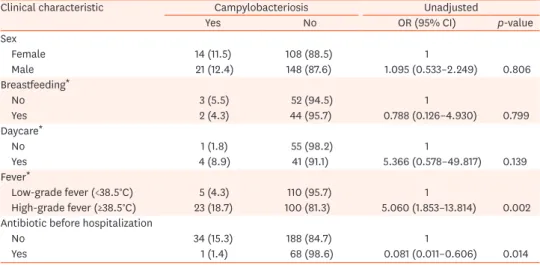
Among the predictors for Campylobacter infection shown in Table 4, sex, breastfeeding, and daycare appeared to have no significant association with Campylobacter culture positivity. In contrast, children with high-grade fever (≥38.5°C) were 5-folds more likely to be positive for Campylobacter than those with low-grade fever (odds ratio [OR]=5; confidence interval [CI]=1.8–13.8; p=0.002). In addition, children who had been treated with antibiotics prior to hospitalization were less likely to be tested positive for Campylobacter (OR=0.08; CI=0.01–0.6; p=0.014) compared to non-treated children.
DISCUSSION
Acute gastroenteritis is the second major cause of pediatric morbidity and mortality following acute lower respiratory tract infections in Lebanon and other developing countries [17]. Identifying specific enteropathogens responsible for acute gastroenteritis is essential for the application of appropriate and suitable clinical measures to manage the disease. It has been shown that infection with Campylobacter species is the leading cause of common bacterial gastroenteritis [4]. This study aimed to investigate Campylobacter infection over a period of 6 months in hospitalized children in a rural area of South Lebanon and to examine the relationship between campylobacteriosis, its severity, and routine laboratory tests. To the best of our knowledge, this study is one of the few to investigate the prevalence and occurrence of campylobacteriosis in Lebanon [13,15,18], and the first study conducted on children in South Lebanon.
Table 4. Predictors of Campylobacter infection among the 291 pediatric patients enrolled in the study
Clinical characteristic Campylobacteriosis Unadjusted
Yes No OR (95% CI) p-value
Sex
Female 14 (11.5) 108 (88.5) 1
Male 21 (12.4) 148 (87.6) 1.095 (0.533–2.249) 0.806
Breastfeeding*
No 3 (5.5) 52 (94.5) 1
Yes 2 (4.3) 44 (95.7) 0.788 (0.126–4.930) 0.799
Daycare*
No 1 (1.8) 55 (98.2) 1
Yes 4 (8.9) 41 (91.1) 5.366 (0.578–49.817) 0.139
Fever*
Low-grade fever (<38.5°C) 5 (4.3) 110 (95.7) 1
High-grade fever (≥38.5°C) 23 (18.7) 100 (81.3) 5.060 (1.853–13.814) 0.002 Antibiotic before hospitalization
No 34 (15.3) 188 (84.7) 1
Yes 1 (1.4) 68 (98.6) 0.081 (0.011–0.606) 0.014
Values are presented as number (%).
OR: odds ratio, CI: confidence interval.
*Missing data.
We previously reported causative enteropathogens for acute gastroenteritis that are routinely tested in children [17,19,20], and showed that E. histolytica, rotavirus, and adenovirus are the main agents. The present study shows an additional diagnostic tool for the detection of Campylobacter, which is not routinely performed in gastroenteritis cases in Lebanese hospitals.
The prevalence of campylobacteriosis in pediatric patients with acute gastroenteritis enrolled in our study was 12.02%, which was very close to that reported by Dabboussi et al.
[15] in 2012 (11.1%), who evaluated the frequency of Campylobacter infection in children in the region of North Lebanon. Moreover, our results are similar to those reported in other countries, such as Papua New Guinea (Oceania) [21] and Kolkata, India [22], where the prevalence of Campylobacter among hospitalized children with gastroenteritis was 12% and 10%, respectively. Our findings were also similar to those of previous studies, such as those in Mexico (15.7%) and China (14.9%) [3].
Since Campylobacter is recognized as an essential foodborne pathogen associated with human gastroenteritis, mainly from contaminated poultry, our findings could be supported by several studies previously conducted in Lebanon and revealed a high degree of contamination in broiler chickens collected from Lebanese slaughterhouses [13,23-25].
Among the Campylobacter-positive patients, there were more male (60%) than female (40%), which was in agreement with the results reported by Sadiq et al. [26] in 2019; the study reported that the prevalence of C. jejuni was statistically higher in men than in women [26].
Moreover, our results revealed a more severe status of acute gastroenteritis in patients infected with Campylobacter than in those infected with other enteropathogens. This severe profile was characterized by a higher percentage of patients with high-grade fever, diarrhea episodes occurring more than six times per day, diarrhea lasting for more than five days, and dehydration. In addition, our results revealed that the mean body temperature of Campylobacter- positive patients was significantly higher by almost 1°C than that of Campylobacter-negative patients. Although this increase might not seem clinically relevant, it is important to report it as it could define the severity of high-grade fever. Notably, a high proportion of Campylobacter- negative patients tested positive for viral infection (adenovirus and rotavirus). In accordance, Shim et al. [27] reported that bacterial gastroenteritis is associated with more episodes of diarrhea and longer-lasting diarrhea than common viral infections. Consequently, our data resulted in a higher VSS score for Campylobacter-positive patients, showing a severe profile of acute gastroenteritis compared to a moderate profile for Campylobacter-negative patients, which is consistent with our previous results on rotavirus and adenovirus cases showing a lower severity of acute gastroenteritis in children when compared to other enteropathogens [17].
Infectious markers, such as the ANC and CRP levels, were also measured using the patients' peripheral blood. Although previous data have shown that the accumulation of neutrophils is part of the classical response of the innate immune system to campylobacteriosis [28], our findings demonstrated that the ANC was significantly lower in Campylobacter-positive patients. This could be explained by the fact that Campylobacter-negative patients were, as mentioned above, tested positive for other enteropathogens, namely rotavirus and adenovirus, that might have been more invasive, leading to a more pronounced acute inflammation. Our results also showed that children with campylobacteriosis presented with higher levels of CRP compared to Campylobacter-negative patients; however, the difference was not significant. The significance of the statistical tests was more pronounced with the analysis of the VSS parameters discussed above, compared to ANC and CRP level analysis,
which is in agreement with previous findings showing an advantage of the VSS over CRP and ANC in the detection of bacterial acute gastroenteritis [27].
Our study had several limitations. First, it was conducted within a period of six months, hence it lacked the seasonality parameter that had been previously shown to affect acute gastroenteritis cases when evaluated over a complete year [13,17]. Second, the antibiogram test was not performed to evaluate the susceptibility of Campylobacter strains to commonly prescribed antibiotics. Third, identification tests were not performed to discriminate between Campylobacter species. This could have been achieved by a standardized hippurate hydrolysis test and PCR technique. Fourth, some data were missing from the medical records of the patients (marked using asterisks in the corresponding tables). Fifth, the percentage of other enteropathogens causing non-Campylobacter gastroenteritis was not identified. Our group is currently conducting another study on a larger cohort of pediatric patients covering a one-year period to assess the non-Campylobacter group separately and more thoroughly to identify viral, bacterial, and parasitic infections causing gastroenteritis in this subgroup.
In conclusion, the present study highlights that Campylobacter infection is frequent among children with acute gastroenteritis. We believe that the detection of Campylobacter should be tested for the diagnosis of human gastroenteritis in Lebanon, along with routine enteropathogens.
ACKNOWLEDGEMENTS
We would like to thank all the parents of the children enrolled in this study who accepted to give us the requested information. Second, we would like to express our gratitude to the healthcare center and Lebanese University for their support in conducting this study.
REFERENCES
1. Thompson SA, Blaser MJ. Pathogenesis of Campylobacter fetus infections. In: Nachamkin I, Blaser MJ, eds. Campylobacter. 2nd ed. Washington, DC: ASM Press, 2000:321-47.
2. European Food Safety Authority and European Centre for Disease Prevention and Control (EFSA and ECDC). The European Union summary report on trends and sources of zoonoses, zoonotic agents and food-borne outbreaks in 2017. EFSA J 2018;16:e05500.
PUBMED
3. Kaakoush NO, Castaño-Rodríguez N, Mitchell HM, Man SM. Global epidemiology of Campylobacter infection. Clin Microbiol Rev 2015;28:687-720.
PUBMED | CROSSREF
4. Fischer GH, Paterek E. Campylobacter. In: StatPearls. Treasure Island (FL): StatPearls Publishing, 2020.
5. Liu F, Ma R, Wang Y, Zhang L. The clinical importance of Campylobacter concisus and other human hosted Campylobacter species. Front Cell Infect Microbiol 2018;8:243.
PUBMED | CROSSREF
6. Igwaran A, Okoh AI. Human campylobacteriosis: a public health concern of global importance. Heliyon 2019;5:e02814.
PUBMED | CROSSREF
7. Hameed A. Human immunity against Campylobacter infection. Immune Netw 2019;19:e38.
PUBMED | CROSSREF
8. Humphries RM, Linscott AJ. Practical guidance for clinical microbiology laboratories: diagnosis of bacterial gastroenteritis. Clin Microbiol Rev 2015;28:3-31.
PUBMED | CROSSREF
9. Kulkarni SP, Lever S, Logan JM, Lawson AJ, Stanley J, Shafi MS. Detection of campylobacter species: a comparison of culture and polymerase chain reaction based methods. J Clin Pathol 2002;55:749-53.
PUBMED | CROSSREF
10. Wagenaar JA, French NP, Havelaar AH. Preventing Campylobacter at the source: why is it so difficult? Clin Infect Dis 2013;57:1600-6.
PUBMED | CROSSREF
11. Silva J, Leite D, Fernandes M, Mena C, Gibbs PA, Teixeira P. Campylobacter spp. as a foodborne pathogen:
a review. Front Microbiol 2011;2:200.
PUBMED | CROSSREF
12. Same RG, Tamma PD. Campylobacter infections in children. Pediatr Rev 2018;39:533-41.
PUBMED | CROSSREF
13. Ibrahim JN, Eghnatios E, El Roz A, Fardoun T, Ghssein G. Prevalence, antimicrobial resistance and risk factors for campylobacteriosis in Lebanon. J Infect Dev Ctries 2019;13:11-20.
PUBMED | CROSSREF
14. Ruuska T, Vesikari T. Rotavirus disease in Finnish children: use of numerical scores for clinical severity of diarrhoeal episodes. Scand J Infect Dis 1990;22:259-67.
PUBMED | CROSSREF
15. Dabboussi F, Alam S, Mallat H, Hlais S, Hamze M. [Preliminary study on the prevalence of Campylobacter in childhood diarrhoea in north Lebanon]. East Mediterr Health J 2012;18:1225-8. French.
PUBMED
16. Faul F, Erdfelder E, Lang AG, Buchner A. G*Power 3: a flexible statistical power analysis program for the social, behavioral, and biomedical sciences. Behav Res Methods 2007;39:175-91.
PUBMED | CROSSREF
17. Salami A, Fakih H, Chakkour M, Salloum L, Bahmad HF, Ghssein G. Prevalence, risk factors and seasonal variations of different Enteropathogens in Lebanese hospitalized children with acute gastroenteritis. BMC Pediatr 2019;19:137.
PUBMED | CROSSREF
18. Talhouk RS, el-Dana RA, Araj GF, Barbour E, Hashwa F. Prevalence, antimicrobial susceptibility and molecular characterization of Campylobacter isolates recovered from humans and poultry in Lebanon. J Med Liban 1998;46:310-6.
PUBMED
19. Ghssein G, Salami A, Salloum L, Chedid P, Joumaa WH, Fakih H. Surveillance study of acute
gastroenteritis etiologies in hospitalized children in south Lebanon (SAGE study). Pediatr Gastroenterol Hepatol Nutr 2018;21:176-83.
PUBMED | CROSSREF
20. Zaraket R, Salami A, Bahmad M, El Roz A, Khalaf B, Ghssein G, et al. Prevalence, risk factors, and clinical characteristics of rotavirus and adenovirus among Lebanese hospitalized children with acute gastroenteritis. Heliyon 2020;6:e04248.
PUBMED | CROSSREF
21. Howard P, Alexander ND, Atkinson A, Clegg AO, Gerega G, Javati A, et al. Bacterial, viral and parasitic aetiology of paediatric diarrhoea in the highlands of Papua New Guinea. J Trop Pediatr 2000;46:10-4.
PUBMED | CROSSREF
22. Mukherjee P, Ramamurthy T, Bhattacharya MK, Rajendran K, Mukhopadhyay AK. Campylobacter jejuni in hospitalized patients with diarrhea, Kolkata, India. Emerg Infect Dis 2013;19:1155-6.
PUBMED | CROSSREF
23. Greige S, Rivoal K, Osman M, Safadi DE, Dabboussi F, Hage RE, et al. Prevalence and genetic diversity of Campylobacter spp. in the production chain of broiler chickens in Lebanon and its association with the intestinal protozoan Blastocystis sp. Poult Sci 2019;98:5883-91.
PUBMED | CROSSREF
24. Rafei R, Al Kassaa I, Osman M, Dabboussi F, Hamze M. Molecular epidemiology of Campylobacter isolates from broiler slaughterhouses in Tripoli, North of Lebanon. Br Poult Sci 2019;60:675-82.
PUBMED | CROSSREF
25. Semaan EH, Dib H, Mrad R, Chami C, Jalkh R. Dynamic of Campylobacter species contamination along a poultry slaughtering chain. Ital J Food Saf 2014;3:2246.
PUBMED
26. Sadiq A, Bokhari H, Noreen Z, Asghar RM, Bostan N. Magnitude of Rotavirus A and Campylobacter jejuni infections in children with diarrhea in Twin cities of Rawalpindi and Islamabad, Pakistan. BMC Infect Dis 2019;19:978.
PUBMED | CROSSREF
27. Shim DH, Kim DY, Cho KY. Diagnostic value of the Vesikari Scoring System for predicting the viral or bacterial pathogens in pediatric gastroenteritis. Korean J Pediatr 2016;59:126-31.
PUBMED | CROSSREF
28. Janssen R, Krogfelt KA, Cawthraw SA, van Pelt W, Wagenaar JA, Owen RJ. Host-pathogen interactions in Campylobacter infections: the host perspective. Clin Microbiol Rev 2008;21:505-18.
PUBMED | CROSSREF