Urological Oncology
Predictive Preoperative Factors for Renal Insufficiency in Patients Followed for More Than 5 Years After Radical Nephrectomy
Joong Seo Ahn, Hyung Joon Kim, Hwang Gyun Jeon, Byong Chang Jeong, Seong Il Seo, Hyun Moo Lee, Han Yong Choi, Seong Soo Jeon
Department of Urology, Samsung Medical Center, Sungkyunkwan University School of Medicine, Seoul, Korea
Purpose: We assessed the predictive factors for renal insufficiency in patients followed for more than 5 years after radical nephrectomy.
Materials and Methods: Age, gender, history of diabetes, history of hypertension, body mass index, preoperative estimated glomerular filtration rate (eGFR), serum uric acid, urine albumin, normal renal parenchymal volume, tumor size, and ratio of normal pa- renchymal volume of the removed kidney to that of the remaining kidney were eval- uated retrospectively in 89 patients who underwent radical nephrectomy from January 2001 to December 2005. Patients were included whose renal parenchymal volume was measurable by use of perioperative imaging (computed tomography or magnetic reso- nance imaging), whose preoperative eGFR was greater than 60 mL/min/1.73 m2, and who were followed for more than 5 years. To measure renal parenchymal volume from imaging, we integrated the extent of the normal renal parenchyma from axial slides of images.
Results: In univariate and multivariate binary regression analysis, the parenchymal volume of the remnant kidney (p=0.001), a history of diabetes (p=0.035), and pre- operative eGFR (p=0.011) were independent factors for renal insufficiency. By use of a receiver operating characteristic curve, a volume of 170 mL was determined to be an appropriate cutoff value, with sensitivity of 58.7% and specificity of 74.4% for the paren- chymal volume of the remnant kidney for predicting eGFR less than 60 mL/min/1.73 m2 (area under the curve, 0.678). The parenchymal volume of the remnant kidney was also an independent factor for the downgrading of the chronic kidney disease category in the multivariate linear regression analysis (p=0.021).
Conclusions: Preoperative eGFR, a history of diabetes, and the radiologic volume of the remaining kidney parenchyma could be useful factors for predicting postoperative re- nal function. Patients with parenchymal volumes of less than 170 mL have a higher risk of postoperative renal insufficiency, which should be considered carefully when choosing a treatment modality.
Keywords: Kidney; Nephrectomy; Organ size; Renal insufficiency
This is an Open Access article distributed under the terms of the Creative Commons Attribution Non-Commercial License (http://creativecommons.org/licenses/by-nc/3.0) which permits unrestricted non-commercial use, distribution, and reproduction in any medium, provided the original work is properly cited.
Article History:
received 23 January, 2013 accepted 25 March, 2013
Corresponding Author:
Seong Soo Jeon
Department of Urology, Samsung Medical Center, Sungkyunkwan University School of Medicine, 81 Irwon-ro, Gangnam-gu, Seoul 135-710, Korea
TEL: +82-2-3410-3555 FAX: +82-2-3410-6992
E-mail: seongsoo.jeon@samsung.
com
INTRODUCTION
Many studies have reported that patients with renal cell carcinoma (RCC) may have underlying risk factors for chronic kidney disease (CKD) [1-4]. Barlow et al. [1] sug- gested that patients undergoing renal surgery have a high
rate of new-onset renal insufficiency. Understanding the possibility of renal function change in RCC patients before making any treatment decisions is important because it has been proven that CKD leads to anemia, renal osteodys- trophy, uremic malnutrition, hyperlipidemia, and car- diovascular disease [5].
304 Ahn et al For many years, radical nephrectomy has been consid-
ered the treatment of choice for localized renal cortical tumors. However, over the past decade, advances in imag- ing have allowed for the detection of small, asymptomatic renal tumors, which has altered the management of renal tumors less than 4 cm in size to focus on preventing post- operative renal insufficiency.
Many studies have reported the oncological efficacy and functional advantages of nephron-sparing surgery [1,6-8], and it has been a preferred treatment for small renal tumors. However, in cases with large, endophytic renal tu- mors, radical nephrectomy is still the gold standard unless the patient has only one kidney or bilateral renal tumors.
However, cumulative experience with partial neph- rectomy has revealed challenges in such cases. Many re- cent studies have found that nephron-saving surgery ap- plied to tumors greater than 4 cm provides equivalent onco- logic efficacy and superior renal functional outcomes com- pared with radical nephrectomy [9,10]. This has allowed for flexibility in choosing the appropriate surgical proce- dure for patients, especially those who have a higher proba- bility of postoperative renal insufficiency. Therefore, it is important to consider predictive preoperative factors for renal insufficiency before renal surgery and to predict post- operative renal function in patients with RCC.
Recently, some studies have reported that the paren- chymal volume, which is measured by use of computed to- mography (CT), strongly correlates with differential renal function on nuclear renal scanning for normal or chroni- cally obstructed kidneys [11]. A significant correlation was identified between preoperative kidney volume and the glomerular filtration rate (GFR) as well as between post- operative kidney volume and GFR for the unaffected kid- ney after unilateral nephrectomy [12].
We investigated the predictive preoperative factors for renal insufficiency following radical nephrectomy by focus- ing on the association between the measured parenchymal volume of the unaffected and the affected kidneys before radical nephrectomy and the postoperative estimated GFR (eGFR) in RCC patients.
MATERIALS AND METHODS 1. Patients
Two hundred thirty-six patients who underwent radical nephrectomy for RCC between January 2001 and December 2005 at a single center were included in our study. Patient characteristics, including age, gender, and body mass index (BMI), were measured at hospital admission. Contrast-enhanced CT or magnetic resonance imaging (MRI) was performed and serum creatinine levels were measured before and 5 years after surgery. The GFR was estimated by using the Modification of Diet in Renal Disease (MDRD) formula:
MDRD eGFR=186×(serum creatinine)-1.154 × age-0.203 × (0.742 if female)
All individuals with an MDRD eGFR of less than 60
mL/min/1.73 m2 at 5 years after radical nephrectomy were classified as having renal insufficiency.
We excluded those patients whose renal parenchymal volumes were not measurable by use of a perioperative imaging modality (CT or MRI) or whose eGFR was less than 60 mL/min/1.73 m2 before surgery. After patients with the aforementioned conditions were excluded, 89 patients were included in our study.
We categorized patients into 2 groups on the basis of the eGFR 5 years after radical nephrectomy: eGFR <60 mL/min/1.73 m2 (group A) and eGFR ≥60 mL/min/1.73 m2 (group B).
2. Imaging procedures
Kidney volume before radical nephrectomy was measured by using CT or MRI (CT: GE 64 channel VCT, GE Healthcare, Waukesha, WI, USA; MRI: Archieva 3.0T TX, Philips, Best, The Netherlands) by using the standard clin- ical abdominopelvic imaging protocol for CT and the kidney imaging protocol for MRI in our institution. Venous scans of entire abdomens for CT scan were performed with a 70-s delay after starting the 2-mL/kg intravenous injection of iodinated contrast agent through an antecubital vein. In MRI, T1- and T2-weighted scans of the kidney were per- formed with a 180-s delay after starting the 0.1-mL/kg in- travenous injection of gadolinium contrast agent through an antecubital vein.
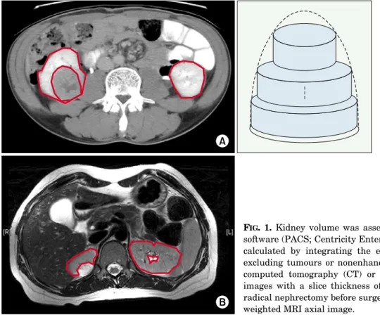
All axial images were checked by one urologist who was blinded to patient characteristics in order to distinguish normal-functioning tissue excluding tumor tissue or non- enhanced areas from the axial side of the delayed CT or MRI images with a slice thickness of 5 mm (Fig. 1). The process of distinguishing the tissue depended only on the gross ap- pearance of the images.
The two-dimensional extent of normal-functioning tis- sue from each image was measured by hand, and the kidney volume was calculated by integrating that extent of nor- mal-functioning tissue.
3. Statistical analysis
Preoperative factors such as age, gender, history of dia- betes, history of hypertension, body mass index, pre- operative eGFR, serum uric acid, urine albumin, normal renal parenchymal volume, tumor size, and the ratio of the normal parenchymal volume of the removed kidney to that of the remaining kidney were compared between groups A and B. For the comparison, the Mann-Whitney U test was used for continuous variables and the chi-square test was used for categorical variables. Predictive factors for renal insufficiency 5 years after radical nephrectomy were ana- lyzed by using univariate and multivariate logistic binary regression.
Because the eGFR 5 years after radical nephrectomy can be influenced by the preoperative eGFR, we reanalyzed the preoperative predictive factors for the downgrading of CKD stage on the basis of the eGFR (Table 1), which may be a more objective measure of kidney function change. For
TABLE 1. Chronic kidney disease stage based on eGFR
Stage GFR Description
1 ≥90 Normal kidney function but urine findings or structural abnormalities or genetic trait point to kidney disease 2 60–89 Mildly reduced kidney function, and other findings (as for stage 1) point to kidney disease
3A 45–59 Moderately reduced kidney function
3B 30–44
4 15–29 Severely reduced kidney function
5 <15 or on dialysis Very severe or endstage kidney failure (sometimes call established renal failure) Defined by the National Institute for Health and Clinical Excellence, 2008 [13].
eGFR, estimated glomerular filtration rate.
that purpose, 89 patients were categorized into 2 groups:
downgrade in CKD stage (group C) and no change in CKD stage (group D).
A receiver operating characteristic (ROC) curve was plotted to determine the cutoff value of the parameters found to be significant for predicting an eGFR greater than 60 mL/min/1.73 m2 at 5 years after radical nephrectomy.
All calculated p-values were two-sided, and values less than 0.05 were considered statistically significant. All data analyses were performed with IBM SPSS ver. 19.0 (IBM Co., Armonk, NY, USA).
RESULTS
The clinical characteristics of the 89 patients included in the present study are shown in Table 2. On the basis of the eGFR 5 years after radical nephrectomy, 43 patients with
an eGFR of less than 60 mL/min/1.73 m2 (group A) and 46 patients with an eGFR of 60 mL/min/1.73 m2 or higher (group B) were compared. Of these patients, 61 were men and 28 were women. The median age of the patients was 61 years (range, 53 to 68 years) in group A and 48 years (range, 42 to 57 years) in group B. The median parenchymal volume of the remnant kidney was 156.28 mL in group A and 181.82 mL in group B.
Median age (61.0 years vs. 48.0 years, p<0.001), pre- operative eGFR (73.97 vs. 83.95, p<0.001), serum uric acid level (5.5 vs. 4.3, p=0.011), and parenchymal volume of the remnant kidney (156.28 vs. 181.92, p=0.003) were sig- nificantly different between groups A and B.
Table 3 shows the results of the univariate and multi- variate binary regression analysis to identify the pre- dictive factors for renal sufficiency (an eGFR less than 60 mL/min/1.73 m2) 5 years after radical nephrectomy. With FIG. 1. Kidney volume was assessed using personal computer-based software (PACS; Centricity Enterprise Web ver. 3.0). The volume was calculated by integrating the extent of normal functioning tissue, excluding tumours or nonenhanced areas, from the axial side of the computed tomography (CT) or magnetic resonance imaging (MRI) images with a slice thickness of 5 mm for patients who underwent radical nephrectomy before surgery (A) delayed CT axial image (B) T2 weighted MRI axial image.
306 Ahn et al
TABLE 2. Patient characteristics
Parameter eGFR<60 (n=43) eGFR≥60 (n=46) p-value
Age (y) 61.0 (53.0–68.0) 48.0 (42.0–57.0) <0.001
Sex 0.809
Male 30 (69.8) 31 (67.4)
Female 13 (30.2) 15 (32.6)
Body mass index (kg/m2) 24.9 (23.1–26.7) 24.2 (22.9–25.5) 0.305
Tumor size (cm) 4.0 (2.7–5.5) 4.5 (2.7–6.0) 0.340
Preoperative eGFR (mL/min/1.73 m2) 73.97 (69.39–82.34) 83.95 (75.27–93.18) <0.001
Ratioa 0.49 (0.47–0.52) 0.49 (0.46–0.51) 0.290
Hypertension 9 (20.9) 6 (13.0) 0.321
Diabetes mellitus 6 (14.0) 1 (2.2) 0.053
Serum uric acid (mg/dL) 5.5 (4.4–6.2) 4.3 (3.9–5.4) 0.011
Positive urine albumin 5 (11.6) 1 (2.2) 0.103
Parenchymal volume of remnant kidney (mL) 156.28 (128.54–168.92) 181.92 (150.16–193.74) 0.003 Values are presented as median (interquartile range) or number (%).
eGFR, estimated glomerular filtration rate.
a:Parenchymal volume of remnant kidney/total parenchymal volume.
TABLE 3. Predictive factors for eGFR<60 mL/min/1.73 m2 5 years after radical nephrectomy (univariate and multivariate analysis)
Varialble Univariate analysis Multivariate analysis
Odds ratio 95% CI p-value Odds ratio 95% CI p-value
Gender 0.456–2.737 0.809 NS
Female Referent
Male 1.117
Age (y) 1.090 1.042–1.140 <0.001 NS
Size (cm) 0.913 0.218–1.178 0.278 NS
Body mass index (kg/m2) 0.699–3.795 0.259 0.409–4.091 0.661
<25 Referent Referent ≥25 1.628 1.294
Parenchymal volume of 1.870–11.758 0.001 2.497–33.785 0.001
remnant kidney (mL)
≥170 Referent Referent <170 4.689 9.185
Ratioa 0.529–2.865 0.629 NS
<0.5 Referent
≥0.5 1.231
Hypertension 0.570–5.461 0.324 NS
No Referent
Yes 1.765
Diabetes mellitus 0.841–63.353 0.071 1.238–327.368 0.035
No Referent Referent Yes 7.297 20.129
Urine albumin 0.663–52.907 0.111 0.753–85.263 0.085
No Referent Referent Yes 5.921 8.012
Serum uric acid 1.404 1.010–1.952 0.043 1.512 0.973–2.349 0.066
Preoperative eGFR 0.934 0.896–0.974 <0.001 0.929 0.879–0.983 0.011
eGFR, estimated glomerular filtration rate; CI, confidence interval; NS, not significant.
a:Parenchymal volume of remnant kidney/ total parenchymal volume.
control for BMI, urine albumin, and serum uric acid, the parenchymal volume of the remnant kidney (odds ratio [OR], 9.185; p=0.001), a history of diabetes (OR, 20.129;
p=0.035), and preoperative eGFR (OR, 0.929; p=0.011)
showed an association with renal insufficiency.
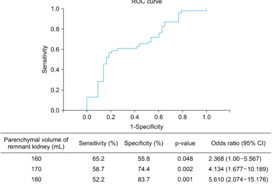
Because eGFR 5 years after radical nephrectomy was found to correlate with the parenchymal volume of the rem- nant kidney, an ROC curve was plotted by using data from
FIG. 2. Receiver operating characteri- stic (ROC) curve based on parenchy- mal volume of remnant kidney volume.
CI, confidence interval.
TABLE 4. Patient characteristics according to CKD category change
Downgrade of CKD No change of CKD
Parameter p-value
category (n=61) category (n=28)
Age (y) 56.0 (46.0–64.0) 55.0 (42.0–60.0) 0.262
Sex 0.374
Male 40 (65.6) 21 (75.0)
Female 21 (34.4) 7 (25.0)
Body mass index (kg/m2) 24.3 (23.0–26.3) 24.5 (23.1–25.5) 0.510
Tumor size (cm) 4.0 (1.5–13.0) 4.0 (2.5–5.7) 0.630
Preoperative eGFR (mL/min/1.73 m2) 80.34 (71.30–93.18) 78.51 (73.58–83.80) 0.327
Ratioa 0.49 (0.47–0.51) 0.49 (0.46–0.51) 0.958
Hypertension 12 (19.7) 3 (10.7) 0.372
Diabetes mellitus 7 (11.5) 0 0.093
Serum uric acid (mg/dL) 4.9 (4.0–6.0) 4.5 (4.1–5.5) 0.627
Positive urine albumin 5 (8.2) 1 (3.6) 0.661
Parenchymal volume of remnant kidney (mL) 159.33 (136.02–178.34) 185.91 (146.90–194.73) 0.054 Values are presented as median (interquartile range) or number (%).
CKD, chronic kidney disease; eGFR, estimated glomerular filtration rate.
a:Parenchymal volume of remnant kidney/ total parenchymal volume.
group A and group B (Fig. 2). The area under the curve was 0.678 (95% CI, 1.677 to 10.189). The optimal cutoff value of the parenchymal volume of the remnant kidney was 170 mL with a sensitivity of 58.7% and a specificity of 74.4% for predicting an eGFR of less than 60 mL/min/1.73 m2.
The clinical characteristic of the 89 patients categorized by change in CKD stage are shown in Table 4. Sixty-one pa- tients showed a downgrade in CKD stage (group C) and 28 patients showed no change in CKD stage (group D). The me- dian parenchymal volume of the remnant kidney was 159.33 mL for group C and 185.91 mL for group D. No pa- rameters were significantly different between groups C and D.
Table 5 provides the results of the univariate and multi- variate binary regression analysis to identify the pre- dictive factors for the downgrading of CKD category 5 years after radical nephrectomy. Only the parenchymal volume of the remnant kidney was statistically significant for the downgrading of CKD category in the multivariate linear re- gression analysis (OR, 3.164; p=0.021). No statistically sig- nificant association was identified for preoperative eGFR (p=0.063).
DISCUSSION
It has been noted that a decrease in the eGFR is associated
308 Ahn et al TABLE 5. Predictive factors for downgrade of CKD category 5 years after radical nephrectomy (univariate and multivariate analysis)
Varialble Univariate analysis Multivariate analysis
Odds ratio 95% CI p-value Odds ratio 95% CI p-value
Gender 0.232–1.735 0.376 NS
Female Referent
Male 0.635
Age (y) 1.020 0.981–1.060 0.316 NS
Size (cm) 1.065 0.890–1.275 0.490 NS
Bddy mass index (kg/m2) 1.086 0.910–1.296 0.362 NS
Parenchymal volume of remnant kidney (mL)
≥170 Referent 1.016–6.348 0.046 Referent 1.191–8.405 0.021
<170 2.540 3.164
Ratioa 0.303–1.846 0.529 NS
<0.5 Referent
≥0.5 0.748
Hypertension 0.527–7.902 0.302 NS
No Referent
Yes 2.041
Urine albumin 0.268–21.661 0.432 NS
No Referent
Yes 2.411
Serum uric acid 1.039 0.746–1.446 0.822 NS
Preoperative eGFR 1.030 0.990–1.073 0.144 1.043 0.998–1.090 0.063
CKD, chronic kidney disease; CI, confidence interval; NS, not significant; eGFR, estimated glomerular filtration rate.
a:Parenchymal volume of remnant kidney/ total parenchymal volume.
with an increased risk of cardiovascular events and many other complications [5,14]. Many studies have found that patients with RCC have a high rate of renal insufficiency and that renal surgery, the treatment of choice for renal tu- mors, can negatively affect postoperative renal function [1-4]. Nephron-sparing surgery is an option that can reduce the chance of postoperative renal insufficiency if it is tech- nically feasible, especially in patients who have more risk factors for postoperative renal insufficiency.
Recently, as experience with partial nephrectomy has accumulated, nephron-saving surgery has been performed even for large (>4 cm), endophytic tumors. Many studies have reported satisfactory results [9,10], which suggests that it is possible to perform nephron-saving surgery for more patients. However, nephron-saving surgery is still technically more difficult than radical nephrectomy for large (>4 cm), endophytic renal tumors and can result in more postoperative complications, a longer operation time, and greater estimated blood loss.
Determining the preoperative risk factors and predict- ing renal insufficiency after radical nephrectomy are im- portant for patients with large, endophytic tumors and could allow nephron-saving surgery to be performed in pa- tients with a higher risk of renal insufficiency, even those at a higher risk of complications after partial nephrectomy.
Several studies have been conducted to identify the pre- dictors of CKD or renal insufficiency after radical nephrec- tomy. Older age, hypertension, smoking, low preoperative renal function, and diabetes have all been identified as po- tential risk factors for renal dysfunction [2].
Recently, functional kidney volume has been identified as an additional predictor for postoperative renal function deterioration. Studies have reported a correlation between the percentage functional volume preservation after renal surgery and the percentage decrease in the GFR [15,16].
Simmons et al. [17] reported that preoperative nephron volume and percentage functional volume preservation are the primary determinants of long-term functional outcomes. Regarding donor nephrectomy, Jeon et al. [18]
reported that preoperative kidney volume measurement could be used to predict delayed kidney function recovery.
These findings suggest that RCC patients with insufficient normal renal parenchymal volume could have a higher rate of renal insufficiency after radical nephrectomy and that surgeons should consider partial nephrectomy in these cases.
In order to use the functional volume of either the af- fected or the normal kidney as a predictor of renal in- sufficiency after the surgery, proper measurement of the parenchymal volume of the kidney is important. Because immediate adaptation, such as hypertrophy and hyper- filtration, occurs in the normal kidney after unilateral nephrectomy, several methods have been used by different groups to identify hypertrophy by use of imaging tech- nologies in donors and patients with renal diseases. For ex- ample, Gomez-Anson et al. [19] used image-directed color Doppler ultrasonography and identified a mean hyper- trophy of approximately 20%. However, the ellipsoid meth- od is less accurate and is affected both by interobserver and intraobserver variability [20,21]. Mullerad et al. [22] eval-
uated renal hypertrophy in 42 patients (mean age of 61.5 years) after they underwent unilateral nephrectomy by us- ing a dimercaptosuccinic acid renal scan. Recently, CT and MRI have been used to accurately assess functional kidney volume, because these techniques can exclude nonfunc- tional tissues such as the great renal vessels, perirenal fat, central sinus fat, and renal mass, which allowed for accu- rate functional kidney parenchymal assessment in our study.
Recent studies have shown that tumor size is a sig- nificant risk factor for new-onset renal insufficiency in pa- tients treated with radical nephrectomy [20]. In a study by Ohno et al. [23], the incidence of renal function deterio- ration (greater than 30% GFR decrease) was higher in pa- tients with tumors sized 7 cm or less than in those with tu- mors greater than 7 cm (74.7% vs 32.8%, p<0.001). In our study, however, no significant association was identified for tumor size in the univariate analysis (OR, 0.913;
p=0.278).
We focused on the influence of the absolute and relative renal parenchymal volume of the remaining kidney on postoperative eGFR. A ratio (parenchymal volume of rem- nant kidney/total parenchymal volume) of more than 0.5 and less than 0.5 had no significant difference in the uni- variate analysis (OR, 1.231; p=0.629). Nonetheless, when the absolute renal parenchymal volume of the remaining kidney is larger, especially when the measured volume was greater than 170 mL, the chance of postoperative renal in- sufficiency (OR, 9.185; p=0.001) or CKD downgrading (OR, 3.164; p=0.021) was significantly lower. We suggest that patients with an unaffected kidney that is already hyper- trophied and hyperfiltrated have a lower chance of post- operative renal insufficiency. The recent study by Funahashi et al. [24] supports this conclusion. They re- ported that a group with an increase in kidney volume of less than 15% at 1 week after surgery as compared to the presurgical volume showed a 4.1-fold percentage increase in their risk of experiencing a 10% reduction in their post- surgical GFR at 6 months compared to the group who expe- rienced an increase in kidney volume of greater than or equal to 15%.
When undergoing renal surgery, patients should be in- formed of the renal functional outcome in addition to the oncological outcome so that they may make an informed preoperative treatment decision. We believe that our pre- dictive model for new-onset renal insufficiency is useful for preoperative patient counseling.
However, our study had several limitations. First, the patients analyzed in this study represent a retrospective, single-institution experience. Furthermore, the lack of randomization introduces a significant selection bias in the observed functional outcomes of patients treated with partial nephrectomy versus radical nephrectomy.
Second, in order to differentiate the nonenhanced area of the kidney from the CT and MRI scans, we depended on one urologist’s opinion about the gross appearance of the images. This could have resulted in bias in our results.
Third, there is no standard method currently available for measuring kidney volume by CT or MRI scan. Conse- quently, the method of measurement could introduce variability.
Last, prospective data are needed to validate our pre- dictive model for renal insufficiency and to analyze post- operative renal function longitudinally.
CONCLUSIONS
The results of this study suggest that preoperative eGFR, history of diabetes, and the radiologic volume of the re- maining kidney parenchyma could be useful factors to pre- dict postoperative renal function. Patients with a remain- ing kidney parenchymal volume of less than 170 mL should be considered candidates for nephron-sparing surgery if technically feasible, because this group has a higher chance of downgrading of CKD category 5 years after radical nephrectomy.
CONFLICTS OF INTEREST The authors have nothing to disclose.
REFERENCES
1. Barlow LJ, Korets R, Laudano M, Benson M, McKiernan J.
Predicting renal functional outcomes after surgery for renal cort- ical tumours: a multifactorial analysis. BJU Int 2010;106:489-92.
2. Huang WC, Levey AS, Serio AM, Snyder M, Vickers AJ, Raj GV, et al. Chronic kidney disease after nephrectomy in patients with renal cortical tumours: a retrospective cohort study. Lancet Oncol 2006;7:735-40.
3. Jeon HG, Jeong IG, Lee JW, Lee SE, Lee E. Prognostic factors for chronic kidney disease after curative surgery in patients with small renal tumors. Urology 2009;74:1064-8.
4. Suer E, Burgu B, Gokce MI, Turkolmez K, Beduk Y, Baltaci S.
Comparison of radical and partial nephrectomy in terms of renal function: a retrospective cohort study. Scand J Urol Nephrol 2011;45:24-9.
5. Thomas R, Kanso A, Sedor JR. Chronic kidney disease and its complications. Prim Care 2008;35:329-44, vii.
6. Belldegrun A, Tsui KH, deKernion JB, Smith RB. Efficacy of nephron-sparing surgery for renal cell carcinoma: analysis based on the new 1997 tumor-node-metastasis staging system. J Clin Oncol 1999;17:2868-75.
7. Fergany AF, Hafez KS, Novick AC. Long-term results of nephron sparing surgery for localized renal cell carcinoma: 10-year followup. J Urol 2000;163:442-5.
8. Lee CT, Katz J, Shi W, Thaler HT, Reuter VE, Russo P. Surgical management of renal tumors 4 cm. or less in a contemporary cohort. J Urol 2000;163:730-6.
9. Simmons MN, Weight CJ, Gill IS. Laparoscopic radical versus partial nephrectomy for tumors >4 cm: intermediate-term onco- logic and functional outcomes. Urology 2009;73:1077-82.
10. Nouralizadeh A, Simforoosh N, Tabibi A, Basiri A, Ziaee SA, Soleimani M, et al. Laparoscopic partial nephrectomy for tu- mours >4 cm compared with smaller tumours: perioperative results. Int Urol Nephrol 2011;43:371-6.
11. Morrisroe SN, Su RR, Bae KT, Eisner BH, Hong C, Lahey S, et al. Differential renal function estimation using computerized to-
310 Ahn et al mography based renal parenchymal volume measurement. J
Urol 2010;183:2289-93.
12. Funahashi Y, Hattori R, Yamamoto T, Kamihira O, Sassa N, Gotoh M. Relationship between renal parenchymal volume and single kidney glomerular filtration rate before and after unilat- eral nephrectomy. Urology 2011;77:1404-8.
13. National Institute for Health and Care Excellence. Chronic kid- ney disease: early identification and management of chronic kid- ney disease in adults in primary and secondary care. September 2008 [Internet]. London: National Institute for Health and Care Excellence; c2013 [cited 2013 Jan 10]. Available from:
http://www.nice.org.uk/nicemedia/live/12069/42117/42117.pdf.
14. Go AS, Chertow GM, Fan D, McCulloch CE, Hsu CY. Chronic kid- ney disease and the risks of death, cardiovascular events, and hospitalization. N Engl J Med 2004;351:1296-305.
15. Jeon HG, Gong IH, Hwang JH, Choi DK, Lee SR, Park DS.
Prognostic significance of preoperative kidney volume for predict- ing renal function in renal cell carcinoma patients receiving a rad- ical or partial nephrectomy. BJU Int 2012;109:1468-73.
16. Sharma N, O'Hara J, Novick AC, Lieber M, Remer EM, Herts BR.
Correlation between loss of renal function and loss of renal volume after partial nephrectomy for tumor in a solitary kidney. J Urol 2008;179:1284-8.
17. Simmons MN, Fergany AF, Campbell SC. Effect of parenchymal volume preservation on kidney function after partial nephrec- tomy. J Urol 2011;186:405-10.
18. Jeon HG, Lee SR, Joo DJ, Oh YT, Kim MS, Kim YS, et al.
Predictors of kidney volume change and delayed kidney function recovery after donor nephrectomy. J Urol 2010;184:1057-63.
19. Gomez-Anson B, Carrero-Lopez V, Diaz-Gonzalez R. Image-di- rected color Doppler ultrasound evaluation of the single kidney after unilateral nephrectomy in adults. J Clin Ultrasound 1997;25:29-35.
20. Bakker J, Olree M, Kaatee R, de Lange EE, Moons KG, Beutler JJ, et al. Renal volume measurements: accuracy and repeat- ability of US compared with that of MR imaging. Radiology 1999;211:623-8.
21. Emamian SA, Nielsen MB, Pedersen JF, Ytte L. Kidney di- mensions at sonography: correlation with age, sex, and habitus in 665 adult volunteers. AJR Am J Roentgenol 1993;160:83-6.
22. Mullerad M, Kastin A, Issaq E, Moskovitz B, Groshar D, Nativ O. The value of quantitative 99M technetium dimercaptosuccinic acid renal scintigraphy for predicting postoperative renal in- sufficiency in patients undergoing nephrectomy. J Urol 2003;169:24-7.
23. Ohno Y, Nakashima J, Ohori M, Hashimoto T, Iseki R, Hatano T, et al. Impact of tumor size on renal function and prediction of renalinsufficiency after radical nephrectomy in patients with re- nal cell carcinoma. J Urol 2011;186:1242-6.
24. Funahashi Y, Hattori R, Yamamoto T, Kamihira O, Moriya Y, Gotoh M. Change in contralateral renal parenchymal volume 1 week after unilateral nephrectomy. Urology 2009;74:708-12.