On account of an increasing understanding of femoroac- etabular impingement (FAI), hip arthroscopy has gained considerable popularity in the past decade.1) And there has been an increasing body of literature regarding ar-
throscopic management of FAI and most of them were intra-articular pathologies about cam- and pincer-type of FAI.2-5) Recent sports medicine literature has focused mainly on these hip conditions, but advances in diagnostic imaging techniques now allow for the precise diagnosis of various intra-articular conditions including labral tears, chondral lesions, hip osteoarthritis, ruptured ligamentum teres, osteonecrosis, problems after hip resurfacing or arthroplasty, hip instability, synovial conditions, and prob- lems associated trauma.6,7) Among them, nonconcentric reduction after hip dislocation or intra-articular fragments
Usefulness of Arthroscopic Treatment of Painful Hip after Acetabular Fracture or Hip Dislocation
Jung-Taek Hwang, MD, Woo-Yong Lee, MD*, Chan Kang, MD*, Deuk-Soo Hwang, MD*, Dong-Yeol Kim, MD*, Long Zheng, MD
†Department of Orthopaedic Surgery, Hallym University Chuncheon Sacred Heart Hospital, Hallym University College of Medicine, Chuncheon,
*Department of Orthopaedic Surgery, Chungnam National University School of Medicine, Daejeon, Korea
†Department of Orthopaedic Surgery, Yanbian University School of Medicine, Yanji, China
Background: Painful hip following hip dislocation or acetabular fracture can be an important signal for early degeneration and progression to osteoarthritis due to intraarticular pathology. However, there is limited literature discussing the use of arthroscopy for the treatment of painful hip. The purpose of this retrospective study was to analyze the effectiveness and benefit of arthroscop- ic treatment for patients with a painful hip after major trauma.
Methods: From July 2003 to February 2013, we reviewed 13 patients who underwent arthroscopic treatment after acetabular fracture or hip dislocation and were followed up for a minimum of 2 postoperative years. The degree of osteoarthritis based on the Tonnis classification pre- and postoperatively at final follow-up was determined. Clinical outcomes were evaluated using visual analogue scale for pain (VAS) and modified Harris hip score (MHHS), and range of motion (ROM) of the hip pre- and postoperatively at final follow-up.
Results: There were nine male and four female patients with a mean age at surgery of 28 years (range, 20 to 50 years). The mean follow-up period of the patients was 59.8 months (range, 24 to 115 months), and the mean interval between initial trauma and ar- throscopic treatment was 40.8 months (range, 1 to 144 months). At the final follow-up, VAS and MHHS improved significantly from 6.3 and 53.4 to 3.0 and 88.3, respectively (p = 0.002 and p < 0.001, respectively). However, there were no significant differences in hip flexion, abduction, adduction, external rotation, and internal rotation as minor improvements from 113.1°, 38.5°, 28.5°, 36.5°, and 22.7° to 118.5°, 39.0°, 29.2°, 38.9°, and 26.5° were observed, respectively (p = 0.070, p = 0.414, p = 0.317, p = 0.084, and p = 0.136, respectively). None of the patients exhibited progression of osteoarthritis of the hip at the final follow-up.
Conclusions: Arthroscopic treatment after acetabular fracture or hip dislocation is effective and delays the progression of trau- matic osteoarthritis.
Keywords: Hip, Wounds and injuries, Osteoarthritis, Arthroscopy
Copyright © 2015 by The Korean Orthopaedic Association
This is an Open Access article distributed under the terms of the Creative Commons Attribution Non-Commercial License (http://creativecommons.org/licenses/by-nc/4.0) which permits unrestricted non-commercial use, distribution, and reproduction in any medium, provided the original work is properly cited.
Clinics in Orthopedic Surgery • pISSN 2005-291X eISSN 2005-4408 Received June 2, 2015; Accepted August 9, 2015
Correspondence to: Deuk-Soo Hwang, MD
Department of Orthopaedic Surgery, Chungnam National University School of Medicine, 266 Munwha-ro, Jung-gu, Daejeon 35015, Korea Tel: +82-42-338-2480, Fax: +82-42-338-2482
E-mail: dshwang@cnu.ac.kr
due to fracture around hip joint are possible candidates for hip arthroscopy.8-12)
Fractures or dislocations around the hip joint are caused by high-energy trauma and mainly occur in young adults as a result of traffic accidents, such as motor vehicle accidents.13,14) The rate of coxarthrosis after hip dislocation is 24% for simple dislocations and 88% for those associ- ated with acetabular fractures.9) Hip pain after acetabular fracture and hip dislocation is associated with anatomical reduction, presence of intra-articular loose fragments, in- stability of the hip joint, and concomitant injury. The most common painful intra-articular pathologies after acetabu- lar fracture or hip dislocation major hip joint trauma are ligamentum teres injury, loose body, cartilage injury, and labral tear.8) The hip pain due to intra-articular pathol- ogy can potentially be a signal for early degeneration and progression to osteoarthritis.10) However, there is limited literature discussing the usefulness of arthroscopy for the treatment of hip pain after high-energy acetabular fracture or traumatic hip dislocation.15,16)
We hypothesized that arthroscopic treatment of painful hip after acetabular fracture or hip dislocation is
effective and delays the progression of traumatic osteoar- thritis. This retrospective study was carried out to evaluate clinical outcomes and radiological outcome of arthroscop- ic treatment for painful hip with intra-articular patholo- gies after acetabular fracture or hip dislocation.
METHODS
Patient Selection
This study was approved by the Chungnam National Uni- versity Hospital Institutional Review Board and informed consent was waived from all patients. From July 2003 to February 2013, we performed retrospective review of 13 patients, who underwent hip arthroscopy due to persistent pain and intraarticular pathology after major hip trauma such as acetabular fracture and hip dislocation. The exclu- sion criteria was as follows: (1) a history of low-energy hip trauma (i.e., contusion or sprain); (2) history of surgery involving the femur or pelvis before the major trauma; (3) Tonnis grade 2 or above;17) (4) proliferative disease of the hip (i.e., synovial chondromatosis, pigmented villonodular synovitis); (5) neuromuscular disease (i.e., cerebral palsy);
A B
C D
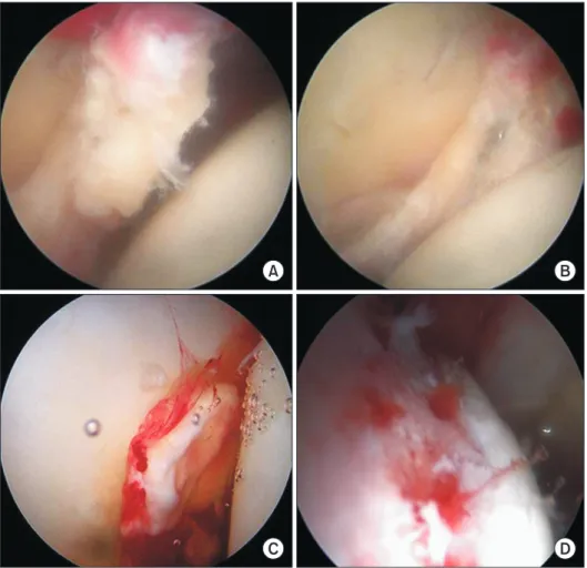
Fig. 1. A 50-year-old male who had traumatic hip dislocation underwent arthro scopic treatment. (A) Arthroscopic view shows posterior labral tear. (B) Arthroscopic view shows arthroscopic partial labrectomy. (C) Arthroscopic view shows intra-articular loose body on ace- tabular articular surface. (D) Arthro scopic view shows removal of loose body and microfracture.
(6) Legg-Calve-Perthes deformity; (7) ankylosing spondy- litis or diffuse idiopathic skeletal hyperostosis; and (8) de- velopmental dysplasia of the hip. Arthroscopic treatment was planned based on persistent or aggravating hip pain and intra-articular pathologies such as loose fragments, labral tears, and ligamentum teres injury on hip three-di- mensional computed tomography (3D CT) and magnetic resonance imaging (MRI).18)
Surgical Technique and Postoperative Care
All arthroscopic procedures were performed by a single senior surgeon. After general anesthesia, the patient was placed in a supine position, the hip joint was abducted by 10° to 15°, and the lower extremity was placed in a neutral position and fixed to the fracture table. For procedures on the central compartment of the joint, the hip joint was extended about 8 to 10 mm under monitoring with an image amplifier. Three portals (anterior, anterolateral, and posterolateral) were made, mainly using a 70°-angled arthroscope. After the intra-articular pathologies were identified subsequent to appropriate anterolateral capsu- lotomy, loose fragments were removed using a grasper via the anterior or posterolateral portal, and labral tears were debrided using an arthroscopic shaver or repaired using a 2.9-mm Bioraptor suture anchor (Smith & Nephew, London, UK) (Fig. 1A–C). Ligamentum teres injury was debrided using a shaver or was shrunk by Vulcan (Smith
& Nephew) through the anterior or posterolateral portal, and cartilage injuries of the femoral head and acetabulum were treated by microfracture (Fig. 1D). After traction was released, the arthroscope was moved towards the periph-
eral compartment through the same portal while the hip joint was flexed by about 40°, at which time a 30° angled arthroscope was introduced. Pathologies of the peripheral compartment, such as a femoral head neck spur or a trau- matic bony deformity, were treated by femoroplasty.13,18)
All cases underwent 3D CT on the second day post- operatively to check the postoperative state of the acetabu- loplasty and femoroplasty and removal of loose bodies (Fig. 2). All patients were discharged on the third day after the procedure. Partial weight-bearing with crutches was permitted for four weeks in case of patients who had un- dergone osteoplasty and labral repair. Continuous passive motion and passive pendulum exercise were started after the procedure to avoid postoperative capsular adhesion.
Hyperflexion of the hip over 90° was forbidden for four weeks.
Radiological and Clinical Evaluation
We identified the degree of osteoarthritis based on the Tonnis classification pre- and postoperatively at final follow-up.17) Clinical outcomes were evaluated using visual analogue scale for pain (VAS) and modified Harris hip score (MHHS), and range of motion (ROM) of the hip pre- and postoperatively at final follow-up.19)
Statistical Analysis
The paired t-tests were used to assess the difference in out- comes before surgery and at the time of the final follow- up. The IBM SPSS ver. 19.0 (IBM Co., Armonk, NY, USA) was used for all statistical analyses, with the α level set at 0.05.
A B
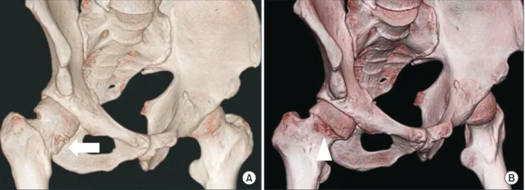
Fig. 2. Pre- and postoperative three-dimensional computed tomography (3D CT) images. (A) Preoperative 3D CT image shows loose body on femoral head and neck (arrow). (B) Postoperative 3D CT image shows removal of loose body and femoroplasty (arrowhead).
RESULTS
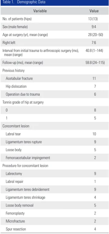
There were 13 patients (nine males and four females) with a mean age of 28 years (range, 20 to 50 years) at the index operation. The mean follow-up period was 59.8 months (range, 24 to 115 months). The mean interval from initial trauma to arthroscopic surgery was 40.8 months (range, 1 to 144 months) (Table 1). The cause of injury was traffic
accident in ten patients and sports injury in three patients, of them, six patients had undergone hip surgery after ac- etabular fracture or hip dislocation. Arthroscopically, we performed loose bony fragment removal, debridement or repair of a torn acetabular labrum, debridement or shrink- age of a torn ligament teres remnant, excision of osteo- phytes, synovectomy, or microfractures (Table 1).
At the final follow-up, VAS and MHHS improved significantly from 6.3 and 53.4 to 3.0 and 88.3, respectively (p = 0.002 and p < 0.001, respectively). However, there were no significant differences in hip flexion, abduction, adduction, external rotation, and internal rotation as im- provement was seen from 113.1°, 38.5°, 28.5°, 36.5°, and 22.7° to 118.5°, 39.0°, 29.2°, 38.9°, and 26.5° , respectively (p = 0.070, p = 0.414, p = 0.317, p = 0.084, and p = 0.136, respectively) (Table 2).
None of the patients exhibited progression of os- teoarthritis of the hip at the final follow-up. Eight and five patients with preoperative Tonnis grade 0 and 1 were suc- cessful in maintaining their grade, respectively. There were no intra- or perioperative complications such as neural injury and wound infection.
DISCUSSION
Hip arthroscopy provides a clear view of the articular sur- face of the femoral head, the acetabular labrum, the liga- mentum teres and synovium.7) Today, hip arthroscopy is becoming popular for procedures not only inside the hip joint but also surrounding it.6,20,21) Therefore, arthroscopic surgery of the hip joint can facilitate diagnosis and treat- ment of labral tears and other intra-articular and periph- eral pathologies.8,18,22-24) However, few reports have focused on arthroscopic treatment of coxarthrosis after fracture Table 1. Demographic Data
Variable Value
No. of patients (hips) 13 (13)
Sex (male:female) 9:4
Age at surgery (yr), mean (range) 28 (20–50)
Right:left 7:6
Interval from initial trauma to arthroscopic surgery (mo),
mean (range) 40.8 (1–144)
Follow-up (mo), mean (range) 59.8 (24–115)
Previous history
Acetabular fracture 11
Hip dislocation 7
Operation due to trauma 6
Tonnis grade of hip at surgery
0 8
1 5
Concomitant lesion
Labral tear 10
Ligamentum teres rupture 9
Loose body 5
Femoroacetabular impingement 2
Procedure for concomitant lesion
Labrectomy 9
Labral repair 1
Ligamentum teres debridement 9
Ligamentum teres shrinkage 4
Loose body removal 5
Femoroplasty 2
Microfracture 2
Spur resection 4
Table 2. Comparison of the Pre- and Postoperative Clinical Out- comes at the Final Follow-up
Variable Preoperative Final p-value
Visual analogue scale 6.3 ± 1.7 3.0 ± 2.4 0.002 Modified Harris hip score 53.4 ± 21.2 88.3 ± 7.7 < 0.001
Flexion (°) 113.1 ± 13.0 118.5 ± 7.7 0.070
Abduction (°) 38.5 ± 3.2 39.0 ± 2.8 0.414
Adduction (°) 28.5 ± 2.4 29.2 ± 1.9 0.317
External rotation (°) 36.5 ± 6.6 38.9 ± 4.6 0.084 Internal rotation (°) 22.7 ± 9.0 26.5 ± 7.5 0.136 Values are presented as mean ± standard deviation.
or dislocation of the hip joint. In the previous study, ar- throscopic debridement of 23 ligamentum teres ruptures that occurred after major hip trauma showed a satisfactory outcome on the Harris hip score with a 20-point improve- ment or better, and 15 cases had intra-articular patholo- gies such as loose bodies, cartilage injury, and labral tears.8) The most common intra-articular pathologies that occur in athletes after hip dislocation are labral tears, cartilage injury, loose bodies, and ligamentum teres injury.10) In our study, loose bodies, labral tears, ligamentum teres injury, and cartilage injury as intra-articular lesions were checked, and the VAS and MHHS improved significantly at the fi- nal follow-up after surgery. In addition, osteoarthritis did not progress at the final follow-up.
In particular, an intra-articular loose body, frequent- ly observed after hip trauma, could cause progression of traumatic osteoarthritis.25-27) Svoboda et al.11) reported that arthroscopic removal of the loose body in patients with posterior dislocation of the hip was safe and effective. Ya- mamoto et al.12) reported that among 11 patients with frac- ture and/or dislocation of the hip joint, who underwent ar- throscopic treatment, nine patients were treated with loose body removal, which safely prevented traumatic arthritis.
In our study, five patients underwent arthroscopic removal with effective relief of their symptoms. Furthermore, it has been reported that labral tear results in hip pain and osteoarthritis.2,3,28) Ilizaliturri et al.13) reported that among 17 patients, who underwent arthroscopic treatment after posterior dislocation of the hip, 14 patients had anterior labral tears and six patients had posterior labral tear and most of the patients had significant improvement after hip arthroscopy. In our study, ten patients had labral tear on arthroscopy and experienced relief of symptoms after treatment with debridement and/or repair (Table 3).
Our study had several limitations. First, the number
of patients was not large enough to generate significant results. However, patients, who underwent arthroscopic treatment due to acetabular fracture or hip dislocation, were exceptional. Second, we could not design a prospec- tive comparative study. Nevertheless, our study is associ- ated with significant features. First, there exist only few studies discussing the use of arthroscopy for the treatment of hip pain following high-energy acetabular fracture or traumatic hip dislocation. Second, we demonstrated that arthroscopic treatment for coxarthrosis after acetabular fracture or hip dislocation effectively prevents progression of traumatic arthritis on short-term follow-up.
In conclusion, arthroscopic treatment for painful hip after acetabular fracture or hip dislocation could result in satisfactory outcomes including relief of symptoms and prevention of the progression of traumatic arthritis on a minimum follow-up of 2 years. Consequently, the hypoth- eses that arthroscopic treatment of painful hip after ac- etabular fracture or hip dislocation is effective and delays the progression of traumatic osteoarthritis were supported through the present study. However, future study demands mid- and long-term results in more patients, which will provide detailed insights to this area of study.
CONFLICT OF INTEREST
No potential conflict of interest relevant to this article was reported.
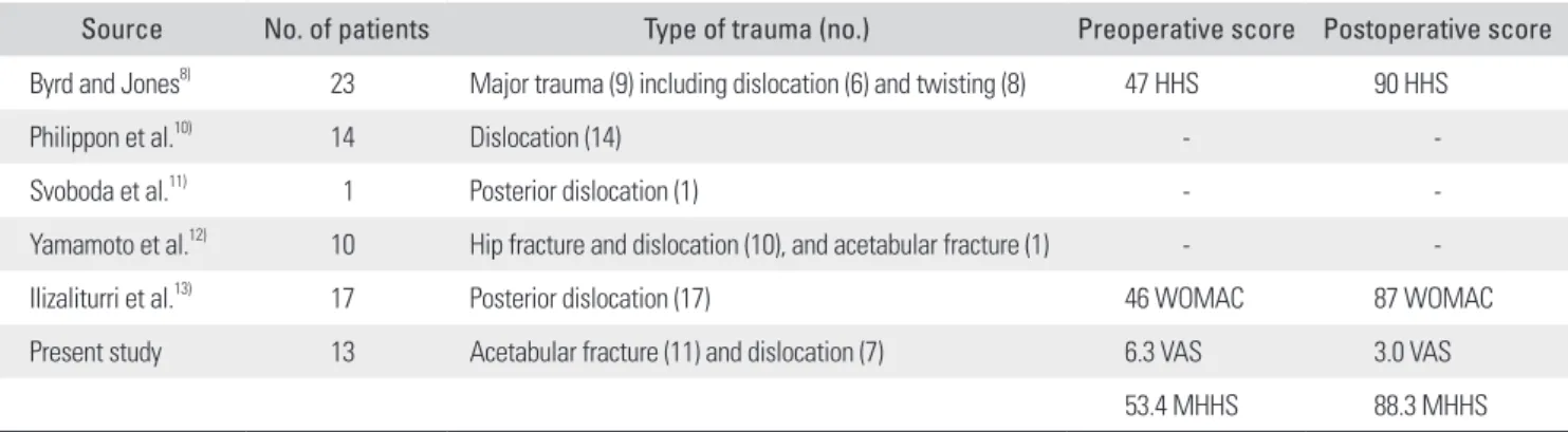
Table 3. Summary of Arthroscopic Treatment of Trauma around Hip Joint
Source No. of patients Type of trauma (no.) Preoperative score Postoperative score
Byrd and Jones8) 23 Major trauma (9) including dislocation (6) and twisting (8) 47 HHS 90 HHS
Philippon et al.10) 14 Dislocation (14) - -
Svoboda et al.11) 1 Posterior dislocation (1) - -
Yamamoto et al.12) 10 Hip fracture and dislocation (10), and acetabular fracture (1) - -
Ilizaliturri et al.13) 17 Posterior dislocation (17) 46 WOMAC 87 WOMAC
Present study 13 Acetabular fracture (11) and dislocation (7) 6.3 VAS 3.0 VAS
53.4 MHHS 88.3 MHHS
HHS: Harris hip score, WOMAC: Western Ontario and McMaster Universities Osteoarthritis Index, VAS: visual analogue scale for pain, MHHS: modified HHS.
REFERENCES
1. Larson CM, Guanche CA, Kelly BT, Clohisy JC, Ranawat AS. Advanced techniques in hip arthroscopy. Instr Course Lect. 2009;58:423-36.
2. Wagner S, Hofstetter W, Chiquet M, et al. Early osteoarthrit- ic changes of human femoral head cartilage subsequent to femoro-acetabular impingement. Osteoarthritis Cartilage.
2003;11(7):508-18.
3. Ganz R, Parvizi J, Beck M, Leunig M, Notzli H, Siebenrock KA. Femoroacetabular impingement: a cause for osteoar- thritis of the hip. Clin Orthop Relat Res. 2003;(417):112-20.
4. Beck M, Kalhor M, Leunig M, Ganz R. Hip morphology influences the pattern of damage to the acetabular cartilage:
femoroacetabular impingement as a cause of early osteoar- thritis of the hip. J Bone Joint Surg Br. 2005;87(7):1012-8.
5. Sutter R, Zanetti M, Pfirrmann CW. New developments in hip imaging. Radiology. 2012;264(3):651-67.
6. Aprato A, Jayasekera N, Bajwa A, Villar RN. Peri-articular diseases of the hip: emerging frontiers in arthroscopic and endoscopic treatments. J Orthop Traumatol. 2014;15(1):1- 11.
7. Bagaria V, Sapre V. Arthroscopic removal of intraarticular fragments following fracture dislocation of the hip. Indian J Orthop. 2008;42(2):225-7.
8. Byrd JW, Jones KS. Traumatic rupture of the ligamentum teres as a source of hip pain. Arthroscopy. 2004;20(4):385- 91.
9. Foulk DM, Mullis BH. Hip dislocation: evaluation and man- agement. J Am Acad Orthop Surg. 2010;18(4):199-209.
10. Philippon MJ, Kuppersmith DA, Wolff AB, Briggs KK. Ar- throscopic findings following traumatic hip dislocation in 14 professional athletes. Arthroscopy. 2009;25(2):169-74.
11. Svoboda SJ, Williams DM, Murphy KP. Hip arthroscopy for osteochondral loose body removal after a posterior hip dis- location. Arthroscopy. 2003;19(7):777-81.
12. Yamamoto Y, Ide T, Ono T, Hamada Y. Usefulness of ar- throscopic surgery in hip trauma cases. Arthroscopy. 2003;
19(3):269-73.
13. Ilizaliturri VM Jr, Gonzalez-Gutierrez B, Gonzalez-Ugalde H, Camacho-Galindo J. Hip arthroscopy after traumatic hip dislocation. Am J Sports Med. 2011;39 Suppl:50S-57S.
14. Clegg TE, Roberts CS, Greene JW, Prather BA. Hip disloca-
tions: epidemiology, treatment, and outcomes. Injury. 2010;
41(4):329-34.
15. Kashiwagi N, Suzuki S, Seto Y. Arthroscopic treatment for traumatic hip dislocation with avulsion fracture of the liga- mentum teres. Arthroscopy. 2001;17(1):67-9.
16. Mullis BH, Dahners LE. Hip arthroscopy to remove loose bodies after traumatic dislocation. J Orthop Trauma. 2006;
20(1):22-6.
17. Tonnis D. Normal values of the hip joint for the evaluation of X-rays in children and adults. Clin Orthop Relat Res.
1976;(119):39-47.
18. Kang C, Hwang DS, Cha SM. Acetabular labral tears in pa- tients with sports injury. Clin Orthop Surg. 2009;1(4):230-5.
19. Wilkin G, March G, Beaule PE. Arthroscopic acetabular labral debridement in patients forty-five years of age or old- er has minimal benefit for pain and function. J Bone Joint Surg Am. 2014;96(2):113-8.
20. Ayeni OR, Levy BA, Musahl V, Safran MR. Current state- of-the-art of hip arthroscopy. Knee Surg Sports Traumatol Arthrosc. 2014;22(4):711-3.
21. Stevens MS, Legay DA, Glazebrook MA, Amirault D. The evidence for hip arthroscopy: grading the current indica- tions. Arthroscopy. 2010;26(10):1370-83.
22. Byrd JW. Labral lesions: an elusive source of hip pain case reports and literature review. Arthroscopy. 1996;12(5):603- 12.
23. Ide T, Akamatsu N, Nakajima I. Arthroscopic surgery of the hip joint. Arthroscopy. 1991;7(2):204-11.
24. Kelley B, Anderson R, Miles K. Acetabular labrum tear in a 15-year-old male: diagnosis with correlative imaging. Aus- tralas Radiol. 1997;41(2):157-9.
25. Letournel E. Acetabulum fractures: classification and man- agement. Clin Orthop Relat Res. 1980;(151):81-106.
26. Matta JM, Anderson LM, Epstein HC, Hendricks P. Frac- tures of the acetabulum: a retrospective analysis. Clin Or- thop Relat Res. 1986;(205):230-40.
27. Stewart MJ, McCarroll HR Jr, Mulhollan JS. Fracture-dislo- cation of the hip. Acta Orthop Scand. 1975;46(3):507-25.
28. Fitzgerald RH Jr. Acetabular labrum tears: diagnosis and treatment. Clin Orthop Relat Res. 1995;(311):60-8.