99m Tc-Pertechnetate Scintigraphy Predicts Successful
Postoperative Ablation in Differentiated Thyroid Carcinoma Patients Treated with Low Radioiodine Activities
Luca Giovanella, Gaetano Paone, Teresa Ruberto, Luca Ceriani, Pierpaolo Trimboli
Clinic for Nuclear Medicine and Competence Center for Thyroid Diseases, Oncology Institute of Southern Switzerland, Bellinzona, Switzerland
Background: Postoperative routine radioiodine (RAI) treatment is currently debated for patients with low-risk differentiated thyroid carcinoma (DTC) patients. If performed, a low 131I activity (i.e., 1 to 2 GBq) is recommended with the aim to ablate thyroid remnant and facilitate subsequent follow-up by thyroglobulin measurement. The purpose of this study was to evaluate the relationship be- tween postsurgical technetium-99m (99mTc)-pertechnetate scintigraphy and the rate of successful remnant ablation after low activity radioiodine ablation in patients with DTC.
Methods: Enrolled were 193 patients with low risk DTC who underwent total thyroidectomy and RAI ablation with a fixed 1.1 GBq activity of 131I. 99mTc-pertechnetate scans were done and thyrotropin stimulated thyroglobulin (sTg) levels measured just before abla- tion. Ablation effectiveness was assessed 6 to 12 months later by sTg measurement, neck ultrasound and diagnostic whole body scan.
Results: A negative 99mTc-perthecnetate scans was the best predictor of successful ablation (P<0.001) followed by preablative sTg levels <0.8 ng/mL (P=0.008) and 99mTc-pertechnetate uptake rate values <0.9% (P=0.065). Neither sex nor age of the patient at the time of ablation or tumor histology and size showed a significant association with the rate of successful ablation.
Conclusion: The 99mTc-pertechnetate scintigraphy is a simple and feasible tool to predict effectiveness of low activity 131I thyroid to ablate thyroid remnants in patients with DTC.
Keywords: Differentiated thyroid carcinoma; Radioiodine; Radionuclide imaging; Sodium pertechnetate Tc 99m
INTRODUCTION
Postoperative routine radioiodine (RAI) treatment is currently debated for low-risk differentiated thyroid carcinoma (DTC) patients. However, consideration of specific features of the indi- vidual patient that could modulate recurrence risk, disease fol- low-up implications, and patient preferences are relevant to the
decision-making [1]. If performed, a low 131I activity (i.e., 1 to 2 GBq) is recommended with the aim to ablate thyroid remnant and facilitate subsequent follow-up by thyroglobulin (Tg) mea- surement [2]. Post-surgical diagnostic scintigraphy with 131I can give very useful information to properly inform RAI prescrip- tion and questions regarding its potentially negative impact on the efficacy for successful remnant ablation (i.e., ‘‘stunning’’)
Received: 29 September 2018, Revised: 27 December 2018, Accepted: 28 December 2018
Corresponding author: Pierpaolo Trimboli
Clinic for Nuclear Medicine and Competence Center for Thyroid Diseases, Oncology Institute of Southern Switzerland, Via Ospedale 12, Bellinzona 6500, Switzerland
Tel: +41-91-8118672, Fax: +41-91-8118250, E-mail: [email protected]
Copyright © 2019 Korean Endocrine Society
This is an Open Access article distributed under the terms of the Creative Com- mons Attribution Non-Commercial License (http://creativecommons.org/
licenses/by-nc/4.0/) which permits unrestricted non-commercial use, distribu- tion, and reproduction in any medium, provided the original work is properly cited.
can be mitigated by using either low-activity of 131I (37-111 MBq) or alternative tracers as 123I or technetium-99m (99mTc)- pertechnetate [3]. The latter is cheap, widely available and was previously proved to be accurate in predicting successful RAI ablation [4]. However, previous data were mostly obtained in patients treated with RAI activities ≥3.7 GBq [4-6] while data are lacking with currently recommended low activities. The present study was carried out to evaluate the role of 99mTc- pertechnetate scintigraphy to predict the effectiveness of low- activities of 131I administered to ablate postoperative thyroid remnants in low-risk DTC. Finally, the relationship between
99mTc-pertechnetate scan results and corresponding preablation Tg values was assessed.
METHODS
Ethics
The study was approved by the Institutional Review Board (CE- 2013-00014/LG/MN) of Oncology Institute of Southern Swit- zerland and Ethics Committee of Canton Ticino, Bellinzona, Switzerland. An informed consensus was obtained before en- rolling patients, records were anonymized and de-identified pri- or to analysis and all data were fully anonymized prior to any access by the authors.
Standard of care in our center
As per European Association of Nuclear Medicine guidelines all patients with DTC >10 mm in the largest diameter under- went RAI ablation in our centers. In most cases ablation is per- formed 3 to 6 weeks after surgery (i.e., without prescribing thy- roid hormone in between) and 1.1, 2 to 3.7, and 3.7 to 7.4 GBq of 131I are administered in low-, intermediate-, and high-risk DTC patients, respectively. A blood sample is obtained just be- fore RAI treatment and thyrotropin (TSH), Tg and Tg autoanti- bodies (TgAb) are measured. A post-treatment (PT) whole body scan (WBS) with additional single photon emission computed tomography/computed tomography (SPECT-CT) is obtained 3 to 7 days after treatment as previously reported [7-9]. Response to treatment is assessed at 6 to 12 months after ablation by neck ultrasound (US), diagnostic WBS (DxWBS), and recombinant human TSH (rhTSH)-stimulated Tg (sTg) testing. A successful ablation is defined by negative neck US and DxWBS with a corresponding sTg values <1 ng/mL. Ablated patients are fol- lowed-up by basal Tg measurement and clinical examination with additional neck US when indicated by the attending physi- cian. Non-ablated patients are managed according to decisions
of our multidisciplinary thyroid board.
Patients enrollment and study design
This study involved 372 patients with DTC >10 mm in largest diameter who underwent total thyroidectomy followed by low- activity RAI ablation in our center between January 2014 and June 2018. Among them patients who belonged to the low-risk group according to American Thyroid Association 2015 criteria were enrolled [1]. Demographic and clinical data, pathological parameters (tumor size, lymph node involvement, pathologic TNM stage) and pre-ablation TSH-stimulated serum Tg values were recorded. For the purpose of the present study a preabla- tion 99mTc-pertechnetate scan was obtained before RAI ablation and compared to PT-WBS/SPECT-CT, preablation Tg levels and response to treatment, respectively. Patients were prepared by thyroid hormone withdrawal and placed on a low iodine diet 2 weeks before ablation. Pregnancy was excluded in each wom- an of child-bearing age. Before 131I administration a blood sam- ple was obtained and TSH, free thyroxine, Tg, and TgAb were measured in all patients. PT WBS/SPECT-CT images were ex- amined by two experienced nuclear medicine physicians (L.C., G.P.) unaware of the results of 99mTc-pertechnetate scan and classified as (1) negative (i.e., physiologic RAI distribution), (2) remnant tissue (i.e., median foci localized either in the thyroid bed and thyroglossal duct), and (3) positive (i.e., focal uptake localized in front of a focal lesion discernable on morphological CT imaging).
Technetium-99m pertechnetate scintigraphy
Fifteen minutes after intravenous administration of about 185 MBq of 99mTc-pertechnetate, anterior images of the neck were acquired using a gamma-camera equipped with high-resolution parallel-hole collimator (Symbia, Siemens, Erlangen, Germany) with an acquisition time of 10 minutes, using a 20% window centered around the 140 KeV peak of 99mTc and a 128×128 computer matrix. Images were analyzed separately by two ex- perienced board-certified nuclear medicine physicians unaware of PT-WBS/SPECT-CT results (L.G., A.P.). Scans were report- ed as negative (i.e., physiological uptake within the neck and upper mediastinum) or positive (i.e., any uptake above back- ground within the neck and upper mediastinum). Additionally, the 99mTc-pertechnetate uptake rate was determined using the re- gion-of-interest technique as previously described [4]. For sta- tistical analysis, the uptake value was arbitrarily set at 0.1% in patients with no visually discernable thyroid remnant. In case of disagreeing interpretation, a consensus was reached.
TSH was measured on a UniCel DxI 800 immunochemilumi- nescence automated platform (Beckman Coulter SA, Nyon, Switzerland; normal reference range, 0.40 to 4.00 mIU/L). Se- rum Tg and TgAb were measured on the Kryptor compact Plus instrument (BRAHMS Thermo Fisher Scientific, Henningsdorf, Germany). This method uses two fluorophores, Lumi4 and Cya- nin5.5, detected separately or combined in the same antigen-an- tibody complex, a method known as the time-resolved ampli- fied cryptate emission (TRACE). The Kryptor hTg sensitive as- say is calibrated against BCR457 international reference stan- dard. The functional sensitivity (corresponding to inter-assay imprecision of 20%) had been estimated at 0.15 ng/mL by the manufacturer (Instruction For Use, ThermoFisher). The Kryptor TgAb is standardized against 1st International Reference Prepa- ration 65/93 and the functional sensitivity had been estimated at 33 IU/mL by the manufacturer (Instruction For Use, Thermo- Fisher). Then, serum TgAb levels <33 IU/mL were considered as negative (i.e., not interfered) [8,10,11].
Statistical analysis
Descriptive statistics included mean±standard deviation, median and percentiles, minimum and maximum of continuous factors and scores; in the case of categorical factors, number and per- centage distribution. We used Pearson’s chi-square and Kruskal- Wallis or Mann-Whitney U tests to compare categorical and con- tinuous factors, respectively. The Spearman’s rho was adopted to search for correlations. Finally, the nonparametric association coefficient qu (q-phi) was used to evaluate predictive factors for successful ablation. The receiver operating characteristic (ROC) curve analysis was used to identify optimal cut-off levels of se- rum Tg level for predicting PT-WBS/SPECT-CT results (i.e., presence of metastases). In patients without metastases ROC curve analysis was used to identify optimal cut-off levels of se- rum Tg level and 99mTc-pertechnetate uptake rate for predicting successful ablation after low-activity RAI treatment, respective- ly. All analyses were conducted by means of STATA version 14 (StataCorp., College Station, TX, USA) software. A P<0.05 was considered to define nominal statistical significance.
RESULTS
Among 431 DTC patients, 59 had positive TgAb and were ex- cluded from the present study. Among the remaining 372 en- rolled were 193 patients (52%) belonged to the low-risk group:
their demographic, clinical, and pathological data are summa- rized in the Table 1. The PT-WBS/SPECT-CT revealed thyroid
remnants in all cases with additional lymph-nodes metastasis in 24 (12%), respectively. None of our patients had RAI-avid dis- tant metastases. Thyroid remnants and lymph node metastases were visually detected by 99mTc-pertechnetate scan in 99/193 (51%) and 2/24 (8%) of cases, respectively. Neither Tg concen- trations (2.45 ng/mL [<0.15 to 27.2] vs. 3.30 ng/mL [<0.15 to 78.9]) nor the rate of Tg levels exceeding 1 ng/mL (15/24 [62%]
vs. 96/169 [57%]) differed significantly in patients with and without metastases, respectively. Then it was not possible to set any Tg level to accurately detect the presence of DTC metasta- ses (area under the ROC curve 0.520; curve not shown). Further analysis was conducted on the 169 patients without metastases as the present study was specifically designed to assess thyroid remnant ablation. As reported in the Table 2 neither sex nor age of the patient at the time of ablation or tumor histology and size showed a significant association with the rate of successful ab- lation. Vice versa, visually positive scan was obtained in 96% of non-ablated and 52% of ablated patients (P<0.001); additional- ly, significantly higher Tg levels (P<0.01) and 99mTc-pertechne- tate uptake rates (P=0.05) were observed in non-ablated pa- tients. Significantly higher Tg levels were found in 99 patients with visually discernable 99mTc-pertechnetate uptake compared to 70 patients without any discernable uptake (1.8 ng/mL [<0.1 to 16.5] vs. 3.2 ng/mL [0.4 to 78.9], P<0.001). Interestingly, however, the proportions of patients having preablation sTg val- ues exceeding 1 or 2 ng/mL were similar in ablated and non-ab- lated patients, respectively. As can be expected, a correlation between 99mTc-pertechnetate uptake and Tg levels could only be established when solely regarding the patients with a visually positive scintigraphy. Among them the median 99mTc-perthecne- tate uptake was 0.6% (range, 0.2% to 5.6%) and a significant relationship was found with corresponding Tg levels (Spear- man’s rho 0.265, P=0.002).
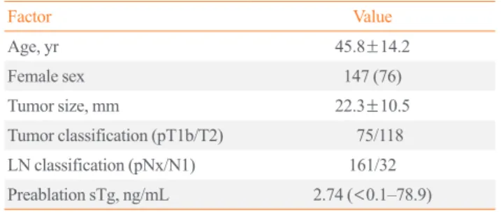
Table 1. Demographic, Clinical, Pathological, and Biochemical Data of Enrolled Patients
Factor Value
Age, yr 45.8±14.2
Female sex 147 (76)
Tumor size, mm 22.3±10.5
Tumor classification (pT1b/T2) 75/118 LN classification (pNx/N1) 161/32 Preablation sTg, ng/mL 2.74 (<0.1–78.9) Values are expressed as mean±SD, number (%), or median (range).
sTg, stimulated thyroglobulin.
Predictors of successful ablation
As shown in Table 2, visually positive 99mTc-perthecnetate scan,
99mTc-pertechnetate uptake values exceeding 0.9% were signifi- cantly associated with unsuccessful ablation (Figs. 1, 2). The optimal cut-off values of sTg and 99mTc-pertechnetate uptake were settled at 0.8 ng/mL (sensitivity 65%, specificity 67%;
area under the curve, 0.620; P=0.025) and 0.9% (sensitivity 70%, specificity 55%; area under the curve, 0.710; P=0.020) (Fig. 3). In order of significance, the best predictive factors for successful ablation were visually negative scan (ρφ=0.91, P<
0.001), preablative sTg level <0.8 ng/mL (ρφ=0.55, P<0.008),
and 99mTc-pertechnetate uptake rate values <0.9% (ρφ=0.44, P=0.065).
DISCUSSION
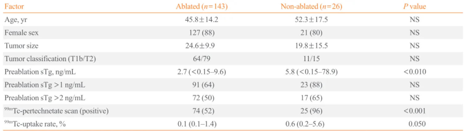
The present study proves the value of 99mTc-pertechnetate scin- tigraphy to assess DTC patients before RAI ablation, in that pa- tients with a negative 99mTc-pertechnetate scintigraphy have a much higher chance of successful 131I remnant ablation. Differ- ences in administered activity, 131I uptake and retention time in the remnants, the mass of the thyroid remnant, different TSH Table 2. Preablative 99mTc-Pertechnetate Scintigraphy and Thyroglobulin Results in Ablated and Non-Ablated Differentiated Thyroid Carcinoma Patients
Factor Ablated (n=143) Non-ablated (n=26) P value
Age, yr 45.8±14.2 52.3±17.5 NS
Female sex 127 (88) 21 (80) NS
Tumor size 24.6±9.9 19.8±15.5 NS
Tumor classification (T1b/T2) 64/79 11/15 NS
Preablation sTg, ng/mL 2.7 (<0.15–9.6) 5.8 (<0.15–78.9) <0.010
Preablation sTg >1 ng/mL 91 (64) 23 (88) NS
Preablation sTg >2 ng/mL 72 (50) 17 (65) NS
99mTc-pertechnetate scan (positive) 74 (52) 25 (96) <0.001
99mTc-uptake rate, % 0.1 (0.1–1.4) 0.6 (0.2–5.6) 0.050
Values are expressed as mean±SD, number (%), or median (range).
99mTc, technetium-99m; NS, not significant; sTg, stimulated thyroglobulin.
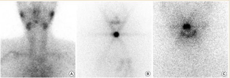
Fig. 1. Postoperative technetium-99m (99mTc)-pertechnetate scintigraphy (A) showing multiple uptake foci (arrows) (uptake rate 1.3%), whole body scan (WBS) with additional single photon emission computed tomography/computed tomography, (B) showing a correspond- ing intensely iodine-avid remnants (arrows), and (C) diagnostic WBS performed 6 months later showing two areas of persisting faint uptake (arrows) in a 47 years old woman with papillary thyroid carcinoma pT2pN0 (preablation stimulated thyroglobulin, 4.4 ng/mL; response as- sessment: basal [0.4 ng/mL] and stimulated [1.2 ng/mL] thyroglobulin, respectively). Response assessment: unsuccessful ablation.
A B C
levels, may account for differences in successful ablation rates between and within DTC patients, most of these factors are not likely to play a role in the present study. In facts, treatment pro- tocol was strictly standardized and fixed low 131I activities were administered; consequently, the mass of the thyroid remnants (which largely depends on the quality of surgery) most likely accounts for the majority of variation in success of ablation in the present patient population. Accordingly, as successful abla- tion is prognostically relevant, it could be argued that patients with visually positive 99mTc-pertechnetate scintigraphy should
receive a higher 131I activity to maximize the chance of success- ful ablation, especially if 99mTc-pertechnetate uptake ratio and sTg levels exceed 0.4% and 0.8 ng/mL, respectively.
Residual lymph node metastases were found on PT-WBS/
SPECT-CT in 24 patients and can be assumed to be not distin- guishable preoperatively via US as a careful examination was always performed before surgery by an experienced thyroidolo- gist. Then, unfortunately, neither 99mTc-pertechnetate scintigra- phy nor serum Tg would be accurate in detecting lymph node metastases in our series. In contrast, Lee et al. [12] recently Fig. 2. Postoperative technetium-99m (99mTc)-pertechnetate scintigraphy (A) with no visually discernable neck uptake, whole body scan (WBS) with additional single photon emission computed tomography/computed tomography, (B) showing a iodine-avid remnant, and (C) diagnostic WBS performed 6 months later showing complete ablation in a 32 years old female with papillary thyroid carcinoma pT2pNx (stimulated thyroglobulin: preablation, 1.2 ng/mL; response assessment <0.15 ng/mL). Response assessment: successful ablation.
A B C
100
50
0
100
50
0
Sensitivity (%) Sensitivity (%)
100-Specificity (%) 100-Specificity (%)
Preablation stimulated Tg Preablation 99mTc-pertechnetate uptake rate
20 40 60 80 100 20 40 60 80 100
Fig. 3. Receiving operator characteristics curve analysis for preablation technetium-99m (99mTc)-pertechnetate uptake rate (A) and stimulat- ed thyroglobulin (sTg) (B) to predict successful ablation. The optimal cutoffs (arrows) were 0.9% (sensitivity 70%, specificity 55%; area under the curve, 0.710; P=0.020) and 0.8 ng/mL (sensitivity 65%, specificity 67%; area under the curve, 0.620; P=0.025) for predicting successful routine radioiodine ablation.
A B
found no RAI-avid lymph-node metastases at PT-WBS/SPECT- CT in low-risk DTC patients with rhTSH-sTg values below 0.5 ng/mL. However, their patients were operated on by two experi- enced thyroid surgeons who performed more than 300 cases of thyroid cancer surgery per year while in Switzerland, due to dif- ferent reasons, thyroid operations are mostly performed by low- volume general surgeons [13]. Schneider et al. [14] conducted a retrospective review of 223 DTC patients treated with total thy- roidectomy and subsequent RAI ablation. Among them 21 pa- tients (9%) experienced a recurrence and had a 10-fold higher RAI uptake compared with those without recurrences (P=
0.001). Significantly lower RAI uptake ratios were recorded in patients treated by high-volume indicating the completeness of resection and properly informing the use of Tg as tumor marker [15]. Such data suggest that the use of serum Tg to predict me- tastases and properly inform RAI treatment cannot be general- ized due to the relevant confounding role of the amount of post- operative residual thyroid tissue [1,16,17]. In this context we proved here, as the main result of our study, that preablation
99mTc-pertechnetate scintigraphy performs better than sTg in predicting successful ablation after administration of fixed low RAI activity (i.e., 1.1 GBq). However, considering that positive scans and high uptake values may occur in patients whose rem- nant was successfully ablated DxWBS and/or sTg measure- ments are still needed 6 to 12 months after ablation to prove successful ablation.
In conclusion, radioiodine remnant ablation is not routinely recommended after thyroidectomy for low risk DTC patients but specific features of the individual patient that could modu- late recurrence risk, disease follow-up implications, and patient preferences are relevant to RAI decision-making. The present study shows that 99mTc pertechnetate scintigraphy is a simple and reliable method to predict the effectiveness of postoperative thyroid ablation with low-activity RAI administration in pa- tients with visually negative scan or positive scan with a 99mTc pertechnetate uptake below 0.9%.
CONFLICTS OF INTEREST
No potential conflict of interest relevant to this article was re- ported.
AUTHOR CONTRIBUTIONS
Conception or design: L.G. Acquisition, analysis, or interpreta- tion of data: L.G., G.P., T.R., L.C., P.T. Drafting the work or revis-
ing: L.G., P.T. Final approval of the manuscript: L.G., G.P., T.R., L.C., P.T.
ORCID
Luca Giovanella https://orcid.org/0000-0003-0230-0974 Luca Ceriani https://orcid.org/0000-0002-6371-097X Pierpaolo Trimboli https://orcid.org/0000-0002-2125-4937
REFERENCES
1. Haugen BR, Alexander EK, Bible KC, Doherty GM, Mandel SJ, Nikiforov YE, et al. 2015 American Thyroid Association management guidelines for adult patients with thyroid nod- ules and differentiated thyroid cancer: the American Thyroid Association guidelines task force on thyroid nodules and dif- ferentiated thyroid cancer. Thyroid 2016;26:1-133.
2. Mallick U, Harmer C, Yap B, Wadsley J, Clarke S, Moss L, et al. Ablation with low-dose radioiodine and thyrotropin alfa in thyroid cancer. N Engl J Med 2012;366:1674-85.
3. Luster M, Clarke SE, Dietlein M, Lassmann M, Lind P, Oyen WJ, et al. Guidelines for radioiodine therapy of differ- entiated thyroid cancer. Eur J Nucl Med Mol Imaging 2008;
35:1941-59.
4. Giovanella L, Suriano S, Ricci R, Ceriani L, Verburg FA.
Postsurgical thyroid remnant estimation by 99mTc-pertech- netate scintigraphy predicts radioiodine ablation effective- ness in patients with differentiated thyroid carcinoma. Head Neck 2011;33:552-6.
5. Jung JS, Lee SM, Kim SJ, Choi J, Han SW. Prediction of the success of thyroid remnant ablation using preablative 99mTc pertechnetate scintigraphy and postablative dual 131I scintigraphy. Nucl Med Commun 2015;36:38-44.
6. Ozdemir D, Cuhaci FN, Ozdemir E, Aydin C, Ersoy R, Turkolmez S, et al. The role of postoperative Tc-99m pertech- netate scintigraphy in estimation of remnant mass and pre- diction of successful ablation in patients with differentiated thyroid cancer. Nucl Med Commun 2016;37:640-5.
7. Giovanella L, Suriano S, Ceriani L, Verburg FA. Undetect- able thyroglobulin in patients with differentiated thyroid carcinoma and residual radioiodine uptake on a postablation whole-body scan. Clin Nucl Med 2011;36:109-12.
8. Trimboli P, Zilioli V, Imperiali M, Ceriani L, Giovanella L.
High-sensitive basal serum thyroglobulin 6-12 months after thyroid ablation is strongly associated with early response to therapy and event-free survival in patients with low-to-inter-
mediate risk differentiated thyroid carcinomas. Eur J Endo- crinol 2017;176:497-504.
9. Piccardo A, Puntoni M, Bottoni G, Treglia G, Foppiani L, Bertoli M, et al. Differentiated thyroid cancer lymph-node relapse. Role of adjuvant radioactive iodine therapy after lymphadenectomy. Eur J Nucl Med Mol Imaging 2017;44:
926-34.
10. Giovanella L, Toffalori E, Ceriani L, Caputo M, Verburg FA. Thyroglobulin and thyroglobulin autoantibodies mea- surement using the automated KRYPTOR® platform in pa- tients with differentiated thyroid carcinoma. Horm Metab Res 2011;43:728-30.
11. Verburg FA, Luster M, Cupini C, Chiovato L, Duntas L, Eli- sei R, et al. Implications of thyroglobulin antibody positivity in patients with differentiated thyroid cancer: a clinical posi- tion statement. Thyroid 2013;23:1211-25.
12. Lee CH, Jung JH, Son SH, Hong CM, Jeong JH, Jeong SY, et al. Risk factors for radioactive iodine-avid metastatic lymph nodes on post I-131 ablation SPECT/CT in low- or intermediate-risk groups of papillary thyroid cancer. PLoS One 2018;13:e0202644.
13. Gourin CG, Tufano RP, Forastiere AA, Koch WM, Pawlik TM, Bristow RE. Volume-based trends in thyroid surgery.
Arch Otolaryngol Head Neck Surg 2010;136:1191-8.
14. Schneider DF, Ojomo KA, Chen H, Sippel RS. Remnant
uptake as a postoperative oncologic quality indicator. Thy- roid 2013;23:1269-76.
15. Giovanella L, Ceriani L, Trimboli P. Letter to the editor:
“postoperative thyroglobulin and neck ultrasound in the risk restratification and decision to perform 131i ablation”. J Clin Endocrinol Metab 2017;102:1783-4.
16. Giovanella L, Avram AM, Clerc J, Hindie E, Taieb D, Ver- burg FA. Postoperative serum thyroglobulin and neck ultra- sound to drive decisions about iodine-131 therapy in patients with differentiated thyroid carcinoma: an evidence-based strategy? Eur J Nucl Med Mol Imaging 2018;45:2155-8.
17. Campenni A, Giovanella L, Pignata SA, Vento A, Alibrandi A, Sturiale L, et al. Undetectable or low (<1 ng/ml) postsur- gical thyroglobulin values do not rule out metastases in early stage differentiated thyroid cancer patients. Oncotarget 2018;
9:17491-500.