총 설
폐가스 처리용 바이오필터에 미생물 군집 분석 기법의 적용
이은희*·박현정*·조윤성*·류희욱**·조경숙*,†
*이화여자대학교 환경공학과 120-750 서울특별시 서대문구 대현동 11-1
**숭실대학교 환경화학공학과 156-743 서울특별시 동작구 상도동 511 (2009년 11월 19일 접수, 2009년 12월 11일 채택)
Application of Methodology for Microbial Community Analysis to Gas-Phase Biofilters
Eun-Hee Lee*, Hyunjung Park*, Yun-Seong Jo*, Hee Wook Ryu** and Kyung-Suk Cho*,†
*Department of Environmental Sciences and Engineering, Ewha Womans University, 11-1 Daehyun-dong Seodaemun-gu, Seoul 120-750, Korea
**Department of Chemical and Environmental Engineering, Soongsil University, 511 Sangdo-dong, Dongjak-gu, Seoul 156-743, Korea (Received 19 November 2009; accepted 11 December 2009)
요 약
폐가스 처리용 바이오필터의 핵심 요소 기술은 생촉매(미생물), 담체, 설계·운전 기술 및 진단·관리 기술이다. 특히, 바이오필터의 성능은 부하 조건과 바이오필터 내 미생물 군집 구조에 의해 영향을 받는다. 지금까지 바이오필터의 미 생물 연구는 대부분 배양법을 기초로 하여 수행되어 왔으나, 최근에 보다 신속하고 정확하게 미생물 군집을 분석할 수 있는 방법들이 제시되고 있다. 본 논문에서는 생리적, 생화학적 및 분자생물학적 미생물 군집 분석 방법과 이를 활용 한 바이오필터의 미생물 군집 특성을 조사한 연구사례를 소개하고, 미생물 군집 분석법의 바이오필터에 적용 가능성 에 대해 고찰하였다. Community-level physiological profile 방법은 시료 중에 포함된 종속영양미생물의 탄소기질 이용 능력을 기반으로 군집 특성을 조사하는 것이며, Phospholipid fatty acid analysis는 미생물 세포막 지방산을 분석하여 군집 특성을 조사하는 방법이다. 환경시료로부터 직접 추출한 DNA를 활용하는 분자생물학적 분석법에는 “partial community DNA analysis”와 “whole community DNA analysis”가 있다. 전자의 방법은 PCR 과정에 의해 증폭시킨 염 기서열을 분석하는 것으로 ribosomal operon 유전자가 가장 많이 활용되었다. 이 방법은 다시 PCR fragment cloning 및 genetic fingerprinting으로 구분되며, genetic fingerprinting 방법으로는 denaturing gradient gel electrophoresis, terminal- restriction fragment length polymorphism, ribosomal intergenic spacer analysis 및 random amplified polymorphic DNA 방법으로 세분화된다. 추출된 전체 군집의 DNA를 분석하는 방법에는 total genomic cross-DNA hybridization, 총 추 출 DNA의 열 변성/재결합 방법 및 밀도구배를 이용하여 추출한 DNA를 분획화하는 방법 등이 있다.
Abstract − There are four key factors for gas-phase biofilters; biocatalysts(microorganisms), packing materials, design/operat- ing techniques, and diagnosis/management techniques. Biofilter performance is significantly affected by microbial com- munity structures as well as loading conditions. The microbial studies on biofilters are mostly performed on basis of culture-dependent methods. Recently, advanced methods have been proposed to characterize the microbial community structure in environmental samples. In this study, the physiological, biochemical and molecular methods for profiling microbial communities are reviewed, and their applicability to biofilters is discussed. Community-level physiological profile is based on the utilization capability of carbon substrate by heterotrophic community in environmental samples. Phospho- lipid fatty acid analysis method is based on the variability of fatty acids present in cell membranes of different micro- organisms. Molecular methods using DNA directly extracted from environmental samples can be divided into “partial community DNA analysis” and “whole community DNA analysis” approaches. The former approaches consist in the analysis of PCR-amplified sequence, the genes of ribosomal operon are the most commonly used sequences. These methods include PCR fragment cloning and genetic fingerprinting such as denaturing gradient gel electrophoresis, terminal-restriction fragment length polymorphism, ribosomal intergenic spacer analysis, and random amplified polymorphic DNA. The whole community DNA analysis methods are total genomic cross-DNA hybridization, thermal denaturation and reassociation of whole extracted DNA and extracted whole DNA fractionation using density gradient.
Key words: Community-Level Physiological Profile, DNA-Based Methods, Gas-Phase Biofilter, Microbial Community Analysis, Phospholipid Fatty Acid Analysis
†To whom correspondence should be addressed.
E-mail: [email protected]
‡이 논문은 KAIST 김상돈 교수님의 정년을 기념하여 투고되었습니다.
화학공학 제48권 제2호 2010년 4월
1. 서 론
지난 수십 년 동안 환경문제는 전 세계적으로 주요 이슈가 되고 있으며, 수질오염, 대기오염, 토양오염, 유해 폐기물 등과 같은 환경 오염문제 해결과 환경복원 및 보전을 위해 매우 다양한 환경공학기 술들이 개발되고 있다. 특히, 엄격하게 강화되고 있는 환경 규제치를 만족하기 위해 개발된 환경공학기술을 “추가비용 없이 최적 활용 가 능한 기술(BATNEEC, Best Available Technology Not Entailing Excessive Costs)”로 혁신시키기 위한 많은 노력들이 진행되고 있다[1].
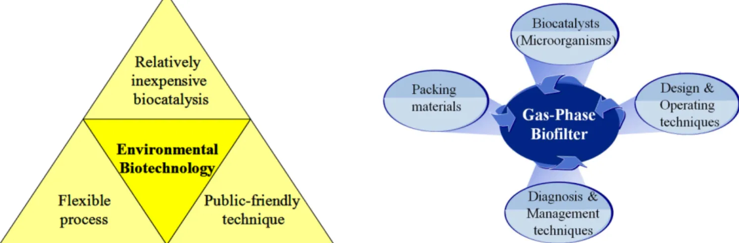
환경공학기술 개발 및 혁신과정에서 생물, 특히 미생물을 활용하 는 환경생물공학기술(Environmental Biotechnology)은 다음과 같은 주요 특성으로 인해 지속적으로 주목받고 있다(Fig. 1)[1]. 생물(주로 미생물)을 촉매로 활용하는 생물학적 환경공학기술은 물리·화학적 환경공학기술에 비교하여 운전비가 매우 저렴한 장점이 있다. 예를 들면, 비교적 저농도의 폐가스를 생물학적 방법으로 처리할 경우 처 리용량이 증가할수록 물리·화학적 방법보다 운전비가 훨씬 절감되는 효과를 얻을 수 있다[2]. 또한, 미생물 공정은 환경적응성이 우수한 매우 유연한 공정이다. 미생물 개체 혹은 군집은 “적응(adaptation)”
을 통해 다양한 환경조건에 내성을 지니거나, 난분해성 물질을 대사 하거나 공대사할 수 있는 능력을 습득할 수 있다[1]. 예를 들면, 바이 오필터를 장기간 운전하게 되면, 어느 정도 범위의 pH, 온도 등의 외 부 변화에 대해 바이오필터의 미생물 군집이 순응하게 되며[3-5], 생 분해가 불가능한 것으로 생각된 인공합성 제초제 및 농약, 다이옥신, 할로겐화합물 등에 대해서도 이들을 분해할 수 있는 신규의 미생물 혹은 미생물 군집이 보고되는 등 미생물의 다양성이 풍부하다[6-8].
환경생물공학기술 원리는 자연의 정화 작용을 활용한 대중 친화 적이며 환경 친화적인 “녹색기술(Green Tech)”이다. 일반적으로 대 중은 콘크리트로 만든 하·폐수처리장보다는 비용이 고가이며 처리 효율도 불안정한 갈대나 습지를 활용한 수질정화시스템을 선호하는 경향이 있다[1].
상기와 같은 주요 특성 때문에 환경생물공학 원리를 활용한 기술 개발에 대한 수요가 점점 증대되고 있다. 환경생물공학기술이 활용 된 대표적인 기술 중 하나가 대기오염물질 처리를 위한 바이오필터 이다[9-11]. 바이오필터는 개발 초기에는 주로 황화수소, 암모니아 등 과 같은 악취 함유 개발 초기에 활용되었으나[12-25], 최근 들어서는 벤젠, 톨루엔, 에틸벤젠, 쟈일렌 등과 같은 휘발성 유기화합물(VOC)
함유 폐가스도 만족할 만한 수준으로 처리가 가능하여 현재 상용화 되고 있다[26-40].
본 논문에서는 악취 및 휘발성 유기화합물 처리용 바이오필터의 4대 핵심 요소 기술에 대해 간략히 고찰하고, 바이오필터의 악취 및 VOC 제거 성능에 가장 큰 영향을 미치는 미생물 군집 거동 특성을 분석하는 최신 방법의 종류, 원리 및 특성과 최신의 미생물 군집 분 석방법을 활용한 바이오필터 미생물 군집 특성에 관한 연구를 소개 하고, 그 중요성에 대해 설명하고자 한다.
2. 폐가스 처리용 바이오필터의 기술 특성 바이오필터의 주요 핵심 요소 기술은 담체, 생촉매(미생물), 설계/
운전, 진단 및 관리 기술이다(Fig. 2). 바이오필터에 사용되는 담체는 생촉매인 미생물의 우수한 부착력, 많은 양의 biomass를 고정할 수 있는 넓은 표면적, 낮은 압력손실과 압밀현상, 뛰어난 내구성을 가져 야 할뿐만 아니라 저렴하여야 한다[12,22,29,30,36,41,42].
바이오필터 성능을 좌우하는 생촉매(미생물)에 대한 연구는 (i) 오 염가스 물질을 분해할 수 있는 특정 미생물을 배양, (ii) 특정 균주의 특성을 조사하여 바이오필터 성능 유지를 위한 운전인자에 관한 정 보를 구축하기 위한 연구가 진행되고 있다[12-16,18,20,21,25,30-32,43].
또 다른 방향의 연구는, 기존의 바이오필터에 의해 분해가 곤란하거 나 분해효율이 저조한 난분해성 오염가스 물질을 대상으로 선정하여, 이를 효율적으로 분해할 수 있는 미생물을 농화배양(enrichment culture) 방법을 통해 선별하여 바이오필터 성능향상을 위한 접종원 으로 활용하고 있다[17,24,26,34].
생물학적 반응은 물리·화학적 반응에 비해 반응속도가 느려 반응 기 부피를 비롯한 제반 설비 규모가 커지는 단점이 있다[2]. 이를 보 완하기 위해 제반 설비를 compact하게 설계하는 기술 개발이 활발 히 진행되고 있다[5,11,20,38,44,45]. 그리고 바이오필터는 미생물이 생육 가능한 조건을 조성하면서 원하는 수준의 처리효율을 얻을 수 있는 조건을 유지하는 운전기술이 매우 중요하다. 지금까지 연구된 운전기술은 주로 폐가스 처리 용량과 농도에 따른 운전 조건, 폐가 스 성상, 수분 및 영양제 공급 주기, pH 및 온도 영향, 그리고 미생 물 농도 등에 대한 데이터베이스가 구축되고 있으며[3-5,13,17,25,28, 31,34,35,46-49], 폐가스 중 오염물질의 성상 및 농도의 일중 혹은 연 중 변화, 휴일 및 휴가 등의 산업 공정 중단에 따른 바이오필터의 장
Fig. 1. Key features of environmental biotechnology. Fig. 2. Key factors demanded for gas-phase biofilter.
·단기 가동 중지 등의 부하 조건 변화에 적합한 설계 및 운전기술에 관한 몇몇 연구도 진행되고 있다[23,44,45,50,51].바이오필터가 최적 활용 가능한 기술로 혁신하기 위해서는 바이오필터 성능을 진단하고, 진단 결과를 토대로 적절한 관리를 취하여 심각한 문제 발생을 사전 에 예방할 수 있는 기술 개발이 필요하지만[51,52], 아직까지 이에 관 련된 정보가 거의 축적되지 못하고 있으며 향후 관련 분야의 연구가 필요하다.
3. 바이오필터의 미생물 군집 분석의 중요성 다양한 산업시설 혹은 환경기초시설로부터 배출되는 폐가스의 성 상과 농도는 매우 다양하고, 불규칙적이며 불예측적으로 배출된다.
불규칙적이고 불예측적인 부하 조건에 대한 바이오필터의 처리 성능 을 보다 증대시키기 위해서는, 바이오필터의 transient response에 대한 연구가 선행되어 Fig. 3에 제시한 관련 정보가 축적되어야 한다[52].
우선, 바이오필터의 transient response에 대한 정보가 축적되기 위 해서는, 부하 조건 변화에 대한 바이오필터의 오염물질 제거 효율을 모니터링하고, pH, 온도, 수분함량, 전도도, 압력손실 등과 같은 물 리·화학적 parameter를 연속적으로 측정할 필요가 있다[2,51]. 또한, 부하 조건 변화에 따라 바이오필터의 성능을 결정적으로 좌우하는 미생물 군집 dynamics에 대해 연구하고, 미생물 군집 dynamics와 바 이오필터 성능간의 상관관계를 종합적으로 분석하는 것은 바이오필 터 성능 향상에 있어 매우 중요한 과제이다[51,52].
바이오필터는 오염물질 분해능을 담당하는 핵심 미생물군을 비롯 하여 다양한 종류의 미생물들이 나름대로의 niche를 가지고 조화롭 게 서식하고 있는 작은 생태계로 볼 수 있다. 그런데, 지금까지의 바
이오필터의 미생물학적 연구의 대다수는 오염물질 분해에 주요 역할 을 할 것으로 기대되는 특정 미생물의 순수 분리에 초점이 맞추어져 진행되고 있다[12-16,18,30,31,25,30-32,43]. 극히 최근에 들어서 바 이오필터 미생물 군집 전체에 대한 연구가 제한적으로 진행되고 있 는 실정이다.
바이오필터에서 오염물질 제거에 주요 역할을 할 것으로 기대되 는 특정 미생물에 대한 연구도 거의 대부분 배양법에 기초로 하여 진 행되어 왔다[12-16,18,20,31,25,30-32,43]. 따라서, 사용하는 배지의 조성 및 배양 조건에 따라, 선택되어지는 미생물종이 달라짐에 따라 미생물학적 정보가 왜곡되거나 제한된 정보만 얻어지는 문제점이 발 생된다[532,5]. 또한, 미생물 분리 단계에서는 오염물질의 분해능에 관한 여부를 알 수 없기 때문에, 순수 분리 후 오염물질의 분해능에 관한 조사를 별도로 수행해야 하므로, 번거롭고 시간이 많이 소요되 는 등의 문제가 있다.
최근 들어 미생물 생태공학 분야에서는 개개의 미생물이 아닌 미 생물 군집 특성을 연구할 수 있는 기법들이 다양하게 개발되고 있고 [53-55], 이 분자생물학적 방법은 비교적 정확하고 신속하게 미생물 군집 특성을 연구할 수 있는 기법이다. 또한, 특정 오염물질을 분해 할 수 있는 미생물을 환경시료로부터 신속하고 정확하게 검출하기 위해서, 오염물질 대사 기작에 관련된 특정 유전자를 검출할 수 있 는 primer를 활용하는 기법이 활용되고 있다[53-55].
4. 미생물 군집 분석 방법
현재 많이 활용되고 있는 최신 미생물 군집 분석은 생리학적 방법, 생화학적 방법 및 분자생물학적 방법으로 구분되며, 각 방법의 개요 및 그 특성을 Table 1에 요약하여 나타내었다[55].
4-1. 생리학적 방법: Community-level physiological profiles(CLPP) Community-level physiological profiles(CLPP) 방법은 각종 환경 시료의 미생물 군집들의 특성을 조사, 비교하기 위해 주로 많이 사 용되고 있는 방법이다[55]. Garland and Mill [56]은 95 종의 탄소 기 질로 96-well microliter plate(한 well에는 탄소기질이 들어있지 않음, 대조군으로 활용)를 이용한 CLPP 방법을 제안하였다(Table 2). 또한, 주로 환경시료 미생물 군집 분석용으로 31 종의 탄소를 이용한 Eco plate가 이용되고 있다[57,58]. 각각의 well은 유일 탄소원과 함께 산화 환원 지시자인 염료 tetrazolium violet이 포함되어 있는데, tetrazolium violet은 호흡 활동을 하는 동안에 감소되어 well의 색변화가 일어나 Fig. 3. Researches for accumulating knowledge of transient response
of biofilter.
Table 1. Summary of commonly used microbial community profiling methods[55]
Methods Main approach Target populations Comments
Community-level physiological profiling (CLPP) using Biolog microplates
Assess sole-carbon source utilization to determine physiological capacity of community
Fast-growing cultivable organisms
Relatively quick and inexpensive.
Difficulties with analysis and interpretation
Phospholipid fatty acid analysis (PLFA)
Assess community structure based on
variability of membrane lipids Entire community
Most variability associated with bacteria not fungi. Overlap among groups for marker PLFAs
Sequence analysis of DNA clone libraries
Catalogue species richness and community composition by sequence analysis of ribosomal RNA (or other) genes
Entire community or portion of (depends on PCR primers)
Bias associated with DNA extraction and PCR. Expensive and time-consuming to sequence full diversity from a single soil sample
DNA-based fingerprints(e.g.
DGGE, T-RFLP, RISA, ITS)
Create visual fingerprints of whole communities based on variation in ribosomal RNA (or other) genes
Entire community or portion of (depends on PCR primers)
Bias associated with DNA extraction and PCR. Resolution may inadequate for diverse communities
화학공학 제48권 제2호 2010년 4월
게 된다. 즉, 시료가 접종된 각각의 well은 시간이 경과함에 따라 미 생물의 호흡 활동으로 인해 특징적으로 색깔 변화가 일어나게 되며, 이 것을 분광광도계를 사용하여 흡광도로 측정하는 것이 microliter plate를 활용한 CLPP 분석법의 원리이다. 각각의 well의 흡광도 값 은 대조군(증류수의 값)으로 보정한 후, 이를 well의 평균 색 발달 정 도 average well colour development(AWCD)로 표시한다[57,58].
CLPP 방법은 저렴하면서도 빠르게 미생물 군집 분석을 할 수 있 다. 그러나 이 기법은 실험실의 일정한 배양 조건 하에서 나타나는 미생물의 활성에 의존하기 때문에, 실제 자연계에서의 미생물의 활 성이 실험실과 동일하게 발휘되는지에 대해서는 불확실하며, 측정된 데이터의 분석과 해석이 어렵다[59].
4-2. 생화학적 방법: Phospholipid fatty acid analysis(PLFA) Phospholipid fatty acid analysis(PLFA) 방법은 미생물의 세포막 지 방산의 다양성을 phenotypic fingerprinting을 이용하여 다양한 환경 시료의 미생물 군집을 분석하는 방법이다[55]. PLFA 방법은 유기 용 매를 첨가한 시료로부터 지질 물질을 추출한 뒤 각 지질이 띠는 극 성을 바탕으로 고체 상태의 추출방법을 이용해 지질로부터 인지질을 분리하는 것이다[60]. Phospholipid fatty acids는 fatty acid methyl
esters(FAME)로 변환되며 이 때 각각의 유형과 양은 기체 크로마토 그래피로 분석된다. 각 시료의 profile은 추출된 PLFA의 풍부도(존 재비율)로 비교 분석되는데, 어떤 PLFA는 특정 미생물 그룹에 대한 표지(marker)로서 사용될 수 있다. Table 3에 특정미생물군의 maker 로 이용될 수 있는 PLFA type을 나타낸 것이다[61]. 몇몇 PLFA는 다양한 종류의 미생물 그룹에 공존하므로 표지로 활용하기 곤란한 점이 있다. 시료들 사이에서 PLFA profile을 비교함으로써 군집 구 조의 차이를 분석할 수 있다[61]. 또한, PLFA 결과로부터 군집 구조 와 환경 변수 사이의 상관성을 검토하기 위해 다양한 통계 기법 (principal components analysis, correspondence analysis, multi- dimensional scaling)을 사용한다.
4-3. 분자생물학적 방법
DNA 추출, PCR 증폭, 및 염기서열분석 등과 같은 분자생물학적 기술들의 발달로 인해, 최신의 미생물 군집 분석 방법은 미생물의 순 수분리 및 배양과정 없이 시료로부터 직접적으로 DNA를 추출하여 미생물 군집을 분석하는 것이 가능하게 되었다[53-55]. 이러한 방법은 Partial community DNA analysis와 whole genomic community DNA analysis로 크게 두 가지로 구분할 수 있다(Fig. 4)[53]. Partial community DNA analysis 방법은 타겟 유전자 부분을 PCR 과정을 거쳐 증폭시 킨 뒤, 그 염기서열을 중점적으로 다룸으로서 미생물 군집을 분석하 는 것이다. Whole genomic community DNA analysis 방법은 미생 물 군집으로부터 추출된 DNA에서 전체적인 유전자 정보를 연구하 여 미생물 군집을 분석하는 것이다.
4-3-1. 부분 군집 DNA 분석(Partial community DNA analysis) 부분 군집 DNA 분석법은 PCR 과정에 의해 증폭시킨 염기서열을 분석하는 것으로, ribosomal operon의 rrs 유전자 (16s rDNA)와 rrs 와 rrl 유전자 사이의 spacer인 intergenic spacer(IGS) 16S-23S가 표 적으로 사용되는 염기서열부분이다[53].
4-3-1-1. PCR fragment cloning 및 characterization
이 방법은 DNA 추출물로부터 PCR 과정을 거쳐 얻은 클론의 rrs 염기서열 정보를 이용하여 박테리아 군집의 다양성을 연구하는 것으 로 염기서열정보를 분석함으로써 난배양성 미생물을 선별해 낼 수 있다[53]. 또한, 환경시료로부터 얻은 염기서열과 이미 알고 있는 미 생물 종의 유전적 정보를 서로 비교함으로써 미생물 종사이의 계통 발생학적 연관성을 예측할 수 있다[62]. 즉, 이 기법은 시료들 사이 Table 2. Carbon sources employed in BIOLOG ECO plate[57,58]
No. Carbon source No. Carbon source
1 β-Methyl-D-glucoside 17 N-Acetyl-D-glucosamine 2 D-Galactonic acid γ-lactone 18 γ-Hydroxybutryric acid
3 L-Arginine 19 L-Threonine
4 Pyruvic acid methyl ester 20 Glycogen
5 D-Xylose 21 D-Glucosaminic acid
6 D-Galacturonic acid 22 Itaconic acid
7 L-asparagine 23 Glycyl-L-glutamic acid
8 Tween 40 24 D-Cellobiose
9 i-Erythritol 25 Glucose-1-phosphate 10 2-Hydroxy benzoic acid 26 α-Ketobutyric acid 11 L-Phenylalanine 27 Phenylethyl amine
12 Tween 80 28 α-D-Lactose
13 D-Mannitol 29 D,L-α-Glycerol phosphate 14 4-Hydroxy benzoic acid 30 D-Malic acid
15 L-Serine 31 Putrescine
16 α-Cyclodextrin
Table 3. Phospholipid fatty acids commonly used as biomarkers for specific groups of microorganisms[55]
PLFA type Indicator for Common PLFA used as marker Comments
Monoenoic unsaturated and
cyclopropane unsaturated Gram-negative bacteria
16:1ω5c, 16:1ω7c, 16:1ω7t, 16:1ω9c, a16:0, cy17:0, 18:1ω5c, 18:1ω7c, 18:1ωtc, 18:1ω9c, cy19:0a
Occur in some Gram-negative (sulphate- reducers)
Terminally branched and 10Me
branched saturated Gram-positive bacteria i14:0, i15:0, a15:0, i16:0, a17:0, i18:0, 10Me16:0
Occur in Gram-positive bacteria and eukaryotes in lesser amounts Saturated straight-chain (<20C) Bacteria (non-specific) 14:0, 16:0, 18:0 -
Methyl branch on 10th C Actinomycetes 10Me16:0, 10Me17:0, 10Me18:0 -
Polyenoic unsaturated Fungi 18:2ω6 (18:1ω9c, 18:3ω3) Occur in other eukaryotes and in bacteria Saturated straight-chain (>20C) Microeukaryotes, higher plants,
mosses 20:0, 21:0, 22:0, 23:0, 24:0, 25:0, 26:0 -
Polyunsaturated Microeukaryotes 20:3ω6, 20:4ω6 Rare in bacteria
Ratio of monoenoic precursors to
cyclopropane PLFA Stress indicator 16:1ω7c/cy17:0, 16:1ω8c/cy19:0 Lower ratios (< 0.5) found in healthy, non- stressed populations
Trans/cis ratio of monoenoic PLFA Stress indicator 16:1ω7t/16:1ω7c, 16:1ω8t/16:1ω8c Lower ratios (< 0.5) found in healthy, non- stressed populations
에서 지리적 환경과 생리 화학적 특징에 의해 나타나는 기능상의 차 이와 관련된 유전적 다양성을 비교하는데 사용된다.
PCR-cloning 방법은 시간이 많이 걸리고 번거로우며 매우 값비싼 실험 장비와 비용이 소요되는 단점이 있다[53]. 또한, primer 종류, PCR 단계의 온도와 cycle 수, 부식질 혼합물에 의한 효소의 억제 효 과 등, 여러 가지 변수들이 PCR로 얻어지는 결과물에 영향을 주어 실험 결과의 분석 및 해석상 오차가 발생하는 문제점이 있다[53].
4-3-1-2. Genetic fingerprint 방법
Genetic fingerprint 방법은 agarose 혹은 polyacrylamide로 만든 젤 상에서 DNA 시료를 전기영동 하였을 때 나타나는 이동 패턴의 차 이를 해석함으로써 증폭된 염기서열의 다양성을 분석하는 원리를 활 용한 것이다. 전기영동시 나타나는 이동 패턴의 차이는 시료유전물 질의 크기 혹은 염기서열에 의해 따라 결정된다[53]. 이 기법은 특히 세균 군집 특성을 비교하는데 적절하며, 각 시료의 genetic fingerprint 정보 사이의 유사도를 계산하여, 각 시료간의 유연관계를 유사도의 계통수로 표시할 수 있다[63]. 또한, 통계기법에 의한 genetic fingerprint 정보 분석은 미생물 군집 구조의 변화에 지리/환경적 인 자가 미치는 영향을 예측할 수 있다[64,65]. 따라서 genetic fingerprint 방법은 실험과정이 용이하고 많은 양의 시료를 신속하게 분석할 수 있다는 장점을 가진다. 그러나 이 방법은 실험 과정에서 오차가 발 생하기 쉽고[66], 전기영동과정에서 DNA 시료 크기 및 분리 등에 있어 제한점이 있으며[67], 유전적 profile이 너무 복잡할 경우 실험 결과 분석과 해석이 어려운 단점이 있다[53].
① 변성제를 이용한 전기영동법에 의한 분리
이 방법은 각기 다른 염기서열을 가진 동일한 크기의 DNA 단편 들을 식별하는데 이용하는 것으로, DNA 변성제(denaturing agent)의 농도를 저농도에서 고농도로 차이가 나도록 만든 polyacrylamide gel 상에서 PCR 증폭산물을 분리하는 것이다[53]. Urea 혹은 formamide 와 같은 화학물을 변성제로 이용하는 것은 DGGE (denaturing gradient gel electrophoresis), 온도 등과 같은 물리적 요인을 변성제
로 활용하면 TGGE (temperature gradient gel electrophoresis)라고 한 다[68,69]. 시료의 염기서열 차이는 DNA denaturation에 영향을 미 쳐 전기영동상에서 나타나는 이동패턴의 차이를 유발하게 되는데, 이 방법은 크기가 500 base pairs보다 큰 시료를 효과적으로 분리할 수 없는 것이 제한점이다[53].
② Amplified ribosomal DNA restriction analysis(ARDRA)와 Terminal-restriction fragment length polymorphism(T-RFLP)
ARDRA 방법은 PCR 과정에 의해 얻어진 16S rDNA 시료를 제 한 효소로 절단한 후, agarose 혹은 polyacrylamide gel에서 DNA 단 편들을 분리하는 것이다. 이 방법은 조작이 매우 편리하고 간단하다.
그러나 최적의 분석 조건을 얻기 위한 제한 효소 선별이 어렵고, profile이 매우 복잡하고 특정한 리보솜 패턴의 검출과 정량화를 추 론하기 위한 정보를 얻을 수 없다는 단점이 있다[53].
이러한 단점을 극복하기 위해, fluorochrome로 라벨링한 primer를 사용하는 방법인 T-RFLP 방법이 개발되었다. T-RFLP의 profile을 비 교함으로서 환경시료의 군집특성 비교가 가능하다. 최근 들어 레이저 를 이용한 검출기술이 개발됨에 따라 해상도와 감도가 향상되었다[53].
③ Ribosomal intergenic spacer analysis(RISA)
RISA 방법은 rrs와 rrl 유전자 사이에 있는 IGS의 다양성을 분석 하는 것이다[53]. IGS 크기는 생물종에 따라 50 bp에서부터 1.5 kb 이상까지 가지각색이며, 생물종에 따라 차이가 있다. IGS를 타깃으 로 하는 primer를 사용하고, 증폭된 IGS 시료는 polyacrylamide gel 상에서 크기에 따라 분리될 수 있다[70]. IGS 크기의 높은 변이성으 로 인해서 군집의 유전적 구조상에서 일어나는 근소한 변화의 검출 이 가능하나, IGS sequence databank가 그리 많지 않으므로 제한점 이 있다[53].
④ Random amplified polymorphic DNA(RAPD)
RAPD 방법은 10bp 정도로 짧은 임의의 primer를 사용하며, 이 primer들은 유전자의 DNA 상에서 각기 다른 위치에서 작용하여 다양 한 길이의 PCR 결과물을 산출하게 된다. 이 PCR 결과물을 agarose Fig. 4. Schematic representation of the different molecular approaches for assessing the genetic diversity and structure of microbial communities[53].
화학공학 제48권 제2호 2010년 4월
혹은 polyacrylamide gel에서 크기별로 분리가 가능하다[53]. 이 방 법을 이용하여 원핵생물 유전자들 사이에서 차이를 고감도로 신속하 게 조사할 수 있으며[71], 군집 레벨에서 세균의 복잡한 군집 구조를 비교·분석하는데 활용가능하다[72]. 이 방법의 단점은 미생물 군집의 구조에 대한 계통발생적인 정보를 제공할 수 없다는 것이다.
4-3-2. 전체 군집 DNA 분석(Whole community DNA analysis) 부분 군집 DNA 분석법과는 달리 미생물 군집으로부터 추출된 DNA의 전체적인 유전자 정보를 연구하여 미생물 군집 분석을 시도 하는 분자생물학적 기법들을 전체 군집 DNA을 분석하는 것이다. 전 체 DNA를 대상으로 한 미생물 군집을 분석하는 방법은 고순도이며 크기가 완전한 고품질 DNA 시료가 대량 필요하며, 세균 이외의 다 른 미생물로부터 함께 추출되는 DNA 물질들은 실험 결과의 분석을 왜곡시킬 수 있으며, 값비싼 실험 설비가 필요한 단점이 있다[53].
Total genomic cross-DNA hybridization 방법은 환경 시료로부터 DNA 추출과 정제, 그리고 각기 다른 시료에서 얻어진 DNA 사이에 서의 cross-hybridization 과정으로 진행된다[73]. 이 기법은 DNA 염 기서열에 있어서 높은 유사성을 지닌 군집 사이들 간의 차이를 검출 하기에는 적절하지 않다[74].
시료로부터 추출한 DNA를 열적으로 변성시킨 후 재결합시키는 방법(thermal denaturation and reassociation of whole extracted DNA)은 시료로부터 추출한 총 DNA 시료에 열을 가해 변성시킨 후 재결합시키는 것이다. 재결합된 DNA product 양은 일반적으로 뉴클 레오티드 농도(moles/L)와 반응시간에 대한 함수로 나타낼 수 있으 며, 유전자의 크기 및 DNA의 복잡성과 비례관계에 있으므로 환경 시료의 미생물 군집의 유전적 다양성에 대한 index로 사용될 수 있 다[75].
시료로부터 추출한 DNA 시료를 밀도 구배를 활용한 분획법 (extracted whole DNA fractionation using density gradient)은 원핵 생물의 DNA가 G+C 함유량이 약 24%에서부터 76%까지 다양함을 활용하는 방법이다[76]. 즉, 세균 염색체의 % G+C 값은 속(屬)의 레 벨에서 세균의 계통 발생 그룹 별로 특정 값을 가지므로, 각각의 % G+C에서 DNA의 상대적인 풍부도는 세균 군집의 계통발생적 그룹 에 대한 상대적인 풍부도를 나타낸다. 환경시료로부터 추출한 DNA 시료를 세슘 클로라이드 농도 구배를 통하여 원심분리기로 분리한 후, % G+C 값의 분포에 대한 DNA 군집 profile을 얻을 수 있다[53].
4-4. 특정 유전자를 표적으로 삼는 primer를 사용한 미생물 군집 특성 연구법
최근 들어 특정한 유전자의 DNA 부분을 표적으로 삼아 미생물 군집의 구조를 분석하는 방법이 활발하게 시도되고 있다[53]. 이는 시료로부터 미생물 군집 구조를 조사할 때 특정 미생물을 검출해낼
수 있는 primer들을 사용하여 그들이 표적으로 삼는 DNA를 지닌 미 생물들을 PCR, real-time PCR(RT-PCR), DGGE 등과 같은 다양한 정성적·정량적인 방법으로 분석하여 미생물 군집 구조를 알아내는 방법이다[54,77-80]. 여기서 중요한 것은 표적으로 삼는 특정 유전자 의 DNA 부분이 해당 미생물 개체군을 다른 종들과 비교하여 탁월 하게 분류식별할 수 있는 것이어야 하며 이는 개발된 primer의 선택 도(감도)와도 직결된다. 또한 primer의 민감도와 특이성도 뛰어나야 한다[54,77-80]. Table 4에 환경시료에서의 중요한 효소 혹은 단백질 을 검출할 수 있는 방법을 요약정리하였다[54].
5. 폐가스 처리용 바이오필터의 미생물 군집 분석 연구 사례
상기에서 소개한 최신 분석방법을 이용하여 폐가스 처리용 바이 오필터의 미생물 군집을 분석한 연구사례를 Table 5에 정리하였다.
Grove 등[81]은 peat compost로 충진한 바이오필터를 이용하여 헥산 과 에탄올을 처리하는 동안 시간에 따른 바이오필터 내의 미생물 군 집 변화를 Eco plate를 이용한 CLPP 방법을 사용하여 조사하였다.
Eco plate는 바이오필터의 미생물 군집 구조에 대한 변화를 평가하 는데 간단하고 신속한 방법임을 밝혔다.
Steele 등[82]은 에탄올 제거 바이오필터의 미생물 군집 분석에 관 한 연구에 분자생물학적 기법을 적용하는데, 이 연구에서는 분자생 물학적 fingerprinting 방법인 ARISA 방법을 활용하였다. ARISA fingerprints 분석결과, 적응기간이 길어질수록 혹은 바이오필터가 성 공적으로 운행될수록 미생물 다양성은 크게 감소하였다. 즉, 다양성 이 낮은 군집구조를 가진 바이오필터가 다양성이 높은 미생물 군집 을 포함한 바이오필터보다 성능이 우수하였다.
Table 4. Molecular tools for the in situ detection of catabolic genes[54]
Enzyme/protein Gene Detection method
Ammonia monooxygenase amoA PCR, cloning, IS-PCR, PCR-DGGE Methane monooxygenase mmoX (sMMO) PCR, probing
pmo (pMMO) PCR, probing
Alkane hydroxylase alkB PCR, probe
Phenol hydroxylase dmp PCR, cPCR
Catechol 2,3-dioxygenase xylE PCR, probing, cPCR Anaerobic toluene degradation nahR qPCR PCR, polymerase chain reaction; IS-PCR, Intergenic spacer PCR; PCR- DEEG, PCR-denaturing gradient gel electrophoresis; cPCR, competitive PCR; pMMO, paticular methane monooxygenase; sMMO, soluble methane monooxygenase; qPCR, quantitative polymerase chain reaction.
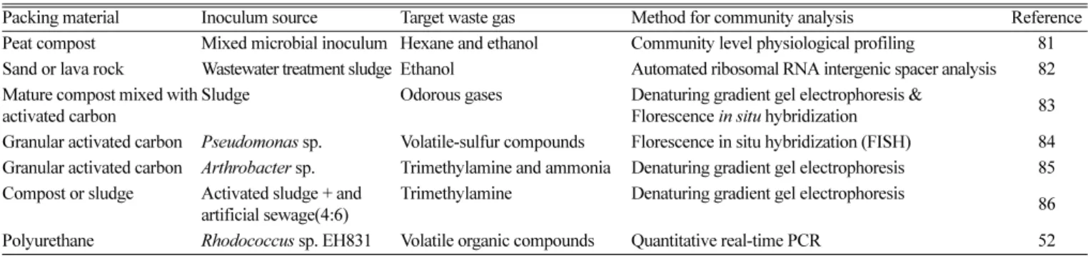
Table 5. Studies on microbial community analysis in biofilters
Packing material Inoculum source Target waste gas Method for community analysis Reference
Peat compost Mixed microbial inoculum Hexane and ethanol Community level physiological profiling 81 Sand or lava rock Wastewater treatment sludge Ethanol Automated ribosomal RNA intergenic spacer analysis 82 Mature compost mixed with
activated carbon
Sludge Odorous gases Denaturing gradient gel electrophoresis &
Florescence in situ hybridization 83 Granular activated carbon Pseudomonas sp. Volatile-sulfur compounds Florescence in situ hybridization (FISH) 84 Granular activated carbon Arthrobacter sp. Trimethylamine and ammonia Denaturing gradient gel electrophoresis 85 Compost or sludge Activated sludge + and
artificial sewage(4:6)
Trimethylamine Denaturing gradient gel electrophoresis
86 Polyurethane Rhodococcus sp. EH831 Volatile organic compounds Quantitative real-time PCR 52
Chung[83]은 compost로부터 발생하는 악취성 가스 제거 바이오필 터의 세균 군집 다양성을 DGGE와 FISH 방법을 사용하여 조사하였 다. 탈취 과정 중 바이오필터 내 세균 군집 구조 분석결과, DGGE profile에서는 최소 16개의 band가 보였고, Proteobacter 문(phylum) 이 가장 우점을 이루었으며, 그 뒤로 Actinobacteria, Bacteroidetes, 및 Firmicutes이었다. 우점종은 바이오필터 환경 상태에 따라 변하였 다. DGGE와 FISH 방법은 바이오필터 내 미생물 군집에서 세균의 기능 혹은 역할에 대한 유용한 정보를 제공함을 보였다.
Ho 등[84]은 Pseudomonas sp.가 접종된 입자상 활성탄을 담체로 활용한 바이오필터를 이용하여 돼지 폐수 처리장에서 발생하는 휘발 성 황화합물 제거 실험을 수행하였고, 담체에 존재하는 세균 군집구 조를 분석하고자 FISH 방법을 사용하였다. 실험결과 운행 31일 후 에 Pseudomonas sp.가 91.9%로 가장 많았고, 그 뒤로 Firmicutes 2.2%, α-Proteobacteria 1.4%, β-Proteobacteria 1.2%, γ-Proteobacteria 1.1%, 그리고 Actinobacteria 0.8% 순으로 나타났다. 비록 초기에 접종한 Pseudomonas sp.의 비율은 바이오필터 운행시간이 늘어날수록 감소하 였지만, 바이오필터 운행 415일의 장기간의 운행 후에도 Pseudomonas sp.가 약 56~70%로 우점한다고 보고하였다.
Ho 등[85]의 또 다른 연구에서는 Arthrobacter sp.가 접종된 granular activated carbon 바이오필터를 이용하여 trimethylamine(TMA)와 암 모니아(NH3)를 제거할 때 바이오필터 내의 미생물 군집 구조를 DGGE 기법을 이용하여 분석하였다. 분석결과 바이오필터의 세균 군 집은 복잡하지만 초기 접종균인 Arthrobacter sp.가 바이오필터 운행 전반에 걸쳐 50% 이상으로 우점하였고, 그 외 Paracoccus denitrificans 가 암모니아의 산화와 nitrite의 환원에 중요한 역할을 한다고 보고하 였다.
Ding 등[86]은 compost 혹은 sludge로 충진된 TMA 제거 바이오 필터의 운행과 세균군집 사이의 상관관계를 DGGE를 이용하여 조 사하였다. 실험결과 바이오필터내의 세균 군집 구조는 시간적 공간 적으로 다르게 변하였는데, 실험 초기(0일)에는 특정한 우점종 없이 α,β,γ-Proteobacteria, Actinobacteria, Bacteroides 그리고 low G+C 그람 양성균 등 다양한 세균이 존재하였다. 그러나 운전 14일 경과 후에는 α-Proteobacteria 인 Rhodococcus sp.와 Agrobacterium sp.가 우점하였지만, 운전 67일 경과 후에는 세균 군집이 더 복잡해졌고 α, β-Proteobacteria에 속하는 세균이 우점을 이룬 반면 나머지 세균은 급격히 감소한다고 보고하였다.
Lee 등[52]은 Rhodococcus sp. EH831이 접종된 폴리우레탄 바이 오필터에서 EH831 균주를 정량화하기 위해 quantitative real-time PCR(qRT-PCR) 방법을 이용하였다. 폴리우레탄 담체에 접종한 EH831 의 탈착률은 100%에 가까웠고, qRT-PCR을 이용한 실험 결과와 담 체에서 EH831을 탈착시켜 배양한 생균수의 수에는 큰 차이를 보이 지 않았다. 이 연구에서는 EH831에 특이성이 있는 TaqMan probe 개 발에 성공하였고, 재현성과 정확성이 높은 standard curve를 작성하 여 향후 바이오필터 내 존재하는 EH831 균주의 정량화에 적용할 수 있는 토대를 마련하였다.
6. 결 론
다양한 미생물 군집 분석방법을 활용하여, 바이오필터 운전 조건 에 따른 미생물 군집구조 변화의 모니터링, 바이오필터 성능과 미생 물 군집간의 상관성 정보를 도출, 생물학적인 인자와 무생물학적 인
자간의 상호관련성을 이해하는 것은 바이오필터 기술 혁신을 위해 매우 중요하다. 본 논문에서 기술한 바와 같이 각 미생물 군집 분석 방법별로 장단점이 있다. 생리학적 방법인 CLPP는 미생물 군집을 신속하고 간단히 분석할 수 있지만, 군집 안에 어떤 미생물이 존재 하는지 알 수 없기 때문에 미생물 구조 분석에는 한계가 있다. 또한, qRT PCR는 정량적으로 미생물을 분석할 수 있는 장점이 있지만, 미 생물이 담체에 부착하여 성장하는 biofilter의 특성상 담체로부터 미 생물을 정확하게 정량적으로 탈착하는데 어려움이 있어 그 적용에 한계가 있다. 현재까지 biofilter 군집 구조 분석에 많이 사용되고 있는 방법은 DGGE이다[83,86,86-90]. DGGE 분석 방법은 biofilter 내 미 생물 군집 차이를 구별할 수 있고, 염기서열 분석을 통해 어떠한 종 의 미생물이 존재하는지 알아 낼 수 있는 장점이 있어 비교적 적은 오차 범위 내에서 biofilter의 미생물 군집 특성을 조사할 수 있는 방 법으로 사료된다. 바이오필터의 미생물 군집 정보를 보다 정확하게 도출하기 위해서는 각 분석법의 장단점을 고려하여 2가지 이상의 분 석법을 동시에 적용하는 것이 바람직하다.
감 사
본 연구는 2008년도 정부(교육과학기술부)의 재원으로 한국과학 재단 지정 차세대바이오환경기술연구센터(AEBRC, R11-2003-006- 06001-0)와 국가지정연구실사업(No. R0A-2008-000-20044-0)의 지 원을 받아 수행된 연구이며 이에 감사드립니다.
참고문헌
1. Grommen, R. and Verstraete, W.,
“Environmental Biotechnol-
ogy: the Ongoing Quest,” J. Biotechnol., 98, 113-123(2002).2. Devinny, J. S., Deshusses, M. A. and Webster, T. S., “Biofiltra- tion for Air Pollution Control,” Florida, Lewis Publishers(1999).
3. Adib, F., Bagreev, A. and Bandosz, T. J., “Effect of pH and Sur- face Chemistry on the Mechanism of H2S Removal by Acti- vated Carbons,” J. Colloid Interface Sci., 216, 360-369(1999).
4. Barona, A., Elias, A., Arias, R., Cano, I. and Gonzalez, R., “Bio- filter Response to Gradual and Sudden Variations in Operating Conditions,” Biochem. Eng. J., 22, 25-31(2004).
5. Vergara-Fernandez, A., Molina, L. L., Pulido, N. A. and Aroca, G., “Effect of Gas Flow Rate, Inlet Concentration and Tempera- ture on the Biofiltration of Toluene Vapors,” J. Environ. Man- age., 84, 115-122(2007).
6. Yanzekontchou, C. and Gschwind, N., “Mineralization of the Her- bicide Atrazine as a Carbon Source by a Pseudomonas Strain,”
Appl. Environ. Microbiol., 60, 4297-4302(1994).
7. Mandelbaum, R. T., Allan, D. L. and Wackett, L. P., “Isolation and Characterization of a Pseudomonas sp. that Mineralizes the s-triazine Herbicide Atrazine,” Appl. Environ. Microbiol., 61, 1451-1457(1995).
8. Radosevich, M., Traina, S. J., Hao, Y. L. and Tuovinen, O. H.,
“Degradation and Mineralization of Atrazine by a Soil Bacterial Isolate,” Appl. Environ. Microbiol., 61, 297-302(1995).
9. Burgess, J. E., Parsons, S. A. and Stuetz, R. M., “Developments in Odour Control and Waste Gas Treatment Biotechnology: a Review,”
Biotechnol. Adv., 19, 35-63(2001).
10. van Groenestijn, J. W. and Hesselink, P. G. M., “Biotechniques
화학공학 제48권 제2호 2010년 4월
for Air Pollution Control,” Biodegradation, 1, 191-206(1993).
11. Wani, A. H., Branion, R. M. R. and Lau, A. K., “Biofiltration: A Promising and Cost-effective Control Technology for Odors, VOCs, and Air Toxics,” J. Environ. Sci. Heal., 32, 2027-2055 (1997).
12. Cho, K. S., Ryu, H. W. and Lee, N. Y., “Biological Deodoriza- tion of Hydrogen Sulfide Using Porous Lava as a Carrier of Thiobacillus thiooxidans,” J. Biosci. Bioeng., 90, 25-31(2000).
13. Cho, K. S., Hirai, M. and Shoda, M., “Enhanced Removal Effi- ciency of Malodorous Gases in a Pilot-scale Peat Biofilter Inoc- ulated with Thiobacillus thioparus DW44,” J. Ferment. Bioeng., 73, 46-50(1992).
14. Chung, Y. C. and Tseng, C. P., “Biotreatment of Ammonia from Air by an Immobilized Arthrobacter oxydans CH8 Biofilter,” Biotech- nol. Prog., 13, 794-798(1997).
15. Kim, N. J., Hirai, M. and Shoda, M., “Removal Characteristics of High Load Ammonia Gas by a Biofilter Seeded with a Marine Bacterium, Vibrio alginolyticus,” Biotechnol. Lett., 22, 1295-1299 (2000).
16. Lee, E. Y., Cho, K. S. and Ryu, H. W., “Degradation Character- ization of Sulfur-containing Malodorous Gases by Acidiothioba- cillus thiooxidans AZ11,” Kor. J. Odor Res. Eng., 2, 46-53(2003).
17. Lee, E. Y., Cho, K. S., Han, H. D. and Ryu, H. W., “Hydrogen Sulfide Effects on Ammonia Removal by a Biofilter Seeded with Earthworm Casts,” J. Environ. Qual., 31, 1782-1788(2002a).
18. Lee, E. Y., Cho, K. S. and Ryu, H. W., “Simultaneous Removal of H2S and NH3 in a Biofilter Inoculated with Acidithiobacillus thiooxidans TAS,” J. Biosci. Bioeng., 99, 611-615(2005).
19. Malhautier, L., Gracian, C., Roux, J. C., Fanlo, J. L. and Cloirec, P. L., “Biological Treatment Process of Air Loaded with an Ammonia and Hydrogen Sulfide Mixture,” Chemoshere, 50, 145- 153(2003).
20. Oyarzun, P., Arancibia, F., Canales, C. and Aroca, G. E., “Biofil- tration of High Concentration of Hydrogen Sulphide Using Thio- bacillus thioparus,” Process Biochem., 39, 165-170(2003).
21. Shinabe, K., Oketani, S., Ochi, T. and Matsumura, M., “Charac- teristics of Hydrogen Sulfide Removal by Thiobacillus thiooxi- dans KS1 Isolated from a Carrier-packed Biological Deodorization System,” J. Ferment. Bioeng., 80, 592-598(1995).
22. Tiwaree, R. S., Cho, K. S., Hirai, M. and Shoda, M., “Biological Deodorization of Dimethyl Sulfide Using Different Fabrics as the Carriers of Microorganisms,” Appl. Biochem. Biotechnol., 32, 135-148(1992).
23. Wani, A. H., Richard, M. R. B. and Anthony, K. L., “Effects of Periods of Starvation and Fluctuating Hydrogen Sulfide Concen- tration on Biofilter Dynamics and Performance,” J. Hazard. Mater., 60, 287-303(1998).
24. Yani, M., Hirai, M. and Shoda, M., “Removal Kinetics of Ammonia by the Biofilter Seeded with Night Soil Sludge,” J.
Ferment. Bioeng., 85, 502-506(1998).
25. Lee, E. Y., Lee, N. Y., Cho, K. S. and Ryu, H. W., “Removal of Hydrogen Sulfide by Sulfate-resistant Acidiothiobacillus thioox- idans AZ11,” J. Biosci. Bioeng., 100(2), 309-314(2006).
26. Delhomenie, M. C., Bibeau, L., Bredin, N., Roy, S., Broussau, S., Brzezinski, R., Kugelmass, J. K. and Heitz, M., “Biofilter- ation of Air Contaminated with Toluene on a Compost-based Bed,” Adv. Environ. Res., 6, 239-254(2002).
27. Jorio, H., Kiared, K., Brzezinski, R., Leroux, A., Viel, G. and Heitz, M.,”"Treatment of Air Polluted with High Concentrations of Toluene and Xylene in a Pilot-scale Biofilter,” J. Chem. Tech- nol. Biotechnol., 73, 183-196(1998).
28. Jorio, H., Bibeau, L. and Heitz, M., “Biofiltration of Air Con- taminated by Styrene: Effect of Nitrogen Supply, Gas Low Rate and Inlet Concentration,” Environ. Sci. Technol., 34, 1764-1771 (2000).
29. Kam, S. K., Kang, K. H. and Lee, M. G., “Removal Character- istics of Ethyl Acetate and 2-butanol by a Biofilter Packed with Jeju Scoria,” J. Microbiol. Biotechnol., 15, 977-983(2005).
30. Kim, J., Ryu, H. W., Jung, D. J., Lee, T. H. and Cho, K. S., “Sty- rene Degradation in a Polyurethane Biofilter Inoculated with Pseudomonas sp. IS-3,” J. Microbiol. Biotechnol., 15, 1207-1213 (2005).
31. Kwon, H. H., Lee, E. Y., Cho, K. S. and Ryu, H. W., “Benzene Biodegradation Using the Polyurethane Biofilter Immobilized with Stenotrophomonas maltophilia T3-c,” J. Microbiol. Biotechnol. 13, 70-76(2003).
32. Lee, E. Y., Jun, Y. S., Cho, K. S. and Ryu, H. W., “Degradation Characteristics of Toluene, Benzene, Ethylbenzene, and Xylene by Stenotrophomonas maltophilia T3-c,” J. Air Was. Manage.
Assoc., 52, 400-406(2002).
33. Lu, C., Lin, M. R. and Lin, W. C., “Removal of BTEX Vapor from Waste Gases by a Trickle Bed Biofilter,” J. Air. Was. Man- age. Assoc., 50, 411-417(2000).
34. Rene, E. R., Murthy, D. V. S. and Swaminathan, T., “Performance Evaluation of a Compost Biofilter Treating Toluene Vapours,” Pro- cess Biochem., 40, 2771-2779(2005).
35. Shim, E. H., Kim, J., Cho, K. S. and Ryu, H. W., “Biofiltration and Inhibitory Interactions of Gaseous Benzene, Toluene, Xylene and Methyl tert-butyl Ether,” Environ. Sci. Technol., 40, 3089- 3094(2006).
36. Singh, R. S., Agnihotri, S. S. and Upadhyay, S. N., “Removal of Toluene Vapour Using Agro-waste as Biofilter Media,” Biores.
Technol., 97, 2296-2301(2006).
37. Zilli, M., Del Borghi, A. and Converti, A., “Toluene Vapour Removal in a Laboratory-scale Biofilter,” Appl. Microbiol. Bio- technol., 54, 248-254(2005).
38. Ryu, H. W., Kim, S. J., Cho, K. S. and Lee, T. H., “Toluene Deg- radation in a Polyurethane Biofilter at High Loading,” Biotech- nol. Bioprocess Eng., 13(9), 360-365(2008).
39. Lee, E. H. and Cho, K. S., “Characterization of Cyclohexane and Hexane Degradation by Rhodococcus sp. EC1,” Chemosphere, 71, 1738-1744(2008).
40. Cho, K. S., Yoo, S. K. and Ryu, H. W., “Thermophilic Biofiltra- tion of Benzene and Toluene,” J. Microbiol. Biotechnol., 17(12), 1976-1982(2007).
41. Hirai M., Mamamoto, M., Yani, M., and Shoda M., “Compari- son of the Biological NH3 Removal Chracteristics Among four Inorganic Packing Materials,” J. Biosci. Bioeng., 91, 428-430(2001).
42. Maestre, J. P., Gamisans, X., Gabriel, D. and Lafuente, J., “Fun- gal Biofilters for Toluene Biofiltration: Evaluation of the pErfor- mance with four Packing Materials Under Different Operating Conditions,” Chemosphere, 67, 684-692(2007).
43. Lee, T. H., Kim, J., Kim, M. J., Ryu, H. W. and Cho, K. S., “Deg- radation Characteristics of Methyl Ethyl Ketone by Pseudomonas.
sp. KT-3 in Liquid Culture and Biofilter,” Chemosphere, 63(2), 315-322(2006).
44. van Lith, C., Leson, G. and Michelsen, R., “Evaluating Design Options for Biofilters,” J. Air Was. Manage. Assoc., 47, 37-48(1997).
45. Yang, Y. and Allen, E. R., “Biofiltration Control of Hydrogen Sulphide, 1: Design and Operational Parameters,” J. Air Waste Manag. Assoc., 44, 863-868(1994).
46. Delhomenie, M. C., Bibeua, L., Gendron, J., Brzezinski, R. and Heitz, M., “A Study of Clogging in a Biofilter Treating Toluene Vapors,” Chem. Eng. J., 94, 211-222(2003).
47. Iliuta, I. and Larachi, F., “Transient Biofilter Aerodynamics and Clogging for VOC Degradation,” Chem. Eng. Sci., 59, 3293-3302 (2004).
48. Kim, D. and Sorial, G. A., “Role of Biological Activity and Bio- mass Distribution in Air Biofilter Performance,” Chemosphere, 66, 1758-1764(2007).
49. Mendoza, J. A., Prado, O. J., Veiga, M. C. and Kennes, C.,
“Hydrodynamic Behavior and Comparison of Technologies for the Removal of Excess Biomass in Gas-phase Biofilters,” Wat.
Res., 38, 404-413(2004).
50. Weber, F. J. and Hart Mans, S., “Use of Activated Carbon as a Buffer in Biofiltration of Waste Gases with Fluctuating Concen- trations of Toluene,” Appl. Microbiol. Biotechnol., 43, 365-369 (1995).
51. Wright, W. F., “Transient Response of Vapor-phase Bio Filters,”
Chem. Eng. J., 113, 161-173(2005).
52. Lee, E. H., Cho, K. S. and Ryu, H. W., “Application of Quanti- tative Real-time PCR for Quantification of Rhodococcus sp.
EH831 in a Polyurethane Biofilter,” J. Environ. Biol., 30, 155- 159(2009).
53. Ranjard, L., Poly, F. and Nazaret, S., “Monitoring Complex Bac- terial Communities Using Culture-independent Molecular Tech- niques: Application to Soil Environment,” Res. Microbiol., 151, 167-177(2000).
54. Watanabe, K. and Hamamura, N., “Molecular and Physiological Approaches to Understanding the Ecology of Pollutant Degrada- tion,” Curr. Opin. Biotechnol., 14, 289-295(2003).
55. Leckie, S. E., “Methods of Microbial Community Profiling and Their Application to Forest Soils,” Forest Ecol. Manag., 220, 88- 106(2005).
56. Garland, J. L. and Mills, A. L., “Classification and Characteriza- tion of Heterotrophic Microbial Communities on the Basis of Patterns of Community-level Sole-carbon-source Utilization,” Appl.
Environ. Microbiol., 57, 2351-2359(1991).
57. Weber, K. P., Gehder, M. and Legge, R. L., “Assessment of Changes in the Microbial Community of Constructed Wetland Mesocosms in Response to Acid Mine Drainage Exposure,”
Water Res., 42, 180-188(2008).
58. Muhammad, A., Xu, J., Li, Z., Wang, H. and Yao, H., “Effects of Lead and Cadmium Nitrate on Biomass and Substrate Utiliza- tion Pattern of Soil Microbial Communities,” Chemosphere, 60, 508-514(2005).
59. Preston-Mafham, J., Boddy, L. and Randerson, P. F., “Analysis or Microbial Community Functional Diversity Using Sole-car- bon-source Utilisation Profilesóa Critique,” FEMS Microbiol.
Ecol., 42, 1-14(2002).
60. Bligh, E. G. and Dyer, W. J., “A Rapid Method of Total Lipid
Extraction and Purification,” J. Biochem. Physiol., 37, 911-917 (1959).
61. White, D. C., Stair, J. O. and Ringelberg, D. B., “Quantitative Comparison of in situ Microbial Biodiversity by Signature Biomarker Analysis,” J. Ind. Microbiol., 17, 185-196(1996).
62. Hugenholtz, P., Goebel, B. M. and Pace, N. R., “Impact of Cul- ture-independent Studies on the Emerging Phylogenetic View of Bacterial Diversity,” J. Bacteriol., 180, 4765-4774(1998).
63. Smit, E., Leeflang, P. and Wernars, K., “Detection of Shifts in Microbial Community Structure and Diversity in Soil Caused by Copper Contamination Using Amplified Ribosomal DNA Restriction Analysis,” FEMS Microbiol. Ecol., 23, 249-261(1997).
64. Ranjard, L., Poly, F., Richaume, A., Gourbiere, F. and Nazaret, S., “Bacterial Community Structure Assess by two DNA Finger- print at Microscale Level in Soil,” in Proceeding of the Interna- tional Congress NATO ASI Molecular Advances in Molecular Ecology, Erice, Italy(1998).
65. Ranjard, L., Nazaretm, S., Gourbiere, F., Thioulouse, J., Linet, P.
and Richaume, A., “A Soil Microscale Study to Reveal the Het- erogeneity of Hg(II) Impact on Indigenous Bacteria by Quantifi- cation of Adapted Phenotypes and Analysis of Community DNA Fingerprints,” FEMS Microbiol. Ecol., 31, 107-115(2000).
66. Wintzingerode, F., Göbel, U. B. and Stackebrandt, E., “Determi- nation of Microbial Diversity in Environmental Samples: Pitfalls of PCR-based rRNA Analysis,” FEMS Microbiol. Rev., 21, 213- 229(1997).
67. Vallaeys, T., Topp, E. and Muyzer, G., “Evaluation of Denatur- ing Gradient Gel Electrophoresis in the Detection of 16S rDNA Sequence Variation in Rhizobia and Methanotrophs,” FEMS Micro- biol. Ecol., 24, 279-285(1997).
68. Felske, A. and Akkermans, A. D. L., “Spatialhomogeneity of Abundant Bacterial 16S rRNA Molecules in Grassland Soils,”
Microbial Ecol., 36, 31-36(1998).
69. Muyzer, G., De Waal, E. C. and Uitterlinden, A. G., “Profiling of Complex Microbial Populations by Denaturing Gradient Gel Electrophoresis Analysis of Polymerase Chain Reaction-ampli- fied Genes Coding for 16S rRNA,” Appl. Environ. Microbiol., 59, 695-700(1993).
70. Borneman, J. and Triplett, E. W., “Molecular Microbial Diver- sity in Soil from Eastern Amazonia: Evidence for Unusual Microorganisms and Microbial Population Shifts Associated with Deforestation,” Appl. Environ. Microbiol., 63, 2647-2653(1997).
71. Hadrys, H., Balick, M. and Schierwater, B., “Applications of Random Amplified Polymorphic DNA(RAPD) in Molecular Ecology,” Mol. Ecol., 1, 55-63(1992).
72. Wikström, P., Andersson, A. C. and Forsman, M., “Biomonitor- ing Complex Microbial Communities Using Random Amplified Polymorphic DNA and Principal Component Analysis,” FEMS Microbiol. Ecol., 28, 131-139(1999).
73. Lee, S. and Furhman, J. A., “DNA Hybridization to Compare Species Composition of Natural Bacterioplankton Assemblages,”
Appl. Environ. Microbiol., 56, 739-746(1990).
74. Xia, X., Bollinger, J. and Ogram, A., “Molecular Genetic Anal- ysis of the Response of Three Soil Microbial Communities to the Application of 2, 4-D,” Mol. Ecol., 4, 17-28(1995).
75. Torsvik, V., Gosk
øyr, J. and Daae, F. L., “High Diversity in
DNA of Soil Bacteria,” Appl. Environ. Microbiol., 56, 782-787화학공학 제48권 제2호 2010년 4월 (1990).
76. Holben, W. E. and Harris, D., “DNA-based Monitoring of Total Bacterial Community Structure in Environmental Samples,”
Mol. Ecol., 4, 627-631(1995).
77. Rotthauwe, J. H., Witzel, K. P. and Liesack, W., “The Ammonia Monooxygenase Structural Gene amoA as a Funtional Marker:
Molecular Fine-scale Analysis of Natural Ammonia-oxidizing Populations,” Appl. Environ. Microbiol., 63, 4707-4712(1997).
78. Auman, A. J. and Lidstrom, M. E., “Analysis of sMMO-contain- ing type I Methanotrophs in Lake Washington Sediment,” Envi- ron. Microbiol., 4, 517-524(2002).
79. Smits, T. H. M., Röthlisberger, M., Witholt, B. and van Beilen, J.
B., “Molecular Screening for Alkane Hydroxylase Genes in Gram-negative and Gram-positive Strains,” Environ. Microbiol., 1, 307-317(1999).
80. Beller, H. R., Kane, S. R., Legler, T. C. and Alvarez, P. J., “A Real-time Polymerase Chain Reaction Method for Monitoring Anaerobic, Hydrocarbon-degrading Bacteria Based on a Cata- bolic Gene,” Environ. Sci. Technol., 36, 3977-3984(2002).
81. Grove, J. A., Kautola, H., Javadpour, S., Moo-Young, M. and Anderson, W. A., “Assessment of Changes in the Microorgan- ism Community in a Biofilter,” Biochem. Eng. J., 18, 111-114(2004).
82. Steele, J. A., Ozis, F., Fuhrmana, J. A. and Devinny, J. S., “Struc- ture of Microbial Communities in Ethanol Biofilters,” Chem.
Eng. J., 113, 135-143(2005).
83. Chung, Y. C., “Evaluation of Gas Removal and Bacterial Com- munity Diversity in a Biofilter Developed to Treat Composting
Exhaust Gases,” J. Hazard. Mater., 144, 377-385(2007).
84. Ho, K. L., Chung, Y. C., Lin, Y. H. and Tseng, C. P., “Microbial Populations Analysis and Field Application of Biofilter for the Removal of Volatile-sulfur Compounds from Swds Wastewater Treatment System,” J. Hazard. Mater., 152, 580-588(2008a).
85. Ho, K. L., Chung, Y. C. and Tseng, C. P., “Continuous Deodor- ization and Bacterial Community Analysis of a Biofilter Treat- ing Nitrogen-containing Gases from Swine Waste Storage Pits,”
Bioresour. Technol., 99, 2757-2765(2008b).
86. Ding, Y., Wu, W., Han, Z. and Chen, Y., “Correlation of Reactor Performance and Bacterial Community Composition During the Removal of Trimethylamine in Three-stage Biofilters,” Biochem.
Eng. J., 38, 248-258(2008).
87. Jun, Y. and Wenfeng, X., “Ammonia Biofiltration and Community Analysis of Ammonia-oxidizing Bacteria in Biofilters,” Bioresour.
Technol., 100, 3869-3876(2009).
88. O-Thong, S., Prasertsan, P. and Birkeland, N. K., “Evaluation of Methods for Preparing Hydrogen-producing Seed Inocula Under Thermophilic Condition by Process Performance and Microbial Community Analysis,” Bioresour. Technol., 100, 909-918(2009).
89. Cai, Z., Sorial, G. A., Zhang, K., Saikaly, P., Zein, M. M. and Oer- ther, D. B., “Effect of Changing VOC Influent Composition on the Microbial Community Structure of TBABs,” Water Air Soil Poll., 8, 311-321(2008).
90. Ait-Benichou, S., Jugnia, L. B., Greer, C. W. and Cabral, A. R.,
“Methanotrophs and Methanotrophic Activity in Engineered Land- fill Biocovers,” Waste Manage., 29, 2509-2517(2009).
![Table 1. Summary of commonly used microbial community profiling methods[55]](https://thumb-ap.123doks.com/thumbv2/123dokinfo/5407049.219878/3.892.67.432.678.866/table-summary-commonly-used-microbial-community-profiling-methods.webp)
![Table 3. Phospholipid fatty acids commonly used as biomarkers for specific groups of microorganisms[55]](https://thumb-ap.123doks.com/thumbv2/123dokinfo/5407049.219878/4.892.67.427.152.460/table-phospholipid-fatty-commonly-biomarkers-specific-groups-microorganisms.webp)